Predisposing factors to severe external root resorption
associated to orthodontic treatment
Gracemia Vasconcelos Picanço1, Karina Maria Salvatore de Freitas2, Rodrigo Hermont Cançado3, Fabricio Pinelli Valarelli4, Paulo Roberto Barroso Picanço5, Camila Pontes Feijão6
How to cite this article: Picanço GV, Freitas KMS, Cançado RH, Valarelli FP, Picanço PRB, Feijão CP. Predisposing factors to severe external root resorption associated to orthodontic treatment. Dental Press J Orthod. 2013 Jan-Feb; 18(1):110-20.
Submitted: June 04, 2010 - Revised and accepted: May 03, 2011
Contact address: Karina Maria Salvatore de Freitas Rua Jamil Gebara, 1-25 – Apto 111 – Brazil
CEP: 17017-150 – Bauru/SP – E-mail: [email protected]
1 MSc in Orthodontics, UNINGA. 2 Post-Doc in Orthodontics, UNINGÁ. 3 PhD in Orthodontics, FOB-USP. 4 PhD in Orthodontics, UNINGÁ. 5 MSc in Orthodontics, UNINGÁ. 6 Specialist in Orthodontics, UVA-CE.
» The authors report no commercial, proprietary or inancial interest in the prod-ucts or companies described in this article.
Objective:The aim of this study was to evaluate predisposing factors among patients who developed moderate or severe
exter-nal root resorption (Malmgren’s grades 3 and 4), on the maxillary incisors, during ixed orthodontic treatment in the perma-nent dentition. Methods: Ninety-nine patients who underwent orthodontic treatment with ixed edgewise appliances were
selected. Patients were divided into two groups: G1 – 50 patients with no root resorption or presenting only apical irregularities (Malmgren’s grades 0 and 1) at the end of the treatment, with mean initial age of 16.79 years and mean treatment time of 3.21 years; G2 – 49 patients presenting moderate or severe root resorption (Malmgren’s grades 3 and 4) at the end of treatment on the maxillary incisors, with mean initial age of 19.92 years and mean treatment time of 3.98 years. Periapical radiographs and lateral cephalograms were evaluated. Factors that could inluence the occurrence of severe root resorption were also recorded. Statistical analysis included chi-square tests, Fisher’s exact test and independent t tests. Results: The results demonstrated
sig-niicant diference between the groups for the variables: Extractions, initial degree of root resorption, root length and crown/ root ratio at the beginning, and cortical thickness of the alveolar bone. Conclusion: It can be concluded that: Presence of root
resorption before the beginning of treatment, extractions, reduced root length, decreased crown/root ratio and thin alveolar bone represent risk factors for severe root resorption in maxillary incisors during orthodontic treatment.
Keywords: Root resorption. Tooth movement. Orthodontics.
Objetivo:avaliar os fatores comuns aos pacientes que desenvolveram reabsorção radicular externa moderada ou severa
(graus 3 e 4 de Malmgren) nos incisivos superiores, durante o tratamento ortodôntico ixo na dentição permanente. Mé-todos: foram selecionados 99 pacientes que iniciaram o tratamento ortodôntico ixo com a técnica Edgewise. Os pacientes
foram divididos em dois grupos: G1 – 50 pacientes que concluíram o tratamento com ausência de reabsorções radiculares ou apresentando apenas irregularidades apicais (graus 0 e 1 de Malmgren), com idade média inicial de 16,79 anos e tempo de tratamento médio de 3,21 anos; G2 – 49 pacientes que inalizaram o tratamento apresentando reabsorção radicular modera-da ou severa (graus 3 e 4 de Malmgren) nos incisivos superiores, com imodera-dade média inicial de 19,92 anos e tempo de tratamen-to médio de 3,98 anos. As radiograias periapicais e telerradiograias foram avaliadas, além de diversos fatratamen-tores que pudessem inluenciar a ocorrência de uma reabsorção severa. A análise estatística incluiu o testes qui-quadrado, teste exato de Fisher e teste t independente. Resultados: foi demonstrada a presença de diferença signiicativa entre os grupos para as variáveis
realização de extrações, grau de reabsorção radicular inicial, comprimento radicular, proporção coroa/raiz e da espessura da cortical óssea alveolar. Conclusão: pode-se concluir que são fatores de risco para reabsorção radicular severa nos incisivos
superiores, durante o tratamento ortodôntico, a presença de reabsorção radicular antes do início do tratamento, a realização de extrações, o comprimento radicular reduzido, a proporção coroa/raiz diminuída e a espessura óssea alveolar ina.
INTRODUCTION
The external root resorption (ERR) is frequently observed by orthodontists and are usually diagnosed in clinical practice when radiographs (panoramic or peri-apical) are performed. ERR are usually asymptomatic, and when the loss of root structure by resorption be-come severe the physiology and retention of the afected
teeth may be compromised.14
The anterior teeth are more afected by root resorp-tion, probably because they are single-rooted with ta-pered roots, conducting the orthodontic force directly to the apex. They are also constantly moved during orthodontic treatment. Moreover, they are more ex-posed to external factors, such as trauma, making these teeth a good reference for the magnitude of root
resorp-tion during orthodontic treatments.22
The literature about root resorption is extensive but very controversial in relation to the factors that actually inluence the occurrence and severity of resorption dur-ing orthodontic treatment. In this context, this study was conducted in order to evaluate which factors are commonly observed in patients who develop moderate or severe degrees of resorption in maxillary incisors dur-ing orthodontic treatment.
MATERIAL AND METHODS
Material
The sample consisted of 99 patients from the Pau-lo Picanço Advanced Orthodontics Center, Fortale-za-CE, who underwent orthodontic treatment with fixed appliance (Edgewise technique) in the perma-nent dentition, and met the following requirements: Good oral and systemic health, no tooth loss in the region from canine to canine in the maxillary arch, absence of vertical bone loss and periodontal disease, and no prosthesis. Patients who developed grade 2
resorption26 at the end of treatment was excluded
from the study. Only patients who had complete treatment records, medical records, initial cephalo-gram and initial and final periapical radiographs of maxillary incisors in good conditions were included in the sample.
Teeth with endodontic treatment, incomplete devel-opment of the root apex, tooth agenesis, supernumerary teeth and patients with incomplete initial records were excluded from the sample. Radiographs with distortion or blur were also eliminated.
Sample division:
» Group 1 (Patients who had degree 0 or 1 of root resorption of the maxillary incisors at the end of treatment): 50 patients, 26 without and 24 with extractions (13 first premolars; 8 second premolars and 3 first molars). The mean age at the beginning of the treatment was 16.79 years, the mean age at the end of treatment was 20.00 years. The mean treat-ment time was 3.21 years.
» Group 2 (patients who had degree 3 or 4 of root resorption in maxillary incisors at the end of treatment): 49 patients, 26 without and 24 with extractions (31 first premolars, 9 second premolars, 2 first molars) The mean age at the beginning of the treatment was 19.92 years and the mean age at the end of treatment was 23.90 years. The mean treatment time was 3.98 years.
Methods
In order to evaluate the variables, initial and final periapical radiographs and initial cephalograms were used of each patient. The radiographs were scanned with a scanner (Microtek ScanMaker i800, Micro-tek International, Inc., Carson, USA) and coupled to a Pentium computer. The images were trans-ferred to the software Dolphin Imaging Premium 10.5 (Dolphin Imaging & Management Solutions, Chatsworth, USA) through which the images were digitized, the points were marked and cephalomet-ric measurements were performed automatically by the software Dolphin.
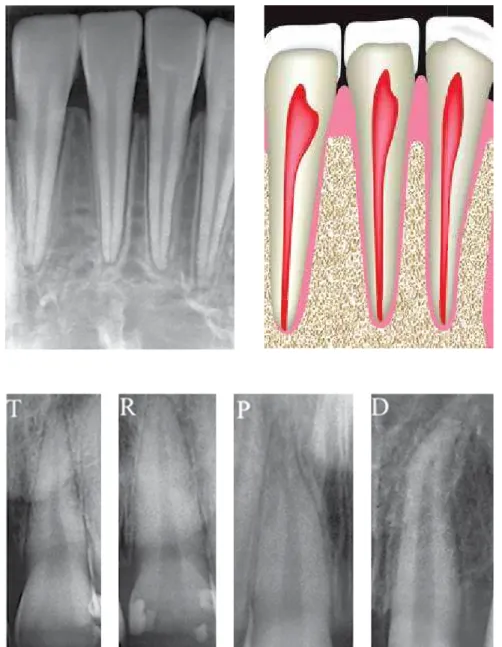
The periapical radiographs were analyzed as fol-lows: The degree of initial and final, based on the classification proposed by Malmgren: Grade 0 (no re-sorption), grade 1 (presence of apical irregularities), grade 2 (presence of resorption by 2 mm), grade 3 (presence of resorption between 2 mm and a third of the original length), grade 4 (the presence of root re-sorption greater than one third of the original length
of the root) (Fig 1).7,8
The evaluation of the root shape was performed
based on the classii cation proposed by Consolaro9 as
follows: Triangular (T), rhomboid (R), pipette (P) and dilacerated (D) (Fig 3).
The root length was obtained by measuring the dis-tance from the apex to the cementoenamel junction, following the long axis of the incisor (Fig 4). This mea-surement was performed in both initial and i nal peri-apical radiographs.
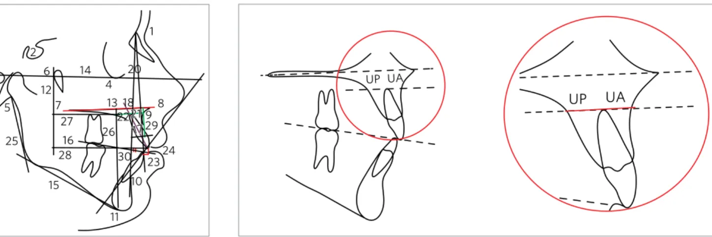
Cephalometric variables used are shown in Figures 5, 6 and 7, and described in Table 1.
Method error
For the evaluation of the intraexaminer error, the measurements were performed on 20 patients ran-domly selected, after a month interval. It was
ap-plied the dependent t test to obtain the systematic
Figure 1 - Malmgren7,8 classii cation.
Figure 2 - Alveolar crest bone morphology.
Figure 4 - Evaluation of the crown/root pro-portion.
Figure 5 - Cephalometric variables used. Figure 6 - UA+UP distance, parallel to the palatal plane (ANS-PNS).
Example of Grade 4 Crown: 11 mm
Root: 10 mm 11 --- 100% 10 --- X X = 0.9
Proportion = 1:0.9
Root/crown proportion
error and Dahlberg11 formula to estimate the
ran-dom error. To evaluate the error of the score of root resorption, the Kappa test was used.
Statistical analysis
It was used the following statistical tests: Chi-square test for intergroup comparison of gender, type
of malocclusion, treatment with or without extrac-tion, shape of the root and bone crest; independent
t test for comparison of the ages, treatment time and
cephalometric variables between the two groups. All tests were performed with Statistica software (Statis-tica for Windows, versão 7.0, Statsoft). Results were considered significant when p < 0.05.
UP
UP
UA
UA
2 3
5
25
15
11 10
23 24 29 9
8
17 21 22
18 13
30 28
26 16 27 7 12
6 14 4
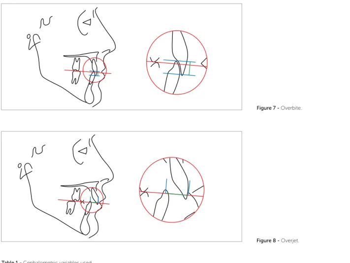
Figure 7 - Overbite.
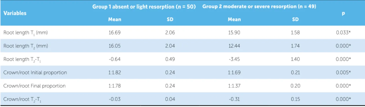
Figure 8 -Overjet.
RESULTS
The largest linear random error found, was for the variable 1-PTV apical (1.36 mm), and angular for the measure 1.NA (1.41°) and the largest systematic error was 0.12 mm of the variable Overjet. The percentage of agreement was substantial (87%) and Kappa coef-icient was 0.794.
Increased age and longer treatment was signiicantly re-lated to the occurrence of severe root resorption (Table 2). Gender, type of malocclusion, morphology of the
root and the bone crest are not risk factors for developing severe root resorption (Table 3). The treatment protocol with extractions increases the risk of severe apical root resorption (Table 3).
The short root length and the decreased proportion of the crown/root ratio at the beginning of treatment increas-es the chance of developing severe rincreas-esorption (Table 4).
Patients with thin maxillary alveolar cortical bone are more likely to develop severe resorption than pa-tients with good bone thickness (Table 5).
1-PTV INC (mm) Distance of the incisal of the maxillary central incisor to the PTV line (line vertical to the pterygoid fossa, perpendicular to Frankfurt). 1-PTV APIC (mm) Distance of the apical root of the maxillary central incisor to the PTV line (line vertical to the pterygoid fossa, perpendicular to Frankfurt). 1.NA (degrees) Angle between the long axis of the maxillary central incisor and the line NA.
FMA (degrees) Angle formed by the horizontal planes of Frankfurt and mandibular (GoMe)
PFH/AFH (mm) Proportion between the posterior face height (S-Go) and the anterior face height (N-Me). ANB (degrees) Angle formed by the lines NA and NB.
Wits (mm) Distance between the points A and B projected perpendicularly to the functional occlusal plane.
Overjet (mm) Distance between the incisal edges of the maxillary and mandibular central incisors projected perpendicularly to the occlusal plane. Overbite (mm) Distance between the incisal edges of the maxillary and mandibular central incisors measured perpendicularly to the occlusal plane. H-11 (mm) Total length of the maxillary central incisor, including crown and root.
UA+UP (mm) Sum of the thickness of the anterior (buccal) and posterior (palatal) alveolar crest bone.
Table 2 - Intergroup comparison of the ages and treatment time (independent t test).
* Statistically signiicant p < 0.05.
Variables
Group 1 absent or light resorption (n = 50) Group 2 moderate or severe resorption (n = 49)
p
Mean SD Mean SD
Initial age (years) 16.79 5.47 19.92 6.94 0.014*
Final age (years) 20.00 5.46 23.90 7.05 0.002*
Treatment time (years) 3.21 0.84 3.98 1.01 0.000*
Table 3 - Intergroup comparison of the gender, type of malocclusion, root morphology, alveolar crest and presence of initial resorption (chi-square).
* Statistically signiicant p < 0.05.
Variables Group 1 absent or light
resorption (n = 50)
Group 2 moderate or
severe resorption (n = 49) λ DF p
Gender
Male 18.18% 13.13%
1.03 1 0.309
Female 32.32% 36.36%
Malocclusion
Class I 8.08% 12.12%
3.45 2 0.179
Class II 29.29% 31.31%
Class III 13.13% 6.06%
Type of treatment
Without extraction 26.26% 9.09%
2.24 1 0.000*
With extraction 24.24% 40.40%
Root morphology
Triangular 4.04% 4.04%
0.97 3 0.806
Rhomboid 34.34% 29.29%
Pipette 10.10% 13.13%
Dilacerated 2.02% 3.03%
Alveolar crest
Flat 29.29% 35.35%
1.95 1 0.162
Sharp 21.21% 14.14%
Initial degree of resorption
0 48.48% 30.30%
17.91 1 0.000*
1 2,02% 19,19%
* Statistically signiicant p < 0.05.
Table 4 - Intergroup comparison of the root length and the crown/root proportion in T1, T2 and T2-T1 (independent t test).
Variables
Group 1 absent or light resorption (n = 50) Group 2 moderate or severe resorption (n = 49)
p
Mean SD Mean SD
Root length T1 (mm) 16.69 2.06 15.90 1.58 0.033*
Root length T2 (mm) 16.05 2.04 12.44 1.74 0.000*
Root length T2-T1 -0.64 0.49 -3.45 1.40 0.000*
Crown/root Initial proportion 1:1.82 0.24 1:1.69 0.21 0.005*
Crown/root Final proportion 1:1.78 0.24 1:1.37 0.20 0.000*
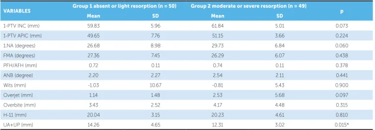
Table 5 - Intergroup comparison of the cephalometric variables (independent t test).
VARIABLES Group 1 absent or light resorption (n = 50) Group 2 moderate or severe resorption (n = 49) p
Mean SD Mean SD
1-PTV INC (mm) 59.83 5.96 61.84 5.01 0.073
1-PTV APIC (mm) 49.65 7.76 51.15 3.66 0.224
1.NA (degrees) 26.68 8.98 29.73 6.84 0.060
FMA (degrees) 27.36 7.45 26.29 6.07 0.438
PFH/AFH (mm) 0.72 0.11 0.74 0.11 0.378
ANB (degree) 2.20 2.27 2.54 2.11 0.441
Wits (mm) -1.03 10.67 -0.81 5.43 0.900
Overjet (mm) 1.14 1.48 2.53 5.68 0.097
Overbite (mm) 3.43 2.52 4.17 4.48 0.315
H-11 (mm) 20.04 3.15 20.23 4.61 0.810
UA+UP (mm) 14.26 4.65 12.31 3.02 0.015*
DISCUSSION
Sample
The sample of this study was obtained from Paulo Picanço Orthodontics Center. Initially, periapical radio-graphs were examined to obtain the degree of resorption and then the patients were divided into two groups. At this time, it was excluded patients with absences in the region from canine to canine in the maxilla, presence of vertical bone loss, patients with resorption grade 2, pa-tients with incomplete, damaged or without inal orth-odontic records. Ater the selection, 165 patients from all subjects had completed iles available. Ater checking the periapical radiographs, the evaluation of the orth-odontic records was made, noting that many patients had incomplete records, absence of inal orthodontic records, lack of signed informed consent, incomplete permanent dentition. At the end, a inal sample of 99 patients attended the inclusion criteria.
Although the control group (group 1, with resorp-tion grade 0 or 1 at the end of treatment) have more patients, this was the most diicult to collect, especially patients with grade 0 who fulilled all the requirements of research, since the literature airms that the
resorp-tion in orthodontically treated patients is 100%.3,4,19,22,36
To collect the ideal sample was the greatest challenge of this study, however, the number of subjects was con-sidered good, because of the strict selection criteria.
Method
In this study only the maxillary incisors were eval-uated, because previous studies showed that these teeth have more susceptibility to develop resorption during orthodontic treatment. Incisors are the more constantly moved, for example, during retraction and
intrusion.8,12,22 Moreover, incisors are single-rooted
el-ements and easier for obtaining images without distor-tion or image overlays.15,18,23,33
The most common radiographic examination used for the detection of resorption is periapical
x-ray. For Sameshima and Asgarifar33 this type of
ra-diograph shows finer detail, allowing visualization of anatomical details such as the cementoenamel junc-tion and have less distorjunc-tion and overlap when
com-pared to panoramic and cephalometric radiographs.26
In this study the standardized technique for periapi-cal radiographs used was the parallelism, and the fact that all patients in the study underwent radiographic follow-up at the Paulo Picanço Orthodontics Cen-ter, ensuring greater standardization of radiographs. The choice for this technique is the fact that it al-lows greater standardization of the image for pre- and post-treatment, what is not possible by the bisection technique, since the average angle of incidence of x-rays is more difficult to reproduce even hindering
over the accuracy of quantitative measurements.32
Age
The results were signiicant in relation to age, because the group 1 presented younger patients when compared to group 2. It can be airmed that older patients have a higher risk of developing moderate or severe root re-sorption during orthodontic treatment (Table 2).
Some authors mentioned that age does not
inlu-ence root resorption.1,9,19 However, corroborating the
results of this study, Sameshima and Sinclair34,35
ob-served that the resorption is most prevalent in adults than in children.
Adults seem more susceptible to resorption be-cause, with aging, the periodontal membrane becomes less vascularized, inelastic, more narrow and the ce-mentum becomes thicker, and also the fact that the apical third of the root is more irmly anchored in adult patients, creating a diiculty in tooth movement and
predisposing to resorption.3,4,6
However, the results of this study must be inter-preted with caution, since the age diference between the groups 1 and 2 was only about three years, and both patients in group 1 and group 2 were considered “young adults” (Table 2).
Treatment time
The results showed that a longer treatment is a risk factor to the occurrence of severe root resorption, as the group 1 showed a signiicantly shorter treatment time compared to group 2 (Table 2).
These results are opposite to those authors who sug-gest no relation between the duration of treatment and
the degree of resorption.2,13,37
According to Sameshima and Sinclair,34,35 the
dura-tion of treatment and the amount of horizontal displace-ment of the apical root of the maxillary incisors had strong correlation with root resorption.
Brin et al5 evaluated the root resorption in patients
with Class II malocclusion treated in only one phase or with treatment divided into two phases, observed that patients undergoing a single phase of treatment had a proportion of moderate to severe resorption slightly larger than the group with two phases of treatment.
Gender
In this study no signiicant relationships were found between severe resorption and the gender (Table 3), agreeing with the results of most authors.9,19,20,31,34,35,36
Type of malocclusion
Regarding the type of malocclusion and orthodontic techniques, many studies have shown that there is no relationship between root resorption and type of
mal-occlusion.6,19,30 The results of this study also found no
signiicant relationship (Table 3).
Type of treatment (with or without extraction) The results of this study showed that patients treated with extraction were more likely to develop severe root resorption than patients whose treatment did not in-clude extraction (Table 3).
Many studies showed that patients treated with ex-tractions showed more resorption and with more severe degree, because mechanical retraction of anterior teeth cause greater movement of the root apex and the need for longer treatment.12,16,29,34,35
Root and bone crest morphology
The results of this study showed no relationship be-tween the morphology of the root and the bone crest to the occurrence of severe resorption during orth-odontic treatment (Table 3).
Most authors consider, regarding the root morphol-ogy, that teeth with atypical root have a higher risk of root resorption.25,29,30,34,35
It is likely that this result is due to the fact that most of the sample presented rhomboid root and tapered bone crest, which according to most authors, reduce the risk of resorption.
Degree of initial resorption
The results have shown that patients with some de-gree of root resorption at the beginning of treatment have a greater predisposition and an increased risk of developing severe root resorption during orthodontic treatment (Table 3).
Some authors believe that patients with minimal or no resorption present little risk to severe resorption, patients with moderate resorption have regular risk to severe and extreme resorption, while patients starting orthodontic treatment with severe resorption has a high
risk of extreme resorption at the end of treatment.6,36
Root length and crown/root proportion
resorption compared to the group with mild resorp-tion (Table 4), as expected, because of the criteri-on for group divisicriteri-on. However, the root length of group 2 was already smaller than the group 1 before the beginning of treatment (Table 4). This can be considered an indication that a smaller root length at the beginning of treatment is a risk factor for in-creased occurrence of root resorption during orth-odontic treatment. Likewise, the crown/root pro-portion at the beginning of treatment was lower in group 2 than in group 1, and continued lower at the end of treatment (Table 4). Furthermore, the crown/ root proportion suffered a greater decrease in group 2 (Table 4), as expected.
The root length and crown/root proportion seem to inluence the tendency to resorption. A large crown will tend to concentrate force on certain focal points, thus short roots tend to sufer more resorption during orthodontic movements.
In cases of patients with severe resorption the pro-fessional should be very careful with the root length, teeth with roots length less than or equal to 9 mm
have a higher risk of teeth mobility.27 According
to Kalkwarf et al,24 the reduction of 3 mm in
api-cal tooth structure corresponds to the loss of alveolar bone crest of 1 mm.
Incisor position
Variables related to the position of the maxillary in-cisor showed no statistically signiicant diference be-tween the groups, indicating no relationship among the protrusion and buccal tipping of the incisors and the oc-currence of severe root resorption (Table 5).
The results showed no statistically signiicant difer-ence between the groups for the protrusion and initial inclination of the incisors, indicating that they are not risk factors to the occurrence of severe root resorption, although group 2 showed a greater buccal inclination of the incisors at the beginning of treatment, but not statis-tically signiicant (Table 5).
FMA and PFH/AFH
The FMA angle showed no statistically signifi-cant difference between the groups, indicating no relationship between the vertical pattern of the patients and the occurrence of severe root resorp-tion (Table 5). The proporresorp-tion PFH/AFH showed
no statistically significant difference between the groups, indicating no relationship between the fa-cial pattern of the patients and the occurrence of severe root resorption (Table 5).
Handelman17 analyzed the variable SN-MP that,
represents the relationship between the cranial base and the mandibular plane and found positive associa-tion between this variable and root resorpassocia-tion.
Harris, Kineret and Tolley19 also conducted a
study to evaluate the relationship between FMA and root resorption and found a relatively high correlation between them.
ANB and WITS
The results of this study demonstrate that the max-illomandibular relationship was not signiicantly dif-ferent between the groups, indicating that this variable is not a risk factor for the occurrence of severe root resorption (Table 5). It is likely that this result is due to the fact that the present sample does not show large maxillomandibular discrepancies.
In a study by Harris, Kineret and Tolley19, these
two variables (ANB and Wits) were evaluated and it was observed that both have strong relationship with the occurrence of resorption, as higher maxilloman-dibular discrepancies tend to require greater retrac-tion of anterior teeth and therefore enhance the risk of resorption.
Overjet and Overbite
The results of this study showed no statistically signiicant diference between the groups for overjet and overbite, indicating that, at the beginning of treat-ment, these variables are not a risk factor to the occur-rence of severe root resorption (Table 5). These ind-ings, however, contradict most of the authors and the justiication for this is the absence of great skeletal dis-crepancies and also the absence of a signiicant number of cases with open bite and/or deep bite.
There is a consensus in considering the overjet as a risk factor for resorption, because the correc-tion requires the retraccorrec-tion of anterior teeth, and the greater the magnitude of this malocclusion, the greater the amount of movement, increasing the risk
and severity of resorption.2,5,28,31 Freitas et al12
H-11
The results showed no signiicant diference be-tween the groups for this variable, indicating that the position of the incisors in relation to the palatal plane at the beginning of treatment is not a risk factor for the occurrence of severe root resorption.
Some studies show that the intrusion with lingual torque movement, horizontal movement of the apex
increase the chance of root resorption.1,10,30,31 For
Park-er and Harris,31 extrusion movements are also risk
fac-tors, but less aggressive than the intrusion. For Freitas
et al,12 anteroposterior movement, is a risk factor found
in 29% of the resorptions observed in patients during orthodontic treatment.
Bone thickness
The bone thickness (UA + UP) showed a statisti-cally significant result (Table 5) confirming the au-thors hypothesis about the increased risk of
resorp-tion in patients with thin cortical.10,17
According to Handelman,17 the dimension of the
alveolus (UA + UP) seems to set limits to orthodon-tic treatment and challenge these limits can accelerate iatrogenic fenestrations and root resorption. Horiuchi,
Hotokezaka and Kobayashi21 observed that the
prox-imity of the apex to the palatal cortex also inluences the resorption. To these authors, the maxillary width insuicient to tooth movement can be considered a risk associated to root resorption.
CONCLUSIONS
1. Baumrind S, Korn EL, Boyd RL. Apical root resorption in orthodontically treated adults. Am J Orthod Dentofacial Orthop. 1996;110(3):311-20. 2. Beck BW, Harris EF. Apical root resorption in orthodontically treated subjects:
analysis of edgewise and light wire mechanics. Am J Orthod Dentofacial Orthop. 1994;105(4):350-61.
3. Brezniak N, Wasserstein A. Root resorption after orthodontic treatment: Part 1. Literature review. Am J Orthod Dentofacial Orthop. 1993;103(1):62-6. 4. Brezniak N, Wasserstein A. Root resorption after orthodontic treatment: Part
2. Literature review. Am J Orthod Dentofacial Orthop. 1993;103(2):138-46. 5. Brin I, Tulloch JF, Koroluk L, Philips C. External apical root resorption in Class
II malocclusion: a retrospective review of 1- versus 2-phase treatment. Am J Orthod Dentofacial Orthop. 2003;124(2):151-6.
6. Capelozza Filho L, Silva Filho OG. Reabsorção radicular na clínica ortodôntica: atitudes para uma conduta preventiva. Rev Dental Press Ortod Ortop Facial. 1998;3(1):104-26.
7. Chiqueto K, Martins DR, Janson G. Efects of accentuated and reversed curve of Spee on apical root resorption. Am J Orthod Dentofacial Orthop. 2008;133(2):261-8.
8. Chiqueto K. Inluência da mecânica intrusiva de acentuação e reversão da curva de spee no grau de reabsorção radicular [dissertação]. Bauru (SP): Universidade de São Paulo; 2005.
9. Consolaro A. Reabsorções dentárias nas especialidades clínicas. 2ª ed. Maringá: Dental Press; 2005.
10. Costopoulos G, Nanda R. An evaluation of root resorption incident to orthodontic intrusion. Am J Orthod Dentofacial Orthop. 1996;109(5):543-8. 11. Dahlberg G. Statistical methods for medical and biological students.
London: George Allen and Unwin; 1940.
12. de Freitas MR, Beltrão RT, Janson G, Henriques JF, Chiqueto K. Evaluation of root resorption after open bite treatment with and without extractions. Am J Orthod Dentofacial Orthop. 2007;132(2):143.e15-22.
13. Dermaut LR, De Munck A. Apical root resorption of upper incisors caused by intrusive tooth movement: a radiographic study. Am J Orthod Dentofacial Orthop. 1986;90(4):321-6.
14. Dudic A, Giannopoulou C, Leuzinger M, Kiliaridis S. Detection of apical root resorption after orthodontic treatment by using panoramic radiography and cone-beam computed tomography of super-high resolution. Am J Orthod Dentofacial Orthop. 2009;135(4):434-7.
15. Furquim LZ. Peril endocrinológico de pacientes ortodônticos com e sem reabsorções dentárias: correlação com a morfologia radicular e da crista óssea. [tese]. Bauru (SP): Faculdade de Odontologia de Bauru; 2002. 16. Gadben JMA, Ribeiro A, Generoso R, Armond MC, Marques LS. Avaliação
radiográica periapical dos níveis de reabsorção radicular de incisivos superiores após tratamento ortodôntico. Arq Odontol. 2006;42(4):257-336. 17. Handelman CS. The anterior alveolus: its importance in limiting orthodontic
treatment and its inluence on the occurrence of iatrogenic sequelae. Angle Orthod. 1996;66(2):95-109.
18. Harris EF, Baker WC. Loss of root length and crestal bone height before and during treatment in adolescent and adult orthodontic patients. Am J Orthod Dentofacial Orthop. 1990;98(5):463-9.
19. Harris EF, Kineret SE, Tolley EA. A heritable component for external apical root resorption in patients treated orthodontically. Am J Orthod Dentofacial Orthop. 1997;111(3):301-9.
REFERENCES
20. Hendrix I, Carels C, Kuijpers-Jagtman AM, Van ‘T Hof M. A radiographic study of posterior apical root resorption in orthodontic patients. Am J Orthod Dentofacial Orthop. 1994;105(4):345-9.
21. Horiuchi A, Hotokezaka H, Kobayashi K. Correlation between cortical plate proximity and apical root resorption. Am J Orthod Dentofacial Orthop. 1998;114(3):311-8.
22. Janson GR, De Luca Canto G, Martins DR, Henriques JF, De Freitas MR. A radiographic comparison of apical root resorption after orthodontic treatment with 3 diferent ixed appliance techniques. Am J Orthod Dentofacial Orthop. 2000;118(3):262-73.
23. Kaley J, Phillips C. Factors related to root resorption in edgewise practice. Angle Orthod. 1991;61(2):125-32.
24. Kalkwarf KL, Krejci RF, Pao YC. Efect of apical root resorption on periodontal support. J Prosthet Dent. 1986;56(3):317-9.
25. Kjaer I. Morphological characteristics of dentitions developing excessive root resorption during orthodontic treatment. Eur J Orthod. 1995;17(1):25-34. 26. Levander E, Malmgren O. Evaluation of the risk of root resorption
during orthodontic treatment: a study of upper incisors. Eur J Orthod. 1988;10(1):30-8.
27. Levander E, Malmgren O. Long-term follow-up of maxillary incisors with severe apical root resorption. Eur J Orthod. 2000;22(1):85-92.
28. Linge L, Linge BO. Patient characteristics and treatment variables associated with apical root resorption during orthodontic treatment. Am J Orthod Dentofacial Orthop. 1991;99(1):35-43.
29. Marques LS, Ramos-Jorge ML, Rey AC, Armond MC, Ruellas AC. Severe root resorption in orthodontic patients treated with the edgewise method: Prevalence and predictive factors. Am J Orthod Dentofacial Orthop. 2010;137(3):384-8.
30. Mirabella AD, Artun J. Risk factors for apical root resorption of maxillary anterior teeth in adult orthodontic patients. Am J Orthod Dentofacial Orthop. 1995;108(1):48-55.
31. Parker RJ, Harris EF. Directions of orthodontic tooth movements associated with external apical root resorption of the maxillary central incisor. Am J Orthod Dentofacial Orthop. 1998;114(6):677-83.
32. Remington DN, Joondeph DR, Artun J, Riedel RA, Chapko MK. Long-term evaluation of root resorption occurring during orthodontic treatment. Am J Orthod Dentofacial Orthop. 1989;96(1):43-6.
33. Sameshima GT, Asgarifar K. O. Assessment of root resorption and root shape: periapical vs panoramic ilms. Angle Orthod. 2011;71(3):185-9. 34. Sameshima GT, Sinclair PM. Predicting and preventing root resorption: Part I.
Diagnostic factors. Am J Orthod Dentofacial Orthop. 2001;119(5):505-10. 35. Sameshima GT, Sinclair PM. Predicting and preventing root resorption: Part
II. Treatment factors. Am J Orthod Dentofacial Orthop. 2001;119(5): 511-5. 36. Silva Filho OG, Berreta EC, Cavassan AO, Capelozza Filho L. Estimativa
da reabsorção radicular em 50 anos casos ortodônticos bem inalizados. Ortodontia. 1993;26(1):24-35.