Anti-inflammatory and Immunomodulatory Effect
of an Extract of
Coccidioides posadasii
in Experimental
Arthritis
Ana Carolina Matias Dinelly Pinto• Rossana de Aguiar Cordeiro•
Jose´ Julio Costa Sidrim•Ana Karine Rocha de Melo Leite• Ana Caroline Rocha de Melo Leite• Virgı´nia Cla´udia Carneiro Gira˜o•Raimunda Saˆmia Nogueira Brilhante•
Marcos Fa´bio Gadelha Rocha•Fernando de Queiroz Cunha• Francisco Airton Castro Rocha
Received: 3 September 2012 / Accepted: 21 January 2013 / Published online: 5 February 2013 ÓSpringer Science+Business Media Dordrecht 2013
Abstract Trying to surpass host defenses, fungal infections alter the immune response. Components from nonpathogenic fungi present therapeutic anti-inflamma-tory and immunomodulating activities. This study reveals that proteins present in aCoccidioides posadasii
extract provide anti-inflammatory benefit in experimen-tal arthritis. Zymosan was given intra-articularly to rats and mice, and groups were pretreated withC. posadasii
extract either per os or intraperitoneally. Controls received the vehicle. Acute hypernociception was eval-uated using articular incapacitation and von Frey methods. Cell influx and cytokine levels were assessed in joint exudates. Joint damage was evaluated by histopathology and determination of glycosaminoglycan
content of the cartilage. Synovia was evaluated for cell death and inducible nitric oxide synthase (iNOS) expression using TUNEL and immunohistochemistry, respectively. Pretreatment with C. posadasii extract significantly inhibited acute and chronic cell influx, hypernociception, and provoked reduction of glycos-aminoglycan loss while reducing chronic synovitis, cell death, and iNOS expression. Reduction/alkylation of
C. posadasiiextract abrogated these effects.C. posadasii
administration did not alter TNF-a, IL-1b, IL-17, and c-interferon levels, whereas IL-10 levels were signifi-cantly reduced. Data reveal that aC. posadasiiextract reduces iNOS expression that is associated with inhibi-tion of synovial apoptosis and decrease in IL-10 levels released into zymosan-inflamed joints. Characterization of active components excluded charged carbohydrates while pointing to a protein as responsible for these effects. In summary, systemic administration of compo-nents from a pathogenic fungus provides anti-inflamma-tory effects, being species-independent and orally active. Besides adding to understand host response against fungi, the results may lead to therapeutic implications.
Keywords CoccidioidesCytokines
Interleukin-10Nitric oxideFungusApoptosis
Introduction
Several studies have shown the anti-inflammatory and immunomodulating potential of fungal components.
A. C. M. D. PintoA. K. R. de Melo Leite
A. C. R. de Melo LeiteF. A. C. Rocha (&)
Department of Internal Medicine, Faculty of Medicine, Federal University of Ceara´, Rua Doutor Jose´ Lourenc¸o 1930, Fortaleza, CE 60115-281, Brazil
e-mail: arocha@ufc.br
R. de Aguiar CordeiroJ. J. C. Sidrim
R. S. N. BrilhanteM. F. G. Rocha
Specialized Medical Mycology Center, Federal University of Ceara´, Fortaleza, CE, Brazil
V. C. C. Gira˜o
Department of Morphology, Faculty of Medicine, Federal University of Ceara´, Fortaleza, CE, Brazil
F. de Queiroz Cunha
Polysaccharides, proteins, lipids, as well as secondary metabolites, such as triterpenes and phenols, have been described as molecules with immunomodulatory properties [1]. While displaying diverse structure, most fungal polysaccharides belong to b-glucans, which can be chemically modified in order to improve their biological potency. Though the exact mechanism of action of bioactive fungal components is unknown, a common pathway involves the activation of T-lym-phocytes and macrophages, thus altering the cytokine repertoire released in the inflammatory response [2].
Differences in the prevalence of allergic and auto-immune diseases between highly industrialized coun-tries and underdeveloped nations have been associated with a lower prevalence of infections [3]. Less exposure to house dust, high ingestion of industrialized food, diminished breast-feeding, and increased vaccination would decrease stimulation to ‘‘real-life’’ germs [4]. In keeping with this assumption, we have demonstrated that the administration of a nematode (Ascaris suum) extract was protective in arthritis models, provoking a decrease in the hypernociceptive response, cell migra-tion, structural damage, and histological changes. These effects were coupled to an alteration in the release of cytokines into the joint exudates [5].
Attempts to demonstrate anti-inflammatory activity of fungi constituents or released products usually focus on nonpathogenic species. However, immuno-modulation is also a strategy employed by pathogens to escape host defenses. The highly virulent fungi
Coccidioides immitis/C. posadasiiare the etiological agents of coccidioidomycosis, a severe disease that occurs in both humans and animals. Coccidioides
species secrete an immunodominant antigen which is able to modulate host immunity in a Th2-biased manner [6]. However,Coccidioides can also secrete endosporulation antigens that stimulate or suppress cell-mediated immunity, depending on the antigen preparation method [7].
Our group is based in Ceara´ state (northeast of Brazil), and we have identified and characterized a
C. posadasiistrain isolated both from the bronchoalve-olar lavage of affected patients and from the soil. In our region, the unprotected practice of armadillo (Dasypus novemcinctus and Euphractus sexcinctus) hunting exposes both humans and dogs to large amounts of dispersedCoccidioidesspp. arthroconidia [8].
Zymosan is derived fromSaccharomyces cerevisae. When injected into joints, zymosan promotes a severe
and chronic arthritis that resembles rheumatoid arthri-tis [9,10]. Coupling to Toll-like receptors, Dectin-1 receptors and also mannose-binding lectins are involved in zymosan inflammation [11], thus opening the possibility of studying both innate and adaptive in vivo immune mechanisms in this arthritis model.
As said above, components from pathogenic fungi and/or their released products may provoke immuno-modulatory changes as they encounter the host immune system. The objective of the present study was to investigate whether the systemic administration of an extract from ourC. posadasiistrain modifies the inflammatory response induced by injection of zymo-san into rat and mouse joints.
Materials and Methods
Animals
All animal procedures and experimental protocols were approved by the local ethics committee on animal experimentation at the Faculty of Medicine of the Federal University of Ceara´, Brazil (protocol number-90/07). All efforts were made to minimize animal suffering and the number of animals used. The animals were housed in temperature-controlled rooms with 12-h light/dark cycles and free access to water and food. Surgical procedure and animal treatments were conducted in accordance with theGuide for the Care and Use of Laboratory Animals (National Institutes of Health Publication).
Induction of the Zymosan Arthritis (ZYA)
Male Wistar rats (180–200 g) or Swiss mice (25–30 g) (n =6 per group) from our own animal facilities were
used throughout the experiments. Rats or mice received an intra-articular (i.a.) injection of either 1 mg (0.05 ml total volume) or 0.1 mg (0.025 ml total volume) zymosan (Sigma, St. Louis, MO, USA), respectively, dissolved in sterile saline, into their right knee joints. Control groups received only saline i.a. [5].
Evaluation of Joint Hypernociception
zymosan injection, rats were put to walk on a steel rotary drum (30 cm wide950 cm diameter) that
rotates at 3 rpm. Specially designed metal gaiters were wrapped around both hind paws. After placement of the gaiters, the animals were allowed to walk freely for habituation. The right paw was then connected via a simple circuit to a microcomputer data input/output port. The paw elevation time (PET) is the time for which, over a 60-s period, the inflamed hind paw is not in contact with the cylinder. This is directly propor-tional to the articular incapacitation. The PET was measured at baseline and then hourly, until killing, at 6 h. Results (s/1 min) are reported as the maximal PET that occurs between 3 and 4 h after injection of the zymosan.
For evaluation of joint hypernociception by the electronic von Frey method, rats were placed in acrylic cages (12910917 cm high) with a wire grid floor
20–30 min before testing. During this adaptation period, the paws were poked two to three times. Before right paw stimulation, the animals were quiet, without exploratory movements or defecation and not resting on their paws. In these experiments, we used a pressure-meter which consisted of a handheld force transducer fitted with a 0.5-mm2 polypropylene tip (Electronic von Frey anesthesiometer, Insight Equipa-mentos Cientı´ficos Ltda., Ribeira˜o Preto, SP, Brasil). The investigator, blinded to the treatment protocol (see below), was trained to apply the polypropylene tip perpendicularly to the central area of the hind paw with a gradual increase in pressure. The test consisted of poking the right hind paw to provoke a flexion reflex followed by a clear flinch response after paw with-drawal. In the electronic pressure-meter test, the intensity of the stimulus was automatically recorded when the paw was withdrawn. The stimulation of the paw was repeated until the animal presented three similar measurements. Results are expressed as the mean of the mechanical threshold (g) after three stimulations (pokes) at each time-point [12].
Collection of Synovial Exudates and Analysis of Cell Influx, Cytokines, and Nitric Oxide (NO) Levels in the Joint Exudates
At 6 h (acute phase) or 7 days (chronic phase) after injection of the zymosan, the animals were anesthetized with chloral hydrate (400 mg/kg intraperitoneal—i.p.),
killed by cervical dislocation, and exsanguinated. The synovial cavity of the knee joints was then washed twice with 0.2 ml (rats) or 0.05 ml (mice) of saline containing 10 mM EDTA. The synovial exudates were collected by aspiration, and total cell counts were performed using a Neubauer chamber. After centrifugation (500 g/10 min), the supernatants were stored at-80°C and
used for the determination of cytokine release. The concentrations of tumor necrosis factor (TNF)-a, inter-leukin (IL)-1b, IL-10, IL-17, andc-interferon (c-IFN) were measured in the synovial exudates obtained 6 h after zymosan injection in rats and mice, using a commercially available ELISA kit (R&D Systems, Sa˜o Paulo, SP, Brasil).
Synovial Histopathology
After killing, the synovia was excised, fixed in 10 % buffered formaldehyde, and processed for routine hematoxylin–eosin (HE) staining. Semiquantitative histopathological grading was performed by one independent pathologist blinded to group allocation according to the presence of edema, synovial prolif-eration, cell infiltration, proliferation of blood vessels, fibrosis, and stage of the disease, ranging from 0 to 3 (0, absent; 1, mild; 2, moderate; 3, severe) for each parameter. The maximal total score was 18. Results are expressed as the median (variation) value for each group of six animals [5].
Immunohistochemistry for Inducible Nitric Oxide Synthase (iNOS) Detection
complex (DAKO, Carpinteria, CA, USA), and the color of the reaction was developed with diamino-benzidine tetrahydrochloride (DAKO, Carpinteria, CA, USA). The slides were counterstained with Mayer’s hematoxylin. The intensity of the staining was analyzed under light microscopy, by counting the number of positive cells/10 randomly selected fields, and was scored as follows: 0=no staining; 1=
low-intensity staining in\50 % of the cells; 2=intense
staining in \50 % of the cells; and 3=intense
staining in[50 % of the cells. Results are expressed
as the median (variation) value for each group of six animals.
Evaluation of Cell Death In Vivo
We used the ApopTag Plus Peroxidase In Situ Detec-tion Kit (Serologicals Corp., Norcross, GA, USA) for TUNEL (terminal deoxynucleotidyltransferase-mediated dUTP-biotin nick end labeling) in order to detect apoptosis in the synovial specimens. The ApopTag Plus Peroxidase In Situ Detection Kit distinguishes apoptosis from necrosis by specifically detecting DNA cleavage and chromatin condensation associated with apoptosis. However, cells that mor-phologically appear to be necrotic may stain lightly. In addition, DNA fragmentation can be absent or incomplete in induced apoptosis. Therefore, the results were presented as TUNEL-positive cells [13]. Paraffin-embedded synovial sample sections were hydrated and incubated with 20lg/ml of proteinase K (Sigma, Brasil) for 15 min at room temperature (RT). Endogenous peroxidase was blocked by treat-ment with 3 % (wt/vol) hydrogen peroxide in PBS for 5 min at RT. Slides were then washed with PBS, and sections were incubated in a humidified chamber at 37°C for 1 h with TdT buffer containing TdT enzyme
and reaction buffer. Samples were then incubated for 10 min at RT with a stop/wash buffer and then incubated in a humidified chamber for 30 min with anti-digoxigenin–peroxidase conjugate at RT. Fol-lowing washing several times in PBS, the slides were covered with peroxidase substrate to develop color and then washed in three changes of distilled H2O and
counterstained in 0.5 % (vol/vol) methyl green for 10 min at RT. Cell apoptosis was measured by counting the number of TUNEL-positive cells in 10 randomly selected fields from each sample, under light
microscopy. Thus, TUNEL-positive cells represent apoptotic cells and possibly some necrotic cells. Data are presented as mean±s.e.m. of stained cells/group.
Assessment of Articular Cartilage Damage
The glycosaminoglycan (GAG) content of the artic-ular cartilage was determined as follows: The cartilage of the distal femoral extremities was excised. The samples were weighed after drying overnight at 80°C.
The material was subjected to proteolysis using Prolav 750 (Prozyn, Sa˜o Paulo, SP, Brasil) and further precipitation in absolute ethanol, followed by dilution in distilled water. This material was separated on a 0.6 % agarose gel electrophoresis. After staining with 0.1 % toluidine blue, quantitation was made by densitometry (525 nm). For comparison, standards of chondroitin 4-sulfate and chondroitin 6-sulfate were subjected to the same protocol. Data were expressed as lg GAG/mg of dried cartilage [14].
Preparation of theC. posadasiiExtract
The antigenic extract was prepared with aC. posadasii
strain (CEMM 01-6-085) isolated in Ceara´ State (Brazil) from a clinical source. The isolate belongs to the Specialized Medical Mycology Center (CEMM) culture collection and was identified by mycological analysis. In brief, cultures of the mycelia phase were grown in a 2 % glucose/1 % yeast extract broth for approximately 30 days at 30°C. Each culture was
killed with 0.2 g thimerosal/l (ethylmercurithiosa-licylic acid sodium salt; Synth, Sa˜o Paulo, SP, Brasil), and the supernatant was collected by paper filtration. Protein was precipitated with solid ammonium sulfate (Sigma-Aldrich, Sa˜o Paulo, SP, Brasil) until the filtrate reached 90 % saturation. The mixture was kept at 4 °C for 24 h, and then the precipitated
proteins were recovered by centrifugation and dia-lysed exhaustively against a 109volume of distilled
water using a dialysis membrane with a 10 kD molecular mass cutoff [15]. As a separate step to denature proteins present in the extract, a sample of the dialysate was subjected to a reducing process, by incubating with 45 mM dithiothreitol (DTT) for 1 h, at 56°C, followed by alkylation with 100 mM
Characterization of Components in the
C. posadasiiExtract
As an initial attempt to identify proteins present in the C. posadasii extract, it was run on a silver-stained polyacrylamide gel (12 %) electrophoresis in glycine buffer. The presence of charged carbohy-drates was investigated by separating the C. posad-asii extract in a 6 % (wt/vol) polyacrylamide gel electrophoresis in diaminopropane acetate buffer (50 mM [pH 9.0]). This gel was stained with a 0.1 % (wt/vol) toluidine blue solution. For compar-ison, high- and low-molecular-weight standards (Bio-rad, Sa˜o Paulo, SP, Brasil) as well as standard chondroitin 4-sulfate and chondroitin 6-sulfate (Sigma, St. Louis, MO, USA) were subjected to the same protocols, as indicated.
Treatments
Test groups received the C. posadasii extract, dis-solved in sterile saline, either i.p. or per os (p.o.) 30 min prior to the i.a. injection of zymosan. The amount of extract to be administered was based on protein content, using the Bradford method [17]. After obtaining a dose–response curve (10–100lg) in mice or rats, groups of animals received the reduced/ alkylated extract in order to test for the activity of protein components, as follows: To reduce disulfide bonds, 100 mM DTT was added to a final concentra-tion of 10 mM in the protein soluconcentra-tions and incubated for 1 h at 56°C in the dark. Free thiol (-SH) groups
were subsequently alkylated with iodoacetamide (50 mM final concentration) for 45 min at room temperature. In order to rule out the effect of an irrelevant protein, a group of animals subjected to ZYA received 100lg bovine serum albumin (BSA), either i.p. or p.o.
Statistical Analysis
Data are presented as mean±standard error of the mean (SEM) or medians (range), as appropriate. Differences between means and medians were ana-lyzed using one-way analysis of variance followed by Tukey’s test or Kruskal–Wallis test, respectively.
p\0.05 was considered significant.
Results
Coccidioides posadasiiExtract Reduced Cell Influx in acute and chronic ZYA in Rats and Mice
Administration of zymosan into the joints of rats and mice induced cell influx into the joint cavity that was most intense at 6 h, with predominance ([85 %) of
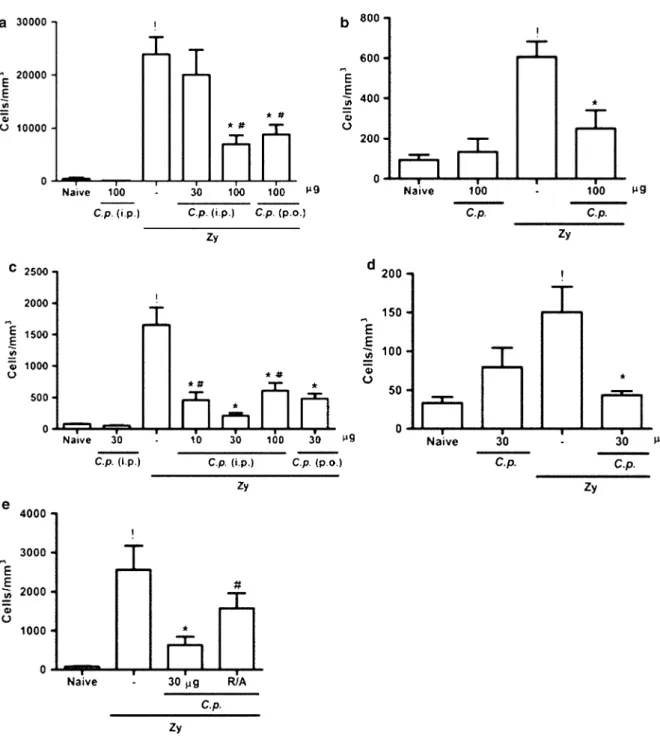
polymorphonuclear cells. At 7 days, cell counts in joint exudates were less pronounced, with mononu-clear cells being predominant [9]. Figure1shows that administration of theC. posadasiiextract, either i.p. or p.o., significantly and dose dependently reduced cell influx in the acute and chronic phase of ZYA in both species, as compared to vehicle-treated animals. Moreover, the isolated administration of the
C. posadasiiextract to naive animals did not alter cell counts. In order to exclude endotoxin contamination, we should stress that both the extract and BSA solution were prepared under sterile conditions and filtered before administration. Additionally, incubation of
C. posadasii extract with polymyxin did not modify its effects on acute cell influx (data not shown). Also, the fact that the extract was effective after being given orally per se excludes the possibility of endotoxin contamination as responsible for the effects observed. In an attempt to demonstrate that a protein component is responsible for the effect observed with the
C. posadasii extract, we subjected the extract to a reducing/alkylating process, a step that denatures proteins containing disulfide bridges. As shown in Fig.1e, administration of the extract following this protein-denaturing process led to a significant reduc-tion in the effect observed with the crude extract. The administration of a 100lg BSA solution given as an irrelevant protein either p.o. or i.p. did not alter hypernociception or cell influx (data not shown).
Coccidioides posadasiiExtract Reduced Hypernociception in ZYA in Rats
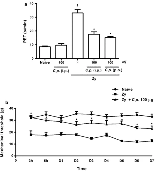
Rats subjected to ZYA displayed an intense hyperno-ciceptive response measured using both the articular incapacitation test and the von Frey methods, as shown in Fig.2a, b, respectively. Similar to what happened with the cell influx, pretreatment of the animals with the
as well as the persistent chronic hypernociceptive response measured from 3 h until 7 days after injection of the zymosan, using the electronic von Frey method
(Fig.2b), as compared to vehicle-treated animals. Administration of the C. posadasii extract to naive animals did not alter the hypernociceptive response.
Fig. 1 Effect of the parenteral and oral administration of the C. posadasii(C.p.) extract on the cell influx in acute and chronic zymosan-induced arthritis (ZYA) in rats and mice. Rats (a,b) and mice (c,d,e) received theC.p.extract or saline (–) either i.p. or p.o. 30 min prior to 1 mg or 0.1 mg zymosan i.a., respectively. (e) Mice received the reduced/alkylated C.p. extract (R/A) i.p. 30 min prior to 0.1 mg zymosan i.a.
Coccidioides posadasiiExtract Effect on Cartilage Damage in ZYA in Rats
The GAG loss in the articular cartilage measured after 7 days of ZYA can be used as an index of structural joint damage in this model. As compared to naive animals, those that received i.a. zymosan displayed a significant reduction in the GAG content [18]. The administration of theC. posadasiiextract provoked a partial, though not reaching statistical significance, reversion of the decrease in the GAG content of the cartilage, as
compared to vehicle-treated rats, as follows: naive (44.1±7.05lg/mg); zymosan (31.2±4.92lg/mg); andC. posadasii?zymosan (36.7±3.65lg/mg).
Coccidioides posadasiiExtract Reduced the Histopathology Changes of the Synovia in ZYA in Mice
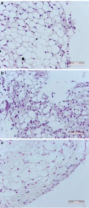
Figure 3illustrates the histopathological appearance of the synovia of mice subjected to ZYA. It can be seen that pretreatment with the C. posadasii extract (Fig. 3c)
Fig. 2 Effect of the parenteral and oral administration of the C.p. extract on the hypernociception in acute (a) or chronic (b) ZYA in rats.aRats received theC.p.extract or saline (–) either i.p. or p.o. 30 min prior to 1 mg zymosan. Naive rats received saline i.a. Articular incapacitation was measured hourly over 4 h as the increase in PET using the rat knee joint incapacitation test. Results show mean±SEM of maximal PET between 3 and 4 h of arthritis.n=6 animals for each group;
almost completely abrogated the synovitis, provoking a marked decrease in the number of infiltrating cells. A synovial sample of a naive mouse is shown for comparison (Fig.3a), as well as the sample from an animal subjected to ZYA treated with the vehicle (Fig.3b). The administration of the extract also pro-voked a decrease in synovial cell hyperplasia, neovas-cular formation, and fibrosis, as shown in Table1.
Coccidioides posadasiiExtract Reduces Cell Death in the Synovial Layer
Injection of the zymosan significantly increased the number of TUNEL-positive cells (apoptosis and possibly necrosis) as compared to synovial samples from naive animals, which display no staining (Fig.4). The number of TUNEL-positive cells was significantly reduced in the samples from the animals that were treated with the C. posadasii extract, as compared to those that received zymosan and the vehicle (cell death is indicated by the brown staining of the cells detected by the TUNEL method).
Coccidioides posadasiiExtract Decreases iNOS Activation in the Synovia
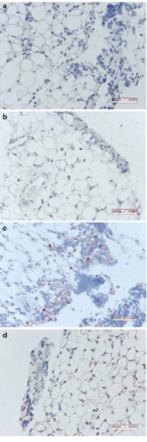
Figure5a–c illustrates photomicrographs of the immunostaining for iNOS activity in the synovia obtained from negative control (5a), naive (5b), zymosan (5c), and 30lg C.p.?zymosan (5d)
groups. There is a clear and significant (p\0.02)
reduction in the immunostaining in the samples of animals that receivedC.p.extract (median score=1;
range 0–2), as compared to those that received zymosan and the vehicle (median score=2; range
1–3).
Effect of theC. posadasiiExtract on the Release of Inflammatory Mediators in acute ZYA
The C. posadasiiextract did not alter the release of IL-1b, TNF-a,c-IFN, and IL-17 into the joints of mice subjected to ZYA (Fig.6a, b, c, d, respectively). The levels of these cytokines were measured at 6 h of arthritis in the joint exudates. On the other hand, Fig.6e shows that IL-10 joint levels, also measured at 6 h, were significantly reduced, as compared to saline-treated animals.
Analogous to what was observed in mice, the
C. posadasiiextract did not alter the release of IL-1b and TNF-a into the joints of rats subjected to ZYA (Fig.7a, b, respectively). The levels of these cyto-kines were measured at 6 h of arthritis in the joint exudates. On the other hand, Fig.7c shows that IL-10 joint levels, also measured at 6 h, were signif-icantly reduced, as compared to saline-treated animals.
Preliminary Characterization of Components in theC. posadasiiExtract
Figure8a, b illustrates a silver-stained 12 % poly-acrylamide electrophoresis gel of the C. posadasii
extract to identify proteins and a toluidine blue-stained 6 % agarose gel electrophoresis aimed to identify charged carbohydrates, respectively. Analysis of the proteins run on lane 2 was done using E-CaptTM(12.7 version for Windows) software aiming to estimate the molecular weight comparing to the Bio-radTM stan-dards run on parallel. Twelve bands were identified, ranging from 4 to 172 kD. There is a wide range of both low- and high-molecular-weight proteins shown in the silver-stained gel. Additional purification is needed in order to try to identify active components implicated in the protective effects of theC. posadasii
extract. On the other hand, the absence of charged carbohydrates, as shown on lane 3 of Fig.8b, virtually excludes such components as responsible for the results achieved.
Discussion
The present study demonstrates that a C. posadasii
extract, administered either parenterally orper os, has dose-dependent in vivo anti-inflammatory properties by inhibiting joint hypernociception and acute neu-trophil migration into the inflamed joints as well as the increase in mononuclear cell counts in the chronic phase—7 days—of zymosan arthritis. At histopathol-ogy, neovascular formation, fibrosis, lymphocyte, and mononuclear cell proliferation together with hyper-plasia of the synovial lining cells were also signifi-cantly reduced by the pre-administration of a single dose of theC. posadasiiextract. In keeping with the pathogenetic relevance of the present results, joint damage, assessed by measuring the glycosaminogly-can content of the cartilage, was reduced, though not significantly, in animals that received the fungal extract, as compared to vehicle-treated rats. The demonstration of efficacy in different species, dose dependence, and activity in the acute and chronic phases both orally and parenterally clearly adds to the bulk of important anti-inflammatory and immuno-modulating properties described with other fungal products [19].
Synovial resident cells, macrophages, and neutro-phils present in the inflamed synovia are main sources of reactive oxygen species during synovitis. Zymosan-induced arthritis is characterized by an acute influx of polymorphonuclear cells that is more prominent at 6 h, while mononuclear cells (mostly lymphocytes
Table 1 Effect of theC. posadasiiextract on joint damage by histopathology in ZYA
Group Cell
infiltration
Stage of the disease
Synovial proliferation
Proliferation of blood vessels
Fibrosis Edema Scores total Naive 0 (0–1.0) 0 (0–1.0) 0 (0–1.0) 0 (0–1.0) 0 (0–0) 0 (0–0) 0 (0–2.0) Zy 2.0 (1.0–3.0) 2.0 (1.0–2.0) 1.0 (0–3.0) 1.0 (1.0–2.0) 2.0 (1.0–2.0) 1.0 (0–2.0) 9.5 (6.0–13.0)
Zy?C.p. 0*
(0–1.0) 1.0* (0–2.0) 0* (0–1.0) 0* (0–1.0) 0* (0–2.0) 1.0 (0–2.0) 3.0* (0–8.0) R/A 2.0 (2.0–3.0) 2.5 (2.0–3.0) 1.5 (0–2.0) 1.0 (1.0–1.0) 0.5* (0–1.0) 0.5 (0–1.0) 8.0 (7.0–9.0)
Mice received 30lg ofC.p.extract (Zy?C.p.), a reduced/alkylated (R/A)C.p.extract or saline given i.p. (Zy), 30 min prior to 0.1 mg zymosan i.a. Naive mice received saline i.a. All animals were killed at 7 days. Results represent medians of synovial histopathology scores
and monocytes) predominate after the first day. At 7 days, there is an intense chronic synovitis with synovial hyperplasia and mononuclear cell prolifera-tion, together with angiogenesis, fibrosis, and, even-tually, giant cell formation [9]. As a result, inflammatory mediators, particularly cytokines, can be directly assessed in the joint exudates, thus allowing the study of mechanisms involved in syno-vitis development.
We have demonstrated that neutrophils contribute to the formation of both nitric oxide (NO) and peroxynitrite in zymosan arthritis [20]. In the present study, administration of the C. posadasii extract markedly decreased the expression of iNOS in the inflamed synovia. Release of NO by macrophages has been proposed to be a protective mechanism linked to innate immunity that prevents disease development in subjects exposed to other fungi, for example, Para-coccidioides brasiliensis [21]. However, we are not aware of in vivo data showing that fungi components given systemically alter NO release either locally or systemically. Mice injected intraperitoneally with viableC. immitisdisplay increased urine nitrate levels, thus reflecting augmented NO production following coccidiomycosis development [22]. On the other hand, macrophages stimulated with zymosan particles in vitro show a decreased release of NO when in the presence ofP. brasiliensis-derived peptides [21]. Our in vivo data suggest that administration of the
C. posadasii extract provoked a decrease in iNOS activation, predominantly in synoviocytes, thereby reducing the generation of reactive oxygen species in the joint.
Joint damage, pain, as well as cell infiltration observed in zymosan arthritis are closely related to the increase in the production of proinflammatory medi-ators such as TNF-aand IL-1blocally [23,24]. Other cytokines, including IL-10 and IL-17, have been described to participate in the pathogenesis of fungi reactions as well as in arthritis. While the former may display anti-inflammatory properties [25], the latter has been implicated in the acute hypernociception in
Fig. 4 Representative illustration of the effect ofC.p.extract on cell death in the zymosan-inflamed synovial detected by TUNEL method. Mice received 30lg ofC.p. extract (c) or saline (b) given i.p. 30 min prior to 0.1 mg zymosan i.a. Naive mice (a) received saline i.a. All animals were killed at 6 h. The results are expressed as the average number of TUNEL-positive cells.p\0.05 when compared to zymosan
experimental arthritis, as we have shown recently [26]. Measuring the level of these mediators in the synovial fluid rather than in serum or using ex vivo strategies may more appropriately reflect what is happening inside the joint. Our results showed no difference between the group that received the C. posadasii
extract and control groups regarding the release of IL-1b, TNF-a,c -IFN, and IL-17 into the joints. We have to stress that although TNF-ajoint levels signif-icantly rise in rats at 6 h of zymosan arthritis, the levels of this cytokine are not significantly elevated in mice joints, possibly due to species variation [5]. Given that the administration of the C. posadasiiextract did not alterc-IFN, IL-1b, TNF-a, and IL-17 joint levels, our data argue against a participation of Th1 cytokines as well as these proinflammatory cytokines to explain the in vivo anti-inflammatory and immunomodulatory effects of theC. posadasiiextract in zymosan arthritis. Interestingly, our results show that IL-10 levels in the joints were significantly reduced in both rats and mice that received the C. posadasii extract. It was shown that experimental coccidiomycosis in suscepti-ble mice is associated with increased levels of IL-10 in the lungs. The same group later reported that in vitro stimulation of macrophages isolated from aC. immitis -susceptible mouse strain led to increased production of IL-10 and reduced levels of TNF-a, as compared to a resistant coccidiomycosis mouse strain. These effects were reduced by an antibody against Dectin-1 [22]. In summary, those data led to a proposal that genetic ablation of the Dectin-1 gene severely impairs IL-10 production [27]. To our knowledge, there are no reports showing alteration of cytokine release following systemic administration of Coccidioides sp. compo-nents. Our data add to these previous in vitro results since we measured in vivo production directly released into inflamed joints. Thus, we speculate that protein components of ourC. posadasiiextract, either isolated or linked to fungal polysaccharides, interfered with zymosan coupling to Dectin-1 receptors, thereby leading to decreased IL-10 production.
Our present results also revealed that apoptosis, predominantly of synovial lining cells, was markedly reduced in the group treated with theC. posadasiiextract. NO is able to promote apoptosis of various cell types, including macrophages, neutrophils, and synoviocytes [28]. It has also been shown that stimulation of monocyte-derived cells with zymosan in the presence of apoptotic
cells enhances IL-10 production [29]. As a possible mechanism to explain the reduced IL-10 levels promoted by theC. posadasiiextract, we propose that the blockade of iNOS activation leads to a reduction in NO levels locally that in turn decreases the number of apoptotic cells, thus reducing IL-10 production by macrophage-like synoviocytes in the zymosan-inflamed joints.
Fig. 6 Effect of the administration ofC.p.extract on the release of inflammatory mediators in ZYA. Mice received 30lg of the C.p.extract or saline (–) i.p. 30 min prior to 0.1 mg zymosan i.a. Naive mice received saline i.a. IL-1b(a), TNF-a(b),c-IFN (c),
IL-17 (d), and IL-10 (e) levels were assessed using ELISA (see text for details). Results represent mean±SEM, measured at 6 h (n=6 animals for each group); !p\0.05 compared to naive mice;*p\0.05 compared to control (–) mice
Fig. 7 Effect of the administration ofC.p.extract on the release of inflammatory mediators in ZYA. Rats received 100lg of the C.p.extract or saline (–) i.p. 30 min prior to 0.1 mg zymosan i.a. Naive rats received saline i.a. (a) IL-1b, (b) TNF-a, and
We are pursuing studies trying to isolate active component(s) present in the C. posadasii extract. Since the extract acted after being given orally, the possibility of lipopolysaccharide contamination to explain our data can be disregarded. The reducing/ alkylating step applied to our C. posadasii extract abrogated the anti-inflammatory/immunomodulating effect, thus indicating that a protein or a polypeptide containing disulfide bridges is responsible for its biological activity. We cannot exclude the possibility that isolated polysaccharides or protein/polypeptide components attached to them are responsible for the anti-inflammatory results achieved. Notwithstanding the complexity of defining a specific active compo-nent, we have to stress that our data are an in vivo original demonstration that either isolated proteins or associated with other fungi components, obtained from a dimorphic pathogenic species, are capable of modifying remote in vivo inflammatory responses. Further characterization of the specific active compo-nents in this extract that we are currently undergoing may unravel mechanistic effects that can be of relevance to understand the host response against fungal components.
Acknowledgments We thank Giuliana Bertozzi for the ELISA work. The work was partially supported by a grant
from Conselho Nacional de Desenvolvimento Cientı´fico e Tecnolo´gico (CNPq)/Fundac¸a˜o Cearense de Apoio ao Desen-volvimento Cientı´fico e Tecnolo´gico (FUNCAP) (PRONEX n 2155-06) and Coordenac¸a˜o de Aperfeic¸oamento de Pessoal de Nı´vel Superior (CAPES), Brazil.
References
1. Lull C, Wichers HJ, Savelkoul HFJ. Antiinflammatory and immunomodulating properties of fungal metabolites. Mediators Inflamm. 2005;2:63–80.
2. Wasser SP. Medicinal mushrooms as a source of antitumor and immunomodulating polysaccharides. Appl Microbiol Biotechnol. 2002;60:258–74.
3. Bach JF. The effect of infections on susceptibility to auto-immune and allergic diseases. N Engl J Med. 2002;347: 911–20.
4. Rook GA, Stanford JL. Give us this day our daily germs. Immunol Today. 1998;19:113–6.
5. Rocha FAC, Leite AKRM, Pompeu MML, Cunha TM, Verri WA Jr, Soares FM, Castro RR, Cunha FQ. Protective effect of an extract from Ascaris suum in experimental arthritis models. Infect Immun. 2008;76:2736–45. 6. Hung CY, Xue J, Cole GT. Virulence mechanisms of
Coccidioides. Ann N Y Acad Sci. 2007;1111:225–35. 7. Brass C, Levine HB, Stevens DA. Stimulation and
sup-pression of cell-mediated immunity by endosporulation antigens ofCoccidioides immitis. Infect Immun. 1982;35: 431–6.
8. Cordeiro RA, Brilhante RS, Rocha MF, Fechine MA, Camara LM, Camargo ZP, Sidrim JJ. Phenotypic charac-terization and ecological features ofCoccidioidesspp. from northeast Brazil. Med Mycol. 2006;44:631–9.
9. Rocha FAC, Araga˜o AGM Jr, Oliveira RC, Pompeu MML, Vale MR, Ribeiro RA. Periarthritis promotes gait distur-bance in zymosan-induced arthritis in rats. Inflamm Res. 1999;48:485–90.
10. Silva FS Jr, Rocha FAC. Artrite induzida por zymosan em ratos-Mecanismos envolvidos na hipernocicepc¸a˜o e na lise da cartilagem articular. Acta Reum Port. 2006;31:143–9. 11. Guerrero ATG, Cunha TM, Verri WA Jr, Gazzinelli RT,
Teixeira MM, Cunha FQ, Ferreira SH. Toll-like receptor 2/MyD88 signaling mediates zymosan-induced joint hy-pernociception in mice: participation of TNF-a, IL-1band CXCL1/KC. Eur J Pharmacol. 2012;674:51–7.
12. Guerrero ATG, Verri WA Jr, Cunha TM, Silva TA, Schivo IRS, Dal-Secco D, Canetti C, Rocha FAC, Parada CA, Cunha FQ, Ferreira SH. Involvement of LTB4in zymosan-induced joint nociception in mice: participation of neutro-phils and PGE2. J Leukoc Biol. 2008;83:122–30.
13. Kroemer G, Galluzzi L, Vandenabeele P, Abrams J, Al-nemri ES, Baehrecke EH, Blagosklonny MV, El-Deiry WS, Golstein P, Green DR, Hengartner M, Knight RA, Kumar S, Lipton SA, Malorni W, Nun˜ez G, Peter ME, Tschopp J, Yuan J, Piacentini M, Zhivotovsky B, Melino G. Nomen-clature Committee on cell death 2009. Classification of cell death: recommendations of the Nomenclature Committee on cell death 2009. Cell Death Differ. 2009;16(1):3–11. 14. Silva FS Jr, Yoshinari NH, Castro RR, Gira˜o VCC, Pompeu
MML, Feitosa JPA, Rocha FAC. Combined glucosamine Fig. 8 Partial identification of proteins and carbohydrates
and chondroitin sulfate provides functional and structural benefit in the anterior cruciate ligament transection model. Clin Rheumatol. 2009;28:109–17.
15. Brilhante RSN, Cordeiro RA, Rocha MFG, Fechine MAB, Furtado FM, Nagao-Dias AT, Camargo ZP, Sidrim JJC. Coccidioidal pericarditis: a rapid presumptive diagnosis by an in-house antigen confirmed by mycological and molec-ular methods. J Med Microbiol. 2008;57:1288–92. 16. Porto IM, Laure HJ, Tykot RH, Sousa FB, Rosa JC, Gerlach
RF. Recovery and identification of mature enamel proteins in ancient teeth. Eur J Oral Sci. 2011;119:83–7.
17. Bradford MM. A rapid and sensitive method for the quan-titation of microgram quantities of protein utilizing the principle of protein-dye binding. Anal Biochem. 1976;72: 248–54.
18. Bezerra MM, Brain SD, Greenacre S, Jeroˆnimo SMB, Melo LB, Keeble J, Rocha FAC. Reactive nitrogen species scavenging, rather than nitric oxide inhibition, protects from articular cartilage damage in rat zymosan-induced arthritis. Br J Pharmacol. 2004;141:172–82.
19. Romani L. Cell mediated immunity to fungi: a reassess-ment. Med Mycol. 2008;46:515–29.
20. Bezerra MM, Brain SD, Gira˜o VCC, Greenacre S, Keeble J, Rocha FAC. Neutrophils-derived peroxynitrite contributes to acute hyperalgesia and cell influx in zymosan arthritis. Naunyn Schmiedebergs Arch Pharmacol. 2007;374: 265–73.
21. Konno AYC, Maricato JT, Konno FTC, Mariano M, Lopes JD. Peptides from Paracoccidioides brasiliensis GP43 inhibit macrophage functions and inflammatory response. Microbes Infect. 2009;11:92–9.
22. del Pilar Jime´nez AM, Viriyakosol S, Walls L, Datta SK, Kirkland T, Heinsbroek SEM, Brown G, Fierer J.
Susceptibility toCoccidioidesspecies in C57BL/6 mice is associated with expression of a truncated splice variant of Dectin-1 (Clec7a). Genes Immun. 2008;9:338–48. 23. Leite ACRM, Cunha FQ, Dal-Secco D, Fukada SY, Gira˜o
VCC, Rocha FAC. Effects of nitric oxide on neutrophil influx depends on the tissue: role of leukotriene B4and adhesion molecules. Br J Pharmacol. 2009;156:818–25. 24. Bombini G, Canetti C, Rocha FA, Cunha FQ. Tumour
necrosis factor-amediates neutrophil migration to the knee synovial cavity during immune inflammation. Eur J Phar-macol. 2004;496:197–204.
25. Moore KW, Malefyt RW, Coffman RL, O’Garra A. Inter-leukin-10 and the interInter-leukin-10 receptor. Annu Rev Immunol. 2001;19:683–765.
26. Pinto LG, Cunha TM, Vieira SM, Lemos HP, Verri WA Jr, Cunha FQ, Ferreira SH. IL-17 mediates articular hyperno-ciception in antigen-induced arthritis in mice. Pain. 2010;148:247–56.
27. Taylor PR, Tsoni SV, Willment JA, Dennehy KM, Rosas M, Findon H, Haynes K, Steele C, Botto M, Gordon S, Brown GD. Dectin-1 is required for b-glucan recognition and control of fungal infection. Nat Immunol. 2007;8:31–8. 28. Chen Q, Casali B, Pattacini L, Boiardi L, Salvarani C.
Tumor necrosis factor-alpha protects synovial cells from nitric oxide induced apoptosis through phosphoinositide 3-kinase Akt signal transduction. J Rheumatol. 2006;33(6):1061–8.


