Submitted10 May 2016 Accepted 3 August 2016 Published31 August 2016
Corresponding author Zhanming Li,
Academic editor Marjorie Longo
Additional Information and Declarations can be found on page 11
DOI10.7717/peerj.2400
Copyright 2016 Yu and Li
Distributed under
Creative Commons CC-BY 4.0
OPEN ACCESS
Influence of droplet coverage on the
electrochemical response of planar
microelectrodes and potential solving
strategies based on nesting concept
Yue Yu1and Zhanming Li1,2
1Department of Biosystems Engineering, Zhejiang University, Hangzhou, China
2Department of Food Science, College of Life Science, China Jiliang University, Hangzhou, China
ABSTRACT
Recently, biosensors have been widely used for the detection of bacteria, viruses and other toxins. Electrodes, as commonly used transducers, are a vital part of electro-chemical biosensors. The coverage of the droplets can change significantly based on the hydrophobicity of the microelectrode surface materials. In the present research, screen-printed interdigitated microelectrodes (SPIMs), as one type of planar microelectrode, were applied to investigate the influence of droplet coverage on electrochemical response. Furthermore, three dimensional (3D) printing technology was employed to print smart devices with different diameters based on the nesting concept. Theoretical explanations were proposed to elucidate the influence of the droplet coverage on the electrochemical response. 3D-printed ring devices were used to incubate the SPIMs and the analytical performances of the SPIMs were tested. According to the results obtained, our device successfully improved the stability of the signal responses and eliminated irregular signal changes to a large extent. Our proposed method based on the nesting concept provides a promising method for the fabrication of stable electrochemical biosensors. We also introduced two types of electrode bases to improve the signal stability.
SubjectsBiochemistry, Bioengineering, Biophysics, Biotechnology, Food Science and Technology
Keywords Droplet coverage, Three dimensional (3D) printing, Signal change, Screen-printed interdigitated microelectrode (SPIM), Nest-like, Electrochemical response
INTRODUCTION
and work in a two-electrode system, thus benefiting the fabrication and performance of electrochemical biosensors (Huey et al., 2012;Park & Beskok, 2008). Microelectrodes exhibit more accurate electrochemical response to low concentrations of electro-active species in solution than large planar electrodes. Excellent flexibility and cycling stability
also promise potential applications in lab-on-a-chip systems (Ch et al., 2006;Huang,
O’Mahony & Compton, 2009;Ueno et al., 2005).
Electrochemical biosensors based on planar microelectrodes have been described in many literatures (Bernalte, Sánchez & Gil, 2011;Taleat, Khoshroo & Mazloum-Ardakani, 2014;Valentini et al., 2014;Wang, Ye & Ying, 2012;Zhu, Zhou & Gao, 1998). The sensitivity and the signal-to-noise ratio of planar microelectrodes can be influenced by many factors, such as the diameter of the electrode, surface coverage, electrode geometry and the electroactivity of analytes (Brett & Thiemann, 2002;Kostecki, Song & Kinoshita, 2000;Liu et al., 2014;Xu et al., 2004;Zhu, Zhou & Gao, 1998). The droplets exhibit different coverage on the microelectrode surface, depending on the hydrophobicity of the microelectrode surface which may trigger irregular signal change. The commonly-used incubation methods for planar microelectrodes include dropping and immersing coatings. The influence of droplet coverage which is important but less concerned, should be evaluated when dropping coating is used for incubation. Screen-printed interdigitated microelectrodes (SPIMs), as one type of planar microelectrode which may integrate the merits of screen printed microelectrodes and interdigitated microelectrodes, have been used to develop sensitive, rapid-responding, cost-effective biosensors (Li et al., 2015a). In this manuscript, we investigate the influence of droplet coverage on the electrochemical response of SPIMs. Three-dimensional (3D) printed devices have captured much attention in many fields, including food safety and analysis (Sun et al., 2015; Xing, Zheng & Duan, 2015). As we know, many birds build nests to incubate their eggs and raise their young in a protective environment (Vilé et al., 2015). In order to further analyze the influence of the droplet coverage quantitatively, 3D printing technology was employed to print smart nest-like devices with different diameters to keep the immobilization and detection within the devices based on nesting concept. Both the theoretical analysis and our experimental results support our conclusion that the droplet coverage has significant influence on the electrochemical response of biosensors.
MATERIALS AND METHODS
Reagents and apparatusE. coliO157:H7 (ATCC43889) was purchased from ATCC (Manassas, MD). Biotin-anti-E.
coliantibodies was obtained from Meridian Life Science (Saco, ME) and dissolved in PBS
solution (pH=7.4). Bovine serum albumin (BSA), streptavidin (SA) and protein A were
purchased from Sangon Biotech (Shanghai, China). MacConkey agar, brain heart infusion (BHI) culture medium were purchased from Becton, Dickinson and Company (Sparks,
NV, USA). PBS solution containing 10 mM K3Fe(CN)6/K4Fe(CN)6(Sangon Biotech.,
Shanghai, China) was used for electrochemical measurements. Ultrapure water (18.2
Mcm) was obtained from a Millipore Milli-Q purification system (Merck Millipore,
Billerica, MA, USA).
The width of a finger and the gap between two fingers for SPIM (AIBIT Biotech
Instrument, Jiangyin, China) are both 200µm. One pair of gold electrodes and two welding
plates were prepared on a ceramic base using screen-printed technology. Two electrodes were connected to the bonding pad. The electrode contained multiple conducting rings
with different diameters and connected by conductive bands.Figure S1showed the details
of the electrodes.
Electrochemical impedance spectroscopy (EIS) and cyclic voltammetry (CV) were performed using ZAHNER electrochemical station (Kronach, Germany). Photosensitive resin was purchased from DSM SOMOS Crop. (Somos imagine 8000; Elgin, IL, USA).
The devices with different diameters (2r=6, 7, 8 and 9 millimeter) were printed by a 3D
printer (Liantai 450, Shanghai, China) with 0.01 millimeter precision.
Experimental methods
Preparation of bacterial samples
E. coliO157:H7 was grown in BHI culture medium at 37 ◦
C for 20 h to the stationary phase. The stationary-phase cultures were diluted to 107–101cfu mL−1in PBS (pH 7.4) and 100
µL
of the diluted solutions were transferred to MacConkey agar plates and incubated at 37 ◦
C for 24 h for enumeration of colonies. At the same time, the dilutions containing approxi-mately 105cfu mL−1of bacteria cells were prepared for evaluation of the proposed devices.
Dropping and immersing coatings
When the planar microelectrodes were incubated, the commonly used incubation methods are dropping and immersing coatings for the incubation of planar microelectrodes. Usually,
the volume of dropping coatings is 10 to 50 µL, and the volume of immersing coating
is more than 1,000µL in order to cover the whole detection area. Generally, the droplet
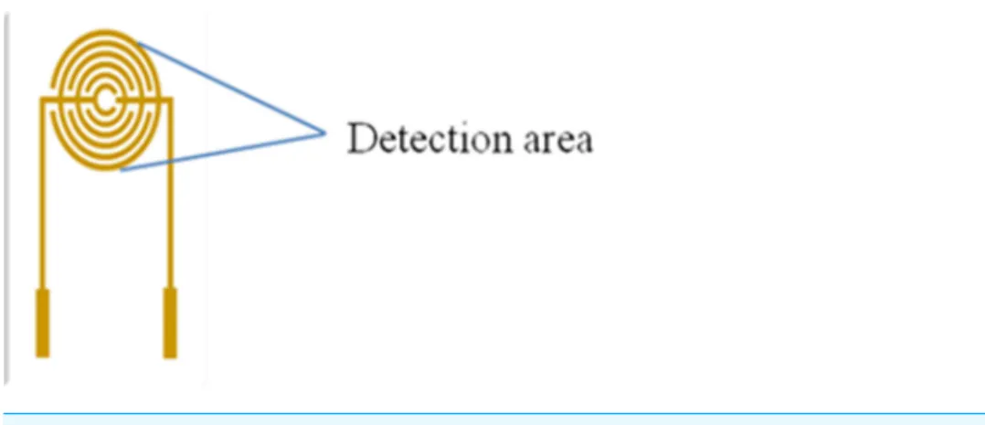
coverage varies due to the discrepancy of the SPIMs surface.Figure 1presents the droplet
coverage of the electrode. The droplet coverage for the bare SPIM is significantly different compared to that for the modified SPIM (Fig. S2).
Figure 1 Detection area (droplets area) of the electrode.
and treatment groups PBS and B were dropping coating, respectively. We used 1,500µL
PBS solution containing 50µL protein A for coating (0.5 mg mL−1) in treatment group
A, 50µL protein A (0.5 mg mL−1) in treatment group B, and 1,500µL protein A (0.5 mg
mL−1) in treatment group C. The signal change was detected with EIS or CV techniques.
Nesting concept for incubation
In order to investigate the difference caused by the droplet coverage, we designed and printed nest-like devices with different diameters. The devices (Fig. 2) possess an equivalent
volume, but with different height and diameter (2r=6, 7, 8 and 9 millimeter). The volume
for these nest-like devices was equivalent, which ensured the same concentration and
quantity of targets. The devices were incubated with BSA solution (2%,w/w) before use
in order to avoid the non-specific absorption. Two devices (2r=6 and 9 millimeter)
were selected to perform the test to investigate the difference introduced by the multistep
modification. The devices were used to incubate SPIMs with SA solution (50µL, 0.5 mg
mL−1) for 45 min, and then biotin-antibody was immobilized according to our previous
research (Li et al., 2015a). After that, the bacteria solution was added into the devices to incubate the SPIMs and the difference caused by multistep modification was evaluated.
3D-printing ring devices
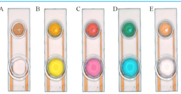
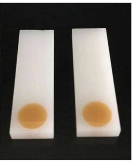
In the present research, both resin and glass were used to investigate the dispersity of the target solution. A smart ring covered with silica gel was fabricated on the surface of the SPIM. We used the nest-like devices with different diameters to cover the detection area and then used silica gel to coat the devices. The caps were removed after the gel was completely dry in order to form a ring area. The ring can stay intact during the incubation and modification processes. In order to prove that the rings are intact, solutions with different colors were added into the devices after each incubation and washing processes (Fig. 3). The influence of NaOH solution was also investigated for the cleaning process.
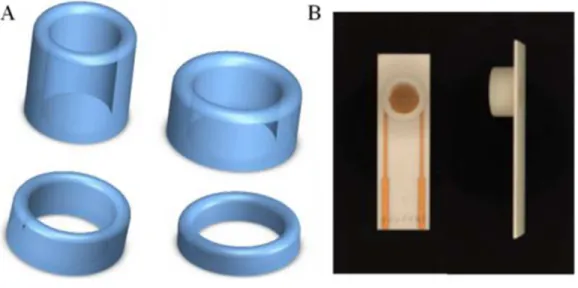
On the basis of silica gel ring, we designed and printed ring devices with different diameters (Fig. 4). We selected two devices (2r=6 and 9 millimeter) to investigate the
Figure 2 The 3D printed nest-like devices (B) with different height (h) and diameter (2r=6, 7, 8 and 9 millimeter) (C) and the photo of the incubation (D). The bird nest picture (A) was cited fromwww. nipic.com/show/7011922.html.
Figure 3 Silica gel ring on the surface of the SPIMs.Solution with different color were used to show the ring concept: (A) no solution, (B) solution with K+
, (C) solution with Co3+
, (D) solution with Cu2+
, (E) ultrapure water.
The same amount of solution was added into the devices to incubate SPIMs for 45 min. Then, the non-specific absorption was washed with ultrapure water. The signal change was evaluated after the incubation.
Statistical analysis
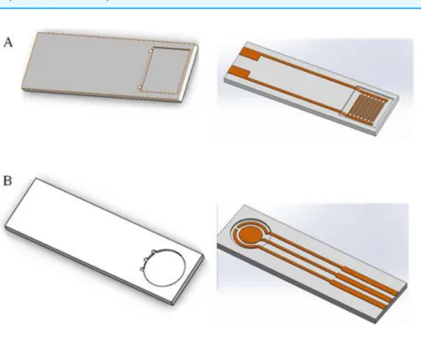
Figure 4 (A) 3D printed ring devices with different diameters and (B) SPIM with ring device to form a nest-like device.
Figure 5 Immersing coating and dropping coating for the microelectrode modification.Treatment groups A and C were immersing coating and treatment groups PBS and B were dropping coating. Treat-ment group A: 1,500µL PBS solution containing 50µL of protein A (0.5 mg mL−1); treatment group B:
50µL of protein A (0.5 mg mL−1); treatment group C: 1,500µL of protein A (0.5 mg mL−1).
nest-like devices with different diameters were collected. Statistical analysis were conducted using SPSS 17.0. The biosensor responses were considered to show significant difference when P-value was less than 0.05 (95% confidence interval).
RESULTS AND DISCUSSION
Dropping and immersing coatingsFour treatment groups were performed conducted and the application of PBS solution
was used as a control (Fig. 5). Performances of treatment A and B were significantly
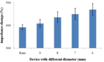
Figure 6 Signal change of the printed nest-like devices with different diameters for protein immobi-lization.Bare was a SPIM without printed devices.
the devices in comparison with treatment B. These results indicates that the signal changes are different between immersing and dropping coatings. Since the two methods required different amount of materials, it is not persuasive to conclude that the difference is caused by droplet coverage.
Performance of nest-like devices
Considering the influence of the droplet coverage on the electrochemical response of SPIMs, a smarter design of devices is necessary. Nest-like devices with different diameters were used for incubation (Fig. 6). The results showed that there was significant difference between 9 millimeter and 6 millimeter devices (p>0.05) and the difference were not significant for other devices. According to the difference in the droplet coverage of these two devices, it can be concluded that the signal response is influenced by droplet coverage.
In order to investigate the influence of the multistep modification, we selected 6 and 9 millimeters devices to perform the test for bacteria detection (Fig. 7). Treatment group A was prepared according to the signal change after SA modification. The results indicated that there was significant difference between the devices (p>0.05). Treatment group B (Fig. 7) was prepared according to the signal change of the bacteria incubation after the multistep modification. The difference between the results collected from these two treatment groups did not disappear. Due to the large dimension of the bacteria cells, not every site was occupied by the bacteria cell. However, only effective absorption can introduce signal response. From the results, we concluded that the influence of droplet coverage was not disappeared by large dimension of cells. Therefore, it is clear that the droplet coverage is an influencing factor for electrochemical response.
Performance of silica gel ring
Figure 7 3D printed nest-like devices (6 and 9 millimeters) for the test.Treatment group A was the per-formance after SA modification. Treatment group B was the perper-formance of the bacteria incubation, after the multi-step modification.
Figure 8 The CV performance of SPIMs with and without silica gel ring device.
situation, we firstly designed a ring was prepared on the surface of SPIM using silica gel. The results indicated that the signal was suppressed significantly compared to the treatment groups without the device, considering that more molecules were immobilized effectively (Fig. 8). Moreover, the washing solution was available for this device without damage, indicating that the material and device are both applicable.
Performance of ring devices
We designed and printed smart ring devices based on the concept of the silica gel ring. The
results inFig. 9show that the immobilization of the protein gave rise to the EIS and CV
Figure 9 The EIS (A) and CV (B) performance of the SPIMs with and without 3D printed ring devices (6 and 9 millimeters).
Figure 10 Two types of electrode bases designed by software.(A) Interdigital microelectrode and (B) screen-printed electrode.
Figure 11 The 3D printed base used to verify the application.
Base design using software
Signal stability can be influenced by the droplet area. Different bases can be designed to reduce the irregular signal. In this paper, we designed two bases using software (Fig. 10) that the droplets can be reserved within the scope of detection area. We used a 3D printer to print the base, verifying the application (Fig. 11).
CONCLUSION
In the present research, SPIMs was employed to evaluate the influence of droplet coverage on the electrochemical response. 3D printing technology was used to print fabricate mini-small smart devices with different diameters based on nesting concept. Nest-like
devices with different diameters (2r=6, 7, 8, 9 millimeter) were designed and printed to
signal and also verify such influence. Detection cost can be greatly reduced by recycling the printed devices. All the devices improve the stability of the signal and successfully eliminate the irregular signal change. Our proposed design and concept shows great potential for application in the field of electrodes fabrication and stable electrochemical biosensors construction.
ACKNOWLEDGEMENTS
The authors thank the Biosensing & Biomodeling Lab at Zhejiang University for their support of the electrodes and electrochemical station. We gratefully acknowledge Muyang Lin and Xi Yu (College of Chemistry, National University of Singapore) for their help.
ADDITIONAL INFORMATION AND DECLARATIONS
Funding
This work was supported by a China Jiliang University Start-Up Grant. The funders had no role in study design, data collection and analysis, decision to publish, or preparation of the manuscript.
Grant Disclosures
The following grant information was disclosed by the authors: China Jiliang University Start-Up Grant.
Competing Interests
The authors declare there are no competing interests.
Author Contributions
• Yue Yu conceived and designed the experiments, performed the experiments, analyzed
the data, contributed reagents/materials/analysis tools, wrote the paper, prepared figures and/or tables, reviewed drafts of the paper.
• Zhanming Li conceived and designed the experiments, performed the experiments,
analyzed the data, contributed reagents/materials/analysis tools, wrote the paper, prepared figures and/or tables.
Data Availability
The following information was supplied regarding data availability:
The raw data has been supplied as aSupplemental File.
Supplemental Information
Supplemental information for this article can be found online athttp://dx.doi.org/10.7717/
peerj.2400#supplemental-information.
REFERENCES
Brett CMA, Thiemann C. 2002.Conducting polymers from aminobenzoic acids and
aminobenzenesulphonic acids: influence of ph on electrochemical behaviour.Journal
of Electroanalytical Chemistry538–539:215–222.
Ch S, Jenke M, Hoffmann P, Brugger J. 2006.Interdigitated 50 nm ti electrode
ar-rays fabricated using xef 2 enhanced focused ion beam etching.Nanotechnology
17(11):2722–2729DOI 10.1088/0957-4484/17/11/002.
Costa R, Pereira CM, Silva AF. 2015.Charge storage on ionic liquid electric dou-ble layer: the role of the electrode material.Electrochimica Acta167:421–428
DOI 10.1016/j.electacta.2015.02.180.
Gerard M, Chaubey A, Malhotra BD. 2002.Application of conducting polymers to biosensors.Biosensors and Bioelectronics17(5):345–359
DOI 10.1016/S0956-5663(01)00312-8.
Gryczan P, Kisiel A, Michalska A, Maksymiuk K. 2015.Electrochemical properties of
polypyrrole doped by alternating polymer micelles.Electroanalysis27(3):752–759
DOI 10.1002/elan.201400582.
Huang X, O’Mahony AM, Compton RG. 2009.Microelectrode arrays for
electrochem-istry: approaches to fabrication.Small5(7):776–788DOI 10.1002/smll.200801593.
Huey E, Krishnan S, Arya SK, Dey A, Bhansali S. 2012.Optimized growth and integra-tion of silica nanowires into interdigitated microelectrode structures for biosensing. Sensors and Actuators B: Chemical175:29–33DOI 10.1016/j.snb.2011.11.056.
Hunt HK, Armani AM. 2010.Label-free biological and chemical sensors.Nanoscale
2(9):1544–1559DOI 10.1039/c0nr00201a.
Kostecki R, Song XY, Kinoshita K. 2000.Influence of geometry on the electrochemical
response of carbon interdigitated microelectrodes.Journal of The Electrochemical
Society147(5):1878–1881DOI 10.1149/1.1393451.
Lepinay S, Staff A, Ianoul A, Albert J. 2014.Improved detection limits of protein optical fiber biosensors coated with gold nanoparticles.Biosensors and Bioelectronics
52:337–344DOI 10.1016/j.bios.2013.08.058.
Li Z, Fu Y, Fang W, Li Y. 2015a.Electrochemical impedance immunosensor based on self-assembled monolayers for rapid detection of Escherichia coli o157:H7 with
sig-nal amplification using lectin.Sensors15(8):19212–19224DOI 10.3390/s150819212.
Li Z, Yu Y, Li Z, Wu T. 2015b.A review of biosensing techniques for detection of trace
carcinogen contamination in food products.Analytical and Bioanalytical Chemistry
407(10):2711–2726DOI 10.1007/s00216-015-8530-8.
Liu J, Wagan S, Dávila Morris M, Taylor J, White RJ. 2014.Achieving reproducible performance of electrochemical, folding aptamer-based sensors on
microelec-trodes: challenges and prospects.Analytical Chemistry86(22):11417–11424
DOI 10.1021/ac503407e.
Park S, Beskok A. 2008.Alternating current electrokinetic motion of colloidal
par-ticles on interdigitated microelectrodes.Analytical Chemistry80(8):2832–2841
DOI 10.1021/ac7024859.
Santoro C, Guilizzoni M, Correa Baena JP, Pasaogullari U, Casalegno A, Li B, Ba-banova S, Artyushkova K, Atanassov P. 2014.The effects of carbon electrode surface
properties on bacteria attachment and start up time of microbial fuel cells.Carbon
67:128–139DOI 10.1016/j.carbon.2013.09.071.
Sun J, Zhou W, Huang D, Fuh JH, Hong G. 2015.An overview of 3d printing
tech-nologies for food fabrication.Food and Bioprocess Technology8(8):1605–1615
DOI 10.1007/s11947-015-1528-6.
Taleat Z, Khoshroo A, Mazloum-Ardakani M. 2014.Screen-printed electrodes
for biosensing: a review (2008–2013).Microchimica Acta181(9–10):865–891
DOI 10.1007/s00604-014-1181-1.
Thévenot DR, Toth K, Durst RA, Wilson GS. 2001.Electrochemical biosensors:
recommended definitions and classification1.Biosensors and Bioelectronics16(1–
2):121–131DOI 10.1016/S0956-5663(01)00115-4.
Ueno K, Hayashida M, Ye J-Y, Misawa H. 2005.Fabrication and electrochemical
char-acterization of interdigitated nanoelectrode arrays.Electrochemistry Communications
7(2):161–165DOI 10.1016/j.elecom.2004.12.002.
Valentini F, Ciambella E, Conte V, Sabatini L, Ditaranto N, Cataldo F, Palleschi G, Bonchio M, Giacalone F, Syrgiannis Z, Prato M. 2014.Highly selective detection of epinephrine at oxidized single-wall carbon nanohorns modified screen printed elec-trodes (spes).Biosensors and Bioelectronics59:94–98DOI 10.1016/j.bios.2014.02.065.
Van Dorst B, Mehta J, Bekaert K, Rouah-Martin E, De Coen W, Dubruel P, Blust R, Robbens J. 2010.Recent advances in recognition elements of food and
envi-ronmental biosensors: a review.Biosensors and Bioelectronics26(4):1178–1194
DOI 10.1016/j.bios.2010.07.033.
Vilé G, Albani D, Nachtegaal M, Chen Z, Dontsova D, Antonietti M, López N, Pérez-Ramírez J. 2015.A stable single-site palladium catalyst for
hydro-genations.Angewandte Chemie International Edition54(38):11265–11269
DOI 10.1002/anie.201505073.
Wang Y, Ye Z, Ying Y. 2012.New trends in impedimetric biosensors for the detection of
foodborne pathogenic bacteria.Sensors12(3):3449–3471DOI 10.3390/s120303449.
Xing J, Zheng M, Duan X. 2015.Two-photon polymerization microfabrication of hydrogels: an advanced 3d printing technology for tissue engineering and drug
delivery.Chemical Society Reviews44(15):5031–5039DOI 10.1039/C5CS00278H.
Xu J, Bao N, Xia X, Peng Y, Chen H. 2004.Electrochemical detection method for nonelectroactive and electroactive analytes in microchip electrophoresis.Analytical Chemistry76(23):6902–6907DOI 10.1021/ac0490595.
Zhu J, Zhou Y, Gao C. 1998.Influence of surfactants on electrochemical behavior of zinc electrodes in alkaline solution.Journal of Power Sources72(2):231–235