Lys-315 at the Interfaces of Diagonal
Subunits of
δ
-Crystallin Plays a Critical Role in
the Reversibility of Folding and Subunit
Assembly
Chih-Wei Huang2,3, Hui-Chen Lin1, Chi-Yuan Chou4, Wei-Chuo Kao1, Wei-Yuan Chou1, Hwei-Jen Lee1*
1Department of Biochemistry, National Defense Medical Center, Taipei, Taiwan,2Pharmacy Division, Kaohsiung Armed Forces General Hospital, Kaohsiung, Taiwan,3Graduate Institute of Medical Sciences, National Defense Medical Center, Taipei, Taiwan,4Department of Life Sciences and Institute of Genome Sciences, National Yang-Ming University, Taipei, Taiwan
Abstract
δ-Crystallin is the major structural protein in avian eye lenses and is homologous to the urea
cycle enzyme argininosuccinate lyase. This protein is structurally assembled as double dimers. Lys-315 is the only residue which is arranged symmetrically at the diagonal subunit interfaces to interact with each other. This study found that wild-type protein had both dimers and monomers present in 2–4 M urea whilst only monomers of the K315A mutant
were observed under the same conditions, as judged by sedimentation velocity analysis. The assembly of monomeric K315A mutant was reversible in contrast to wild-type protein. Molecular dynamics simulations showed that the dissociation of primary dimers is prior to the diagonal dimers in wild-type protein. These results suggest the critical role of Lys-315 in stabilization of the diagonal dimer structure. Guanidinium hydrochloride (GdmCl) denatured wild-type or K315A mutant protein did not fold into functional protein. However, the urea dis-sociated monomers of K315A mutant protein in GdmCl were reversible folding through a multiple steps mechanism as measured by tryptophan and ANS fluorescence. Two partly unfolded intermediates were detected in the pathway. Refolding of the intermediates resulted in a conformation with greater amounts of hydrophobic regions exposed which was prone to the formation of protein aggregates. The formation of aggregates was not pre-vented by the addition ofα-crystallin. These results highlight that the conformational status
of the monomers is critical for determining whether reversible oligomerization or aggregate formation occurs.
Introduction
δ-Crystallin is a taxon-specific eye lens protein. It is the major soluble protein in the eye lens of reptiles and birds and functions as a structural protein to maintain the refraction properties of the lens [1,2].δ-Crystallin and argininosuccinate lyase (ASL) are homologous proteins. ASL is OPEN ACCESS
Citation:Huang C-W, Lin H-C, Chou C-Y, Kao W-C, Chou W-Y, Lee H-J (2016) Lys-315 at the Interfaces of Diagonal Subunits ofδ-Crystallin Plays a Critical Role in the Reversibility of Folding and Subunit Assembly. PLoS ONE 11(1): e0145957. doi:10.1371/ journal.pone.0145957
Editor:Imtaiyaz Hassan, Jamia Millia Islamia, INDIA
Received:October 14, 2015
Accepted:December 10, 2015
Published:January 5, 2016
Copyright:© 2016 Huang et al. This is an open access article distributed under the terms of the
Creative Commons Attribution License, which permits unrestricted use, distribution, and reproduction in any medium, provided the original author and source are credited.
Data Availability Statement:All relevant data are within the paper.
Funding:This work was supported by the National Defense Medical Bureau (MAB104-031), and Tri-Service General Hospital (TSGH-C103-147). The funders had no role in study design, data collection and analysis, decision to publish, or preparation of the manuscript.
Competing Interests:The authors have declared that no competing interests exist.
responsible for the conversion of argininosuccinate into arginine and fumarate in the urea cycle.δ-Crystallin and ASL share about 70% amino acid sequence identity and function as homotetramers, with four identical multi-subunit active sites [1–6].
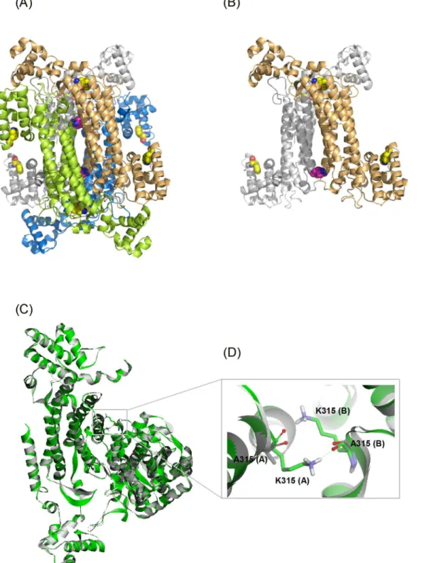
δ-Crystallin and ASL have similar X-ray crystal structures. Each monomer consists of three domains. The helices in domain 2 of each monomer associate to form a central helix bundle, comprising the core structure of the protein (Fig 1A) [4,5,7–10]. The active sites of the enzyme
are located in a cleft between three different monomers [4]. The quaternary structure ofδ -crys-tallin consists of a double dimer. The contact surface between the two dimers is smaller than the interface within the primary dimer of the structure [11]. Hydrogen bonding, salt bridges and hydrophobic interactions are the major forces which stabilize the quaternary structure of the protein.
In the presence of guanidinium chloride (GdmCl), tetramericδ-crystallin is unfoldedviaa multistep pathway involving subunit dissociation into a monomeric molten globule intermedi-ate, followed by denaturation [12,13]. The dimeric form is transiently detected during this unfolding/refolding process. These dimers are unstable and they are prone to self-associate into protein aggregates, and this process competes with the formation of native tetramers [8]. Hence, the assembly of two dimers acts as a kinetic barrier in the refolding pathway [14]. The proper assembly of double dimers is thus important for producing a stableδ-crystallin quater-nary structure.
The N-terminus ofδ-crystallin has been identified as being critical for the proper assembly of the double dimers [8]. In the quaternary structure, the first 25 N-terminal amino acid resi-dues protrude into the neighboring monomer and interact with a hydrophobic cavity. When this sequence of amino acids was deleted the protein became unstable and was prone to form protein aggregates. The salt bridge formed by residues of R302 and E330, located in the helix bundle at the dimer-dimer interface, is also important interaction for stabilization of the qua-ternary structure ofδ-crystallin. When this interaction was disrupted by site-directed mutagen-esis, the exchange rate of subunits was dramatically accelerated [15]. The interactions of E327 with both K299 and R302 and the interaction of E267 with Y158 at the dimer interface were found to stabilize the quaternary structure ofδ-crystallin in a cooperative manner. Mutations of the residues involved in both these interactions caused the majority of dimers to dissociate, whilst only partial dissociation was observed when these interactions were individually dis-rupted, as judged by sedimentation velocity experiments [11].
Inspection of the structure ofδ-crystallin showed that the primary interactions between two symmetrically associated monomers in diagonal positions were provided by residues located at the top and bottom sides of the helical bundles (Fig 1B). K315 is one of the residues symmetri-cal located at this interface, interacting with the same residue in other monomers by hydrogen bonds (Fig 1C and 1D). Substitution of this residue with leucine resulted in part dissociation of the quaternary structure. In contrast, the K315A mutant protein was relatively stable. This pro-tein was unfolded into an intermediate with a stable conformation at 3 M urea [11]. This phe-nomenon was not observed for the K315L mutant protein, which was unfolded under the same conditions. The results lead to the hypothesis that the bulky and charged side-chain of K315 might affect the stability of the intermediate during the process of protein unfolding. When wild-typeδ-crystallin was unfolded in GdmCl, the first transition for subunit dissociation resulted in a monomeric intermediate with a molten globule conformation [12,13]. During the refolding of 5 M GdmCl denaturedδ-crystallin a marked hysteresis was observed, suggesting that the quaternary structure of that inappropriate assembled species might be related to the conformation of the refolded monomers [14,16]. The presence of stable intermediate during unfolding of K315A mutant protein in the presence of urea suggests that the interactions pro-vided by this residue at the interfaces might provide an energy barrier for subunit dissociation. dichroism; AEW, average emission wavelength;λmax,
maximum emission wavelength; Cm, half
Fig 1. The structure of gooseδ–crystallin.(A) The quaternary structure (PDB accession no: 1XWO) and (B) the A-B dimeric pairs from diagonal subunits. Monomers of A, B, C and D are colored as green, blue, yellow and gray, respectively. Residues of K315 and W74/W169 are highlighted as CPK and colored as magenta and yellow, respectively. (C) Superimposition of wild-type and K315A mutant proteins present as green and grey color, respectively. A expand view of the interactions of K315 at the interface of A and B subunit is highlighted in (D). Residues of K315 and A315 are displayed as stick models. The interactions are present by dashed green lines.
In this study, the effects of this interaction on the folding pathway of wild-type and mutant proteins were investigated using urea as a denaturant. The different distributions of dissociated component from wild-type and mutant proteins, as measured by sedimentation velocity exper-iment, suggests the quaternary structure dissociates in different ways for wild-type and mutant proteins. Structural simulation supports different dissociation processes for the two proteins. These results highlight the critical role of K315 in stabilizing the quaternary structure ofδ -crys-tallin. The residue appears to control both the dissociation of dimers into monomers and the stability of the produced monomers. The monomers dissociated from the K315A mutant pro-tein with a stable and compact conformation provided a good model for studying the folding mechanism of theδ-crystallin. This study reveals the conformational status of the monomers, which determines whether functional protein or aggregates are formed.
Materials and Methods
Protein production and purification
The recombinant wild-type and the K315A mutantδ-crystallin orαA-crystallin plasmid were transformed and over-expressed inE.coliBL21 (DE3) with induction by isopropyl-β -D-thio-galactopyranoside (IPTG). Proteins were purified as previously described [8,17]. The superna-tants of crude cell extracts were loaded onto a Q-Sepharose anion exchange column (HiPrep 16/10 Q XL, GE Healthcare) pre-equilibrated in buffer A (50 mM Tris-HCl buffer, pH 7.5) and eluted with a linear gradient of 0 to 0.4 M NaCl in buffer A. Recombinant protein was eluted at approximately 0.15 M NaCl. The eluted protein was pooled and treated with ammonium sul-fate to 1.2 M. After filtration, the sample was loaded onto a hydrophobic interaction column (Source™15PHE) pre-equilibrated in buffer A containing 10% (v/v) glycerol and 1.2 M ammo-nium sulfate and eluted with a linear gradient to the same buffer lacking ammoammo-nium sulfate. The retained proteins were eluted at ~0.3 M ammonium sulfate. Fractions were pooled and loaded onto S-300 Sephacryl column (26 mm x 85 cm) pre-equilibrated in 50 mM Tris-acetic acid buffer, pH 7.5. Fractions were analyzed by SDS-PAGE and protein concentrations deter-mined by the method of Bradford [18]. Proteins possessing a C-terminal His6tag were purified on Ni affinity column (Chelating Sepharose FF, GE Healthcare) then desalted using a Sephadex G-25 column (26 mm x 12 cm) as previously reported [11].
Equilibrium unfolding and refolding experiments
Equilibrium unfolding experiments were carried out by overnight incubation ofδ-crystallin with various concentrations of urea or GdmCl in 50 mM Tris-acetic acid buffer, pH 7.5 at 25°C. The refolding experiments were undertaken by dilution of equilibrium-denaturedδ -crys-tallin to a series of urea or GdmCl concentrations in the same buffer.
The experiments for equilibrium unfolding of monomericδ-crystallin were undertaken by overnight incubation ofδ-crystallin in 1.5 M urea at 25°C followed by addition of various con-centrations of GdmCl to the solution followed by incubation for 2 hrs. The refolding experi-ments were carried out by dilution of the denaturedδ-crystallin into a solution containing 1.5 M urea and 0.8, 3 or 5 M GdmCl in 50 mM Tris-acetic acid buffer, pH 7.5. To analyze the con-formation and quaternary structure of the refoldedδ-crystallin, the refolding experiments were undertaken by 20-fold dilution of the denatured monomericδ-crystallin with buffer.
Data analysis
as a two-state transition for the conversion of the tetramer (T) into monomers (M). The ther-modynamic parameters were obtained by global fitting of the tryptophan fluorescence signal to Eq 1[19]:
y0¼ fðynþmf½DÞ þ ðyuþmu½DÞexpð ðDGu
0
m½DÞ=RTÞg=f1þexpð ðDG
u
0
m½DÞ=RTÞg ð1Þ
whereyis the observed signal from tryptophanfluorescence,ynandyuare the signals in the folded and unfolded states, andmfandmuare the slopes of the baselines preceding and following the transition region.ΔGu0is the free energy difference in the absence of urea andmis the variation in the free energy of unfolding with the urea concentration.
Reversible unfolding of monomeric K315A mutantδ-crystallin were described as a four state process (M$I1$I2$U). The unfolding curve from tryptophan fluorescence was ana-lyzed using a four-state unfolding model described as the transition from N to U with two intermediates, I1and I2, in the process. The thermodynamic parameters were calculated by fit-ting the data toEq 2[8,20]:
y0¼ fyNþyI1expf ðDG1 0
m1½DÞ=RTg þyI2expf ðDG1 0
m1½D þDG2 0
m2½DÞ=RTgþ
yUexpf ðDG1 0
m1½D þDG2 0
m2½D þDG3 0
m3½DÞ=RTgg=f1þexpf ðDG1 0
m1½DÞ=RTg
þexpf ðDG1 0
m1½D þDG2
0 m
2½DÞ=RTg þexpf ðDG1 0
m1½D þDG2 0
m2½D þDG3 0
m3½DÞ=RTgg
ð2Þ
whereyI1andyI2are the signals in the I1and I2states.ΔG10,ΔG20andΔG30are the free energy differences in the absence of GdmCl for N to I1, I1to I2and I2to U transition, respectively, andm1,m2andm3are the variation in the free energy of unfolding with the GdmCl concentration.
Enzymatic activity assay
δ-crystallin was assayed for ASL activity by monitoring the absorption of fumarate at 240 nm in a Perkin-Elmer Lambda 40 spectrophotometer. Assays were performed at least in triplicate in 50 mM Tris-HCl buffer (pH 7.5) with 1 mM sodium argininosuccinate as substrate. A molar absorption coefficient of 2.44 x 10³M-1cm-1was used for all calculations [21].
Fluorescence studies
The fluorescence spectra were measured on a Perkin-Elmer LS-50 luminescence spectropho-tometer at 25°C. Intrinsic tryptophan fluorescence spectra of the protein were recorded with excitation wavelength set at 295 nm and using 5 nm band-width for both excitation and emis-sion wavelength. All spectra were corrected for buffer or denaturant absorption. The average emission wavelength was utilized for data analysis [22].
The ANS (1-anilinonaphthalene-8-sulfonic acid) (Molecular Probes; Eugene, Oregon) was used as probe to bind with proteins and the fluorescence was recorded from 450 to 550 nm Fig 2. The unfolding transition of the wild-type and mutantδ-crystallin.Changes in average emission wavelength (AEW) of tryptophan fluorescence are represented as a function of GdmCl (A) and urea (B) concentration. (C) The integrated area of the fluorescence emission spectrum of ANS at varied urea concentration. The labels of (●) and (), and (▲) and (∆) represent the unfolding and refolding of wild-type and K315A mutant protein, respectively. In (B) and (C), the dark or grey open circles and triangles represent the refolding transition from 3.5 or 4 M urea denatured wild-type and 2.5 or 6 M urea denatured mutant protein, respectively. The (■) and (□) in (B) represent the changes of mean residue of ellipticity (MRE) in the unfolding
of wild-type and mutant protein, respectively. The protein concentrations used in the assays were 0.03, 0.2 and 0.1 mg/mL for tryptophan fluorescence, CD and ANS fluorescence measurements, respectively.
with the excitation wavelength set at 370 nm. The band-widths were set at 5 nm for both the excitation and emission wavelengths. The final concentration of ANS was 0.2 mM.
Circular dichroism studies
The circular dichroism (CD) spectra were measured on a Jasco J-810 spectropolarimeter at 25°C. Experiments were performed in 20 mM Tris-acetic acid buffer (pH 7.5) with a 1 mm path-length over a wavelength range from 200 to 250 nm. All spectra were averaged from three accumulations and were buffer corrected. The observed ellipticity (θ) (degrees) was converted into the mean residue ellipticity [θ] by the equation: [Θ] =Θ×MMRW/10×d×c[23], where MMRWis the mean residue weight,dis the light path (cm), andcis the concentration of protein in g/ml.
Analytical ultracentrifugation studies
The protein sedimentation were performed on a Beckman-Coulter (Palo Alto, CA) XL-A ana-lytical ultracentrifuge (AUC) with an An50 Ti rotor at 20°C with 130 000gin standard double sectors aluminum centerpieces. The radial scans were recorded with a time interval of 7-min and a step size of 0.003 cm. All data were fitted to the continuous c(s) distribution model and a continuous size-distribution with respect to frictional ratio (f/fo) model using the SEDFIT pro-gram [24,25]. The partial specific volume of the protein, solvent density and viscosity were cal-culated using the SEDNTERP program [26]. The solvent density and viscosity of varied urea concentrations were calculated and included in the fitting.
Non-denaturing gradient gel electrophoresis
Electrophoresis was performed using a PhastSystem (GE Healthcare). The samples were sub-jected a PhastGel 4–15% gradient gel which contains the Native Buffer Strip (0.88 M L-alanine,
0.25 M Tris/HCl pH 8.8) attached to the surface of the gel and both electrodes. The electropho-resis was carried out at 10 mA and 15°C for 400 Vh. After electrophoresis, the gel was fixed in
20% (w/v) trichloroacetic acid and stained with Coomassie Brilliant Blue R250.
Protein aggregation measurements
The turbidity of protein solution was measured using a PerkinElmer Lambda 40 spectropho-tometer equipped with a Peltier temperature control accessory to monitor the light scattering at 360 nm. All measurements were carried out at 25°C. Spectra were corrected using measure-ments recorded for native protein in the absence of denaturants. The aggregation rate was cal-culated by fitting the data to the single or double exponential equation:
yt¼y0þ
Xn
i¼1
yi½1 expð kitÞ
Whereytis the signal at time t,y0is the signal of thefinal state,yiis the change in amplitude, andkiis the rate constant for aggregation. Data for the linear increase in signals wasfitted to a linear equation using SigmaPlot 10.
Thioflavin T assay
Structural simulation
The crystal structure of gooseδ–crystallin (PDB code: 1XWO) with all water molecule removed was subjected to the CHARMm force field and energy minimized with the smart minimization algorithm to satisfy (RMS Gradient ~0.1 kcal/mol/Å). The implicit solvent model of General-ized Born was included in the calculation. The structural model was used as a template to build the K315A mutant model using the build mutant protocol (Accelrys Discovery Studio 3.5, Accelrys Inc.). The best scoring model conformation was selected for energy minimization.
Molecular Dynamics (MD) simulations were performed using the standard dynamics cas-cade protocol. The structures of wild-type and mutantδ-crystallin in the CHARMm force field were subjected to initial energy minimization for 500 steps by steepest descent followed by a conjugate gradient for 500 steps. The minimized models were then heated from 50 to 300 K in 2 ps MD simulations followed by equilibration for 2 ps at 300 K in the absence of any structural restraint. Finally, the equilibrated models were submitted to MD simulations for 100 ps at NVT (constant number of particles, volume, and temperature) using the Berendsen coupling algorithm [27].
Results
Conformational reversibility of
δ
-crystallins in the presence of
denaturants
Wild-type and K315A mutantδ-crystallin purified to near homogeneity were used for all anal-ysis. Equilibrium unfolding experiments were conducted by incubation of proteins in buffer supplemented with different GdmCl or urea concentrations overnight. Tryptophan fluores-cence was used to monitor the conformational changes during the unfolding process in the microenvironment around the tryptophan residues (Fig 2A and 2B). There are two tryptophan residues, W74 and W169, in the structure ofδ-crystallin. They are located in the solvent acces-sible domain 1 and the helix bundle of domain 2, respectively (Fig 1A and 1B). Unfolding of the wild-type and K315A mutant protein follows a multistep process in GdmCl solution and was not reversible after 20-fold dilution (Fig 2A). As shown inFig 2A, the dramatic changes in the signal for the first transition were due to subunit dissociation as reported previously, with the GdmCl concentrations for half transition ([D]1/2) at 1 ± 0.05 and 0.5 ± 0.01 M for wild-type and K315A mutant protein, respectively [28]. Unfolding of the two proteins in urea solu-tion followed a three-state process, with the [D]1/2values in the first transition at 3.6 ± 0.1 and 1.7 ± 0.1 M urea, respectively (Fig 2B) [8,12]. The differences in the denaturant concentration required for the half transition clearly show the potency of GdmCl when disrupting of the con-formation ofδ-crystallin [11].
In the presence of urea, the K315A mutant protein was dissociated and stayed in a stable conformation between 2 and 5 M urea. The conformation was more stable than for wild-type protein at urea concentrations exceeding 4 M. When the urea was removed by 20-fold dilution into buffer, the denatured mutant protein was able to refold into a conformation similar to the original state (Fig 2B). In contrast, dilution of 3.5 or 4 M urea denatured wild-typeδ-crystallin or 6 M urea denatured K315A mutant protein did not result in the restoration of the properly folded conformation. These results suggest that the reversible assembly of the quaternary struc-ture ofδ-crystallin is correlated with the conformation of the dissociated monomers.
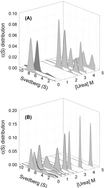
Fig 3. Sedimentation velocity analysis.The continuous sedimentation coefficient distribution of (A) wild-type and (B) K315A mutant protein at different urea concentration were shown. Peaks in grey color represent the unfolded wild-type and mutant protein in 0, 2.5, 3, 3.5 and 4.5 M urea and in 0, 0.25, 0.7, 1.3, 1.5, 2, 2.5, 3.5 and 4.5 M urea, respectively. Wild-type and mutant protein denatured in 3.5 and 2.5 M urea were diluted to final 0.6 and 0.3 M urea, respectively, in 50 mM Tris-acetic acid buffer, pH 7.5, were shown as dark gray peaks. The protein concentration used in all assays was 0.2 mg/mL.
with the observed changes in the tryptophan micro-environment as probed by tryptophan fluo-rescence (Fig 2B). The highest signals occurred around 2 and 5 M urea for the mutant and wild-type protein, respectively. Dilution of the 2.5 M urea denatured K315A mutant protein resulted in restoration of a native-like conformation. However, dilution of 4 or 6 M urea denatured wild-type or mutant protein, respectively, resulted in higher levels exposure of hydrophobic areas.
Sinceα-helices are the major secondary structure inδ-crystallin, the ellipticity at 222 nm was used to analyze the structural changes induced by the presence of urea (Fig 2B). The results showed both proteins retaining relatively stableα-helical structure at concentrations of urea below 2 M. There was about 13% and 30% loss of the structure at 3 M and 4 M urea, respectively.
Effect of urea on the size-and-shape changes of
δ
-crystallin variants
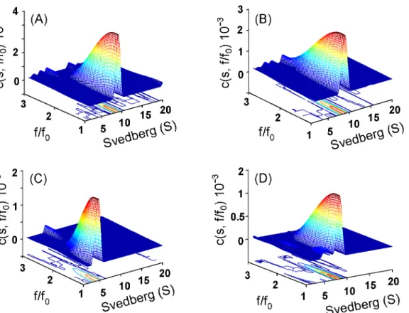
The size and size-and-shape changes of wild-type and K315A mutant protein in different urea concentrations were determined by sedimentation velocity measurements and using continu-ousc(s) distribution andc(s,f/f0) distribution analysis, respectively (Figs3and4) (S1andS2 Figs) [29]. In the absence of urea, the two proteins appeared as one major component with sed-imentation coefficients about 8.5 and 8.4 S, respectively (Fig 3). This peak corresponds to tetra-mericδ-crystallin [11]. They possessed the native conformation as judged from the friction ratio (f/f0) distribution profile (with the centre region below 1.5 as shown by red in the contour) (Fig 4A and 4B).
At 2.5 M urea two components were observed for wild-type protein with sedimentation coefficients about 6.6 and 3.2 S, and these are assumed to be the dissociated dimeric and mono-meric forms. The S values of the two peaks decreased with increasing urea concentration. The proportion of the second (monomeric) peak increased from 6% to 27% to 60% in the presence of 2.5, 3.0 to 3.5 M urea, respectively. Dilution of the denatured wild-type protein at 3.5 M urea resulted in refolding into one major component with an S value of 8.1 (Fig 3A). Measurement of ASL activity showed that around 25% activity was recovered following refolding (Table 1).
Subunit dissociation was observed at about 1.2 M urea for the K315A mutant protein, with S values for the major peak of 6.8 and a shoulder at about 4.5 S (Fig 3B). A single peak with an S value of 4.5 was observed for the mutant protein at ~1.5 M urea. This species is thought to be dissociated monomers possessing the native conformation as judged from the friction ratio (f/ f0) distribution profile (Fig 4C). The monomers were reassembled into a similar quaternary structure of wild-type protein after removing the urea (Fig 4D). When the urea concentration was increased to 4.5 M, the S values for the single component were gradually decreased to about 2.4 (Fig 3B). Dilution of the protein denatured with 2.5 M urea resulted in refolding into one major peak with a S value of 8.1 S. The refolded protein showed around 80% ASL activity was recovered (Table 1).
Conformational reversibility of monomeric K315A mutant
δ
-crystallin
Fig 4. Size-and-shape distribution analysis.The sedimentation velocity experiments were performed and presented as the c(s,f/fo) distribution for (A) wild-type, (B) and (C) K315A protein in the absence and presence of 1.5 M urea, respectively. (D), K315A protein dissociated in 1.5 M urea was diluted to final of 0.26 M urea in 50 mM Tris-acetic acid buffer (pH 7.5).
doi:10.1371/journal.pone.0145957.g004
Table 1. Specific activity ofδ-crystallin under the denaturant effect.
Renaturation conditions Specific activity(nmol/min/mg)
Wild-type
no treatment 4.4±0.1
refolded from 3.5 M ureaa 1.1±0.2
refolded from 5 M GdmClb -d
K315A mutant
no treatment 0.28±0.03
refolded from 1.5 M urea 0.26±0.1
refolded from 2.5 M urea 0.23±0.08
refolded from 5 M GdmCl
-refolded from 1.5 M urea and 5 M GdmClc 0.9±0.01
0.04 M urea and 0.13 M GdmCl 1.5±0.05
aThe proteins unfolded in 3.5, 2.5 or 1.5 M urea were 20-fold diluted with 50 mM Tris-acetic acid buffer (pH
7.5).
bThe proteins unfolded in 5 M GdmCl were 40-fold diluted and incubated for overnight.
cThe mutant protein was unfolded in 1.5 M urea for overnight followed by addition of 5 M GdmCl and
incubation for 2 hrs. Then, the protein was diluted 40-fold and incubated for overnight before activity measurement.
dno detectable activity.
correlated with the exposure of hydrophobic areas, which suggests the possible four-state unfolding model for monomericδ-crystallin.
Dilution of monomeric K315A mutant protein denatured in 5 M GdmCl resulted in refold-ing to a similar conformation as the original monomeric state (Fig 5A and 5B). However, dilu-tion of 1 and 3 M GdmCl denatured monomeric protein resulted in the increasing of the ANS fluorescence, indicating higher exposure of hydrophobic area (Fig 5B). The results suggest the conformation of the partly unfolded intermediate could affect the folding reversibility of the monomeric K315Aδ-crystallin mutant.
Thermodynamic parameters calculation
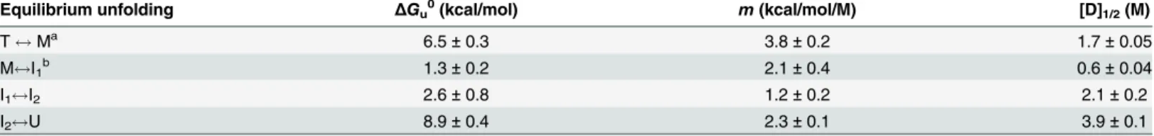
K315A mutantδ-crystallin that denatured in 3 M urea was reversible folded back to the origi-nal conformation after dilution (Fig 2B). Signal changes in the tryptophan fluorescence with different urea concentration were used to calculate the thermodynamic parameters by directly fitted to the two-state mechanism (Eq 1) [19]. The free energy difference in the absence urea (ΔG0) for the transition was determined to be 6.5 ± 0.3 kcal/mol (Table 2).
The changes of tryptophan fluorescence as a function of GdmCl concentration were used to calculate the thermodynamic parameters for the reversible unfolding of the monomeric K315A δ-crystallin mutant (Fig 5A). The unfolding curve was best fitted into a four-state model [8,20]. The [GdmCl]1/2for the transitions from the M to I1, I1to I2and I2to U (denaturation) were about 0.6 ± 0.04 M, 2.1 ± 0.2 M and 3.9 ± 0.1 M, respectively. The thermodynamic parameters determined are summarized inTable 2. The total free energy difference (ΔG0) for folding of monomeric K315Aδ-crystallin mutant was determined to be 12.8 ± 0.7 kcal/mol.
Reversibility of the quaternary structure from denatured monomeric
K315A mutant
δ
-crystallin
To determine the ability about refolding of denatured monomeric K315A mutant followed by reassembly to a tetrameric protein, the monomeric proteins that denatured by GdmCl were diluted with buffer to remove both of the urea and GdmCl. The results showed that the quater-nary structure of K315A mutant protein was recovered from the denatured monomers instantly after dilution. The amount of the assembled protein was increased with the incuba-tion time possessing about 60% of the activity recovered (Fig 5CandTable 1). In contrast, without denaturant treatment, respectively. Samples from refolding of 5 M GdmCl denatured wild-type and mutant protein for time period of zero or overnight are shown in lanes 3–4 and 5–6, respectively. The protein
concentrations used in the assays were 0.03, 0.1 and 0.1 mg/mL for (A), (B) and (C), respectively.
doi:10.1371/journal.pone.0145957.g005
Table 2. Thermodynamic parameters for K315A mutantδ-crystallin.
Equilibrium unfolding ΔGu0(kcal/mol) m(kcal/mol/M) [D]
1/2(M)
T$Ma 6.5±0.3 3.8±0.2 1.7±0.05
M$I1b 1.3±0.2 2.1±0.4 0.6±0.04
I1$I2 2.6±0.8 1.2±0.2 2.1±0.2
I2$U 8.9±0.4 2.3±0.1 3.9±0.1
aThe reversible dissociation process was described as a two-state transition from the conversion of the tetramer (T) to monomeric intermediate (M). [D] 1/2
is the concentration of denaturant at which the transition is half completed. The data was calculated by globalfitting toEq 1.
bThe data were
fitted to a 4-state unfolding model (Eq 2) described as the transition from M to U. Two intermediates, I1and I2, were assumed in the
process before denatured species (U). These data are the mean±SD of at least three independent experiments.
dilution of 5 M GdmCl denatured wild-typeδ-crystallin, the quaternary structure was recov-ered after overnight incubation with no detectable activity (Table 1). The similar result was also shown for 5 M GdmCl denatured K315A mutant protein. These results suggested the dif-ferent pathway for protein folding that seems due to the distinct conformation of the denatured protein caused by different means of denaturation.
Protein aggregate formation from refolding of monomeric intermediate
Since refolding of partly unfolded monomeric mutantδ-crystallin resulted in a conformation with high exposure of hydrophobic regions, the occurrence of protein aggregation in the process was determined using light scattering measurement. No protein aggregation was detected upon dilution of 0.84, 3 and 5 M GdmCl denatured monomeric mutant protein into buffer containing 1.5 M urea. However, when 0.84 and 3 M GdmCl denatured monomeric mutant protein was diluted into buffer, protein aggregation was occurred. The rates for aggregate formation were cal-culated to beca. 0.14 and 0.0004 min-1, respectively (Fig 6A). WhenαA-crystallin, the chaperone protein, was added in a 5:1 ratio to 0.84 M GdmCl denatured monomeric mutant protein in fold-ing buffer, no change in the rate of protein aggregation was observed. Formation of aggregates by αA-crystallin alone did not occur under the same conditions. It is notable that upon dilution of 5 M GdmCl denatured monomeric mutant protein into buffer, no aggregation occurred.The structural features of the protein aggregate were investigated using the thioflavin T assay [30]. An increase in fluorescence intensity resulting from binding of ThT with the aggre-gates over time was observed following dilution of 0.84 and 3 M GdmCl denatured monomeric mutantδ-crystallin into buffer (Fig 6B). The results suggest the possible formation of ordered aggregates. No changes in the signal were observed during the incubation period upon refold-ing of 5 M GdmCl denatured monomeric mutant protein.
Molecular dynamics simulation
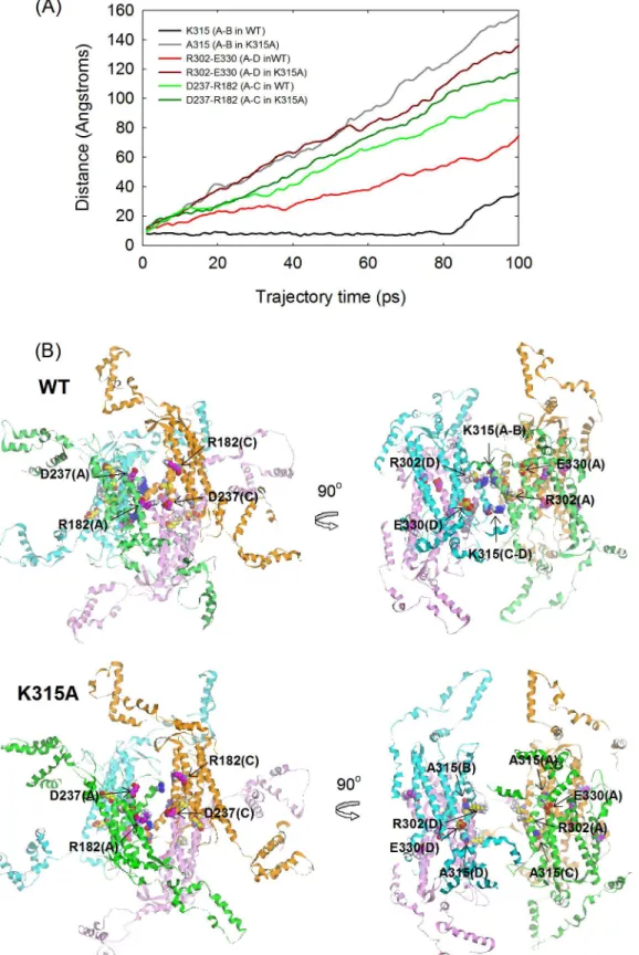
To determine the effect of the interactions provided by K315 at the diagonal subunits in disas-sembly of the quaternary structure, a MD simulation were run for 100 ps for wild-type and mutantδ-crystallin in the absence of any structural restraints. From the simulation trajectory, the dynamic motion for disassembly of the quaternary structure and conformational changes in the tertiary structure were elucidated. The distances between the Cαof D237 and R182, R302 and E330 and the two K315 or A315 residues were measured to evaluate the extent for subunit dissociation between the A-C, A-D and A-B dimeric pairs, respectively (Fig 7A). These residues interact with each other by hydrogen bonding or salt bridges at the dimeric pair interface in the native structure. These interactions are lost on replacement of K315 with A315. The results showed that the distances between D237 and R182, R302 and E330 and the two A315 residues increased linearly at a similar rate, except that the rate of change for the R302-E330 interaction in wild-type protein was about half of that for the mutant protein. In contrast, no changes in the distance between the K315 residues were observed before 80 ps. Inspection of the time-course at 20 ps in wild-type protein showed that the primary dimers of subunit A and C or B and D showed were separated, while the diagonal dimers of subunit A and B or C and D were connected by the interactions of residue K315 (Fig 7B). However, the subunits for both of the primary and diagonal dimers were separated from each other in the mutant protein. The results suggest a different disassembly process for tetrameric wild-type and mutant proteins.
Discussion
double dimers to elucidate their role in the stabilization of the quaternary structure [11]. The unique stable conformation from unfolding of K315A mutant protein in the presence of urea suggests that the interactions provided by this residue at the interfaces may play a critical role in stabilization of the quaternary structure ofδ-crystallin.
Lys-315 is the only residue which is arranged symmetrically at the diagonal subunit inter-faces (Fig 1B). Theε-amino group in the side-chain of this residue forms hydrogen bonds with the carbonyl groups of M312, V313 and K315 within the symmetric subunit (from PISA analy-sis:http://www.ebi.ac.uk/msd-srv/prpt_int/cgi-bin/piserver) (Fig 1D). Substitution of this Fig 6. Aggregates formation from refolding of denatured monomeric K315A mutantδ-crystallin.(A) Light scattering measurement. Monomeric K315A mutantδ-crystallin was incubated in 1.5 M urea before
introducing varied GdmCl concentration. Refolding of 0.84, 3 and 5 M GdmCl denatured monomeric K315A mutant protein by dilution with 50 mM Tris-acetic acid buffer (pH7.5) or the buffer containing 1.5 M urea are shown as (●,▲and■) or (,∆, and□), respectively. The label of (♦) represents refolding of 0.84 M GdmCl
denatured monomers by dilution with buffer includingαA-crystallin.αA-Crystallin alone in 1.5 M urea and 0.84
M GdmCl is shown as (^). The protein concentrations of monomeric K315A andαA-crystallin used in the
assays were 0.1 and 0.5 mg/mL, respectively. (B) The fluorescence spectrum of ThT were shown from refolding of 0.84 (dark), 3 (gray) and 5 M (dark gray) GdmCl denatured monomeric K315A mutant protein by dilution with buffer and incubated for zero (solid-line), 1 (dash-line) or 2 (dot-line) hrs, respectively.
Fig 7. Molecular dynamics simulations.(A) The distances between the Cαof specific residues located at the interfaces along the simulation. (B) The
conformation of wild-type and K315A mutant protein at trajectory frame of 20 ps.
residue by alanine reduces the structural stability of the protein. The results from the previous study showed about a 9°C reduction in the thermal stability of the secondary structure and the changes in the micro-environment surrounding the tryptophan residues [11]. This mutant protein was also more susceptible to chemical denaturation, since about half of the concentra-tion of denaturant was required to disrupt its quaternary structure compared to wild-type pro-tein. Both of the wild-type and mutant protein showed similar and not reversible denaturation in the presence of GdmCl. However, differences in the denaturation pathway were observed when urea was used as the denaturant. The results suggest that the non-covalent interactions between the intra and inter-subunits might be disrupted by the ionic character of GdmCl, with subunit dissociation and denaturation occurring simultaneously for both proteins [31]. The previous studies showing the presence of only a monomeric molten globule intermediate in the dissociation pathway of wild-typeδ-crystallin in GdmCl supports this assumption [12,13]. Thus, the role of K315 in the folding process ofδ-crystallin cannot be distinguished under the strong denaturant.
Around 2~5 M urea, the K315A mutantδ-crystallin is in a stable conformation, as judged by tryptophan fluorescence measurements (Fig 2B). Subunit dissociation occurs under these conditions, resulting in the exposure of hydrophobic regions (Figs2Cand3B). Only mers were identified in this state, as measured by sedimentation velocity analysis. The mono-mers that dissociated from the tetramono-mers at ~1.5 M urea possessed a nearly identical content of secondary structure as native protein and native-like conformation (Figs2Band4C). Mono-mers in this conformation were able to refold and re-associate into tetraMono-mers with a similar conformation as the native protein, and possessed significant ASL activity. When the urea con-centration was increased to 2.5 M, the conformational changes led to further exposure of hydrophobic regions. However, following this conformational change at higher urea concen-trations, protein folding was not reversible. The contrasting result found for wild-type protein was that the dissociation and denaturation were concurrent under the effect of urea (Fig 3A). In this condition, the conformation of the dissociated monomers was partly unfolded, as judged from the reduced level ofα-helix (Fig 2B). The dissociated monomers seem to refold into alternative conformations then re-associate into tetramers with only part of the catalytic activity recovered (Table 1). These results indicated that the conformation of the monomers seems related to the assembly pattern for functional protein.
sedimentation velocity experiments. A possible explanation for this earlier dissociation of the subunit from the primary dimers compared to diagonal dimers is differences in solvent accessi-bility. Unlike the location of the interfaces between the subunits of the primary dimer, the posi-tion of K315 is buried at the interior interfaces away from solvent. Thus, the interacposi-tions of K315 at the interfaces of the protein seem to elevate the stability of the quaternary structure. For wild-type protein, two diagonal dimers were presumed to disassemble initially from the tet-ramers followed by subunit dissociation of the diagonal dimers. However, dissociation at the interfaces of two primary dimers would assume to be the first step in the unfolding process of the K315A mutant protein (Fig 8).
The detail mechanism for folding of monomeric protein remains elusive due to the mono-mers dissociated from wild-typeδ-crystallin were in a molten globule conformation. Thus, the monomers that reversible dissociated from K315A mutantδ-crystallin with a stable conforma-tion and possessing similar level of secondary structure as the original state, and this would be a good model for studying the folding process. The monomeric protein was reversible dena-tured in a four-state mechanism in the presence of GdmCl and two intermediates were detected in the process (Fig 5). Refolding of the partly unfolded intermediate was not reversible which in turn resulted in a conformation with more exposure of hydrophobic regions. Only denaturedδ-crystallin was reversible folded into the monomers with a similar conformation to the original state. It is interesting that the refolded monomers were able to reassemble into tet-ramer instantly upon dilutions, with substantial recovery of activity (Fig 5andTable 1). This contrasts with the slow refolding of GdmCl denatured wild-type protein into its tetrameric form with no detectable activity. The slow recovery of the quaternary structure for the latter protein is due to an energy barrier for the appropriate assembly of double dimers, as reported previously [14]. The results suggest that the conformation of the denatured monomers which Fig 8. A working model proposed for the folding pathway of wild-type and K315A mutantδ-crystallin. The tetrameric wild-type (T) and K315A mutant (T*)δ-crystallin was dissociated through the diagonal dimer (D) and primary dimer (D*) to monomers with partial unfolded (M) and stable (M*) conformation, respectively. Monomers of the wild-type or the mutant protein were then denatured through intermediate (I or I*) into respective unfolded form (U or U*). The monomers (M) of wild-type protein in partial unfolded conformation was associated in alternative pathway to form dimers (D1), and then assembled into tetramers (T1) or
aggregates (A). The aggregation was prevented byαA-crystallin. Refolding followed by assembly of the intermediates (I1*and I2*) of the mutant protein resulted in the aggregates (A1and A2) formation and the
chaperone function ofαA-crystallin was invalid in this process.
was unfolded by stepwise dissociation or directly unfolded with 5 M GdmCl could be different. The consequence of this might be that protein folding occursviadifferent pathways leading to the refolded monomers with different conformations to associate into native structure or alter-native structures without function.
Protein aggregates are prone to form during the reassembly process from refolding of partly unfolded monomeric intermediates ofδ-crystallin. The intermediate with the highest exposure of hydrophobic conformation is particularly prone to aggregate formation (Fig 5). Aggregate formation by monomeric intermediates with defined conformations was also reported for transthyretin under mildly acidic conditions [33]. The result implies that the conformational status of the monomers influences subunit association. It is interesting that the presence ofα -crystallin seems to increase the formation of aggregates from the monomeric intermediates of δ-crystallin with partly unfolded conformation, whileα-crystallin alone was not affected under these conditions (Fig 6). The studies forαA peptide which induces the aggregation of soluble α-crystallin suggested that the mechanism for aggregate formation might due to the changes in the hydrophobicity ofα-crystallin induced by the peptide [34,35]. Our previous study reported that aggregate formation during refolding of GdmCl denatured wild-typeδ-crystallin was due to the improper assembling of double dimers and was prevented by the presence ofα-crystallin [14]. In this study, the aggregate formation was caused by assembly of the refolded monomeric intermediate which facilitated the aggregate formation ofα-crystallin. It thus postulated that the electrostatic interaction with the substrate seems to be key factors to determine the chap-eron-like or anti-chaperone activity of theα-crystallin [34,36]. The underlying mechanism requires further investigation. Nonetheless, the result highlights the conformational status of the monomers which play a critical role in the folding pathway for reversible oligomerization or aggregate formation.
In conclusion, the folding pathways of wild-type and mutantδ-crystallin are summarized as the working models shown inFig 8. This model depicts the key interactions from K315 at the interfaces of diagonal subunits not only to stabilize the quaternary structure ofδ-crystallin but also to act as the energy barrier for dissociation of stable monomers. The stability might be one of the reasons for recruitment of the metabolic enzyme ASL into the lens as a crystallin protein [37]. The single polypeptide chain ofδ-crystallin after translation would be assumed to fold into functional tetramers as the proposed refolding pathway for K315A mutant. However, due to the interactions by K315, the tetrameric protein would be assumed to dissemble in an alter-native manner to form the diagonal dimers, followed by simultaneous subunit dissociation and denaturation. Monomers in this status might associate into dimersviaa different pathway which then assemble slowly into a non-native tetrameric form or self-associate into aggregates which can be prevented by the presence ofα-crystallin. The reversible folding of the monomers that dissociated from the K315A mutant protein with near native conformation provided the folding mechanism of theδ-crystallin. In this process, the ordered aggregate formation from re-association of the partly unfolded intermediate reveals a specific status of the protein to avoid the chaperone function ofα-crystallin. This model proposes a possible mechanism about the aggregate formation for lens protein under stress effect and their interaction withα -crystal-lin. This study reveals the key role of monomers that dissociated from the oligomeric crystallin; their conformational status determines the levels of aggregate formation.
Supporting Information
continuous size distribution model including the solvent using the SEDFIT program [25]. (TIF)
S2 Fig. Sedimentation velocity analysis of K315A mutantδ-crystallin.(A) and (B), the pan-els show the raw sedimentation and theoretical fitted data (solid lines), and the fitting residual, respectively. (C) Grayscale of residual bitmap. The raw sedimentation data were fitted to the continuous size distribution model using the SEDFIT program [25].
(TIF)
Acknowledgments
We thank Dr M. D. Lloyd (University of Bath, U. K.) for reading of this manuscript before publication.
Author Contributions
Conceived and designed the experiments: HJL WYC. Performed the experiments: CWH HCL WCK CYC. Analyzed the data: CWH CYC. Wrote the paper: CWH HJL.
References
1. Wistow G. Lens crystallins: gene recruitment and evolutionary dynamism. Trends Biochem Sci. 1993; 18: 301–306. PMID:8236445
2. Piatigorsky J, Wistow G. The recruitment of crystallins: new functions precede gene duplication. Sci-ence. 1991; 252: 1078–1079. PMID:2031181
3. Piatigorsky J. Lens crystallins. Innovation associated with changes in gene regulation. J Biol Chem. 1992; 267: 4277–4280. PMID:1537817
4. Simpson A, Bateman O, Driessen H, Lindley P, Moss D, Mylvaganam S, et al. The structure of avian eye lensδ-crystallin reveals a new fold for a superfamily of oligomeric enzymes. Nat Struct Biol. 1994;
1: 724–734. PMID:7634077
5. Turner MA, Simpson A, McInnes RR, Howell PL. Human argininosuccinate lyase: a structural basis for intragenic complementation. Proc Natl Acad Sci U S A. 1997; 94: 9063–9068. PMID:9256435
6. Wistow G, Anderson A, Piatigorsky J. Evidence for neutral and selective processes in the recruitment of enzyme-crystallins in avian lenses. Proc Natl Acad Sci U S A. 1990; 87: 6277–6280. PMID:2385592
7. Abu-Abed M, Turner MA, Vallee F, Simpson A, Slingsby C, Howell PL. Structural comparison of the enzymatically active and inactive forms ofδcrystallin and the role of histidine 91. Biochemistry. 1997; 36: 14012–14022. PMID:9369472
8. Lee HJ, Lai YH, Wu SY, Chen YH. The effect of N-terminal truncation on double-dimer assembly of gooseδ-crystallin. Biochem J. 2005; 392: 545–554. PMID:16101585
9. Sampaleanu LM, Vallee F, Slingsby C, Howell PL. Structural studies of duckδ1 andδ2 crystallin
sug-gest conformational changes occur during catalysis. Biochemistry. 2001; 40: 2732–2742. PMID: 11258884
10. Vallee F, Turner MA, Lindley PL, Howell PL. Crystal structure of an inactive duckδII crystallin mutant
with bound argininosuccinate. Biochemistry. 1999; 38: 2425–2434. PMID:10029536
11. Huang CW, Tseng CC, Chen YH, Chen YH, Chou WY, Lee HJ. Substitution of residues at the double dimer interface affects the stability and oligomerization of gooseδ-crystallin. FEBS J. 2009; 276:
5126–5136. doi:10.1111/j.1742-4658.2009.07209.xPMID:19674108
12. Lee HJ, Chang GG. Guanidine hydrochloride induced reversible dissociation and denaturation of duck
δ2-crystallin. Eur J Biochem. 2000; 267: 3979–3985. PMID:10866796
13. Lee HJ, Lu SW, Chang GG. Monomeric molten globule intermediate involved in the equilibrium unfold-ing of tetrameric duckδ2-crystallin. Eur J Biochem. 2003; 270: 3988–3995. PMID:14511381
14. Yin FY, Chen YH, Yu CM, Pon YC, Lee HJ. Kinetic refolding barrier of guanidinium chloride denatured gooseδ-crystallin leads to regular aggregate formation. Biophys J. 2007; 93: 1235–1245. PMID: 17513375
16. Lai Z, McCulloch J, Lashuel HA, Kelly JW. Guanidine hydrochloride-induced denaturation and refolding of transthyretin exhibits a marked hysteresis: equilibria with high kinetic barriers. Biochemistry. 1997; 36: 10230–10239. PMID:9254621
17. Chen YH, Lee MT, Cheng YW, Chou WY, Yu CM, Lee HJ. Distinct interactions ofαA-crystallin with
homologous substrate proteins,δ-crystallin and argininosuccinate lyase, under thermal stress. Biochi-mie. 2011; 93: 314–320. doi:10.1016/j.biochi.2010.10.003PMID:20937351
18. Bradford MM. A rapid and sensitive method for the quantitation of microgram quantities of protein utiliz-ing the principle of protein-dye bindutiliz-ing. Anal Biochem. 1976; 72: 248–254. PMID:942051
19. Pace CN. Determination and analysis of urea and guanidine hydrochloride denaturation curves. Meth-ods Enzymol. 1986; 131: 266–280. PMID:3773761
20. Dignam JD, Qu X, Chaires JB. Equilibrium unfolding of Bombyx mori glycyl-tRNA synthetase. J Biol Chem. 2001; 276: 4028–4037. PMID:11056158
21. Ratner S. Metabolism of amino acids and amines. Methods Enzymol. 1970; 17A: 304–309.
22. Royer CA, Mann CJ, Matthews CR. Resolution of the fluorescence equilibrium unfolding profile of trp aporepressor using single tryptophan mutants. Protein Sci. 1993; 2: 1844–1852. PMID:8268795
23. Kelly SM, Price NC. The application of circular dichroism to studies of protein folding and unfolding. Bio-chim Biophys Acta. 1997; 1338: 161–185. PMID:9128135
24. Brown PH, Schuck P. Macromolecular size-and-shape distributions by sedimentation velocity analyti-cal ultracentrifugation. Biophys J. 2006; 90: 4651–4661. PMID:16565040
25. Schuck P, Rossmanith P. Determination of the sedimentation coefficient distribution by least-squares boundary modeling. Biopolymers. 2000; 54: 328–341. PMID:10935973
26. Laue TM, Shah BD, Ridgeway TM, Pelleter SL. Analytical Ultracentrifugation in Biochemistry and Poly-mer Science. Cambridge, UK: The Royal Society of Chemistry; 1992.
27. Berendsen HJC, Postma PM, DiNola A, Hakk JR. Molecular dynamics with coupling to an external bath. J Chem Phys. 1984; 81: 3684–3690.
28. Hammarstrom P, Sekijima Y, White JT, Wiseman RL, Lim A, Costello CE, et al. D18G transthyretin is monomeric, aggregation prone, and not detectable in plasma and cerebrospinal fluid: a prescription for central nervous system amyloidosis? Biochemistry. 2003; 42: 6656–6663. PMID:12779320
29. Chang HP, Chou CY, Chang GG. Reversible unfolding of the severe acute respiratory syndrome coro-navirus main protease in guanidinium chloride. Biophys J. 2007; 92: 1374–1383. PMID:17142288
30. Khurana R, Coleman C, Ionescu-Zanetti C, Carter SA, Krishna V, Grover RK, et al. Mechanism of thio-flavin T binding to amyloid fibrils. J Struct Biol. 2005; 151: 229–238. PMID:16125973
31. Monera OD, Kay CM, Hodges RS. Protein denaturation with guanidine hydrochloride or urea provides a different estimate of stability depending on the contributions of electrostatic interactions. Protein Sci. 1994; 3: 1984–1991. PMID:7703845
32. Foss TR, Kelker MS, Wiseman RL, Wilson IA, Kelly JW. Kinetic stabilization of the native state by pro-tein engineering: implications for inhibition of transthyretin amyloidogenesis. J Mol Biol. 2005; 347: 841–854. PMID:15769474
33. Lai Z, Colon W, Kelly JW. The acid-mediated denaturation pathway of transthyretin yields a conforma-tional intermediate that can self-assemble into amyloid. Biochemistry. 1996; 35: 6470–6482. PMID: 8639594
34. Fukuhara S, Nishigaki T, Miyata K, Tsuchiya N, Waku T, Tanaka N. Mechanism of the chaperone-like and antichaperone activities of amyloid fibrils of peptides fromαA-crystallin. Biochemistry. 2012; 51:
5394–5401. PMID:22694216
35. Kannan R, Santhoshkumar P, Mooney BP, Sharma KK. TheαA66-80 peptide interacts with solubleα -crystallin and induces its aggregation and precipitation: a contribution to age-related cataract formation. Biochemistry. 2013; 52: 3638–3650. doi:10.1021/bi301662wPMID:23631441
36. Konno T. Amyloid-induced aggregation and precipitation of soluble proteins: an electrostatic contribu-tion of the Alzheimer's beta(25–35) amyloid fibril. Biochemistry. 2001; 40: 2148–2154. PMID: 11329283