Catarina Félix Malta
CONSEQUÊNCIAS DA INDUÇÃO DE C4BP (β
-) EM
MACRÓFAGOS DERIVADOS DE MONÓCITOS
CONSEQUENCES OF C4BP (β
-) INDUCTION OVER
the jury
president Prof. Doutor Ana Luque
Professor associated to Molecular Genetics Laboratory, IDIBELL
Prof. Doutor Cristina Costa
Professor associated to Molecular Oncology Laboratory, IDIBELL
Prof. Joan Torras
Professor associated to Experimental Nephrology Unit, Bellvitge University Hospital
agradecimentos Quando, aos 18 anos, caí desamparada numa cidade desconhecida, bastaram umas semanas para me sentir novamente em casa. Aveiro tem, sem dúvida, um encanto especial, uma luz diferente de todas as outras cidades, que nos faz apaixonar no momento em vemos a ria com os seus moliceiros. Basta este olhar para não querer mais sair. Por alguma razão é a nossa Veneza Portuguesa.
À Universidade de Aveiro, que também me ofereceu a possibilidade de conhecer as pessoas que me acompanharam durante estes 6 anos. Aos colegas de sempre, um grande obrigada por se terem juntado a mim nesta caminhada, fizeram as viagens para a Praça especiais. Fizeram estes anos memoráveis, tão memoráveis que já tenho vontade de repetir esta aventura novamente. É muito gratificante sentir-me rodeado de tantas pessoas importantes que fizeram dos meus anos de estudante universitário os mais divertidos, sonhadores e de aprendizagem que já tive até hoje. Aveiro sentirá a nossa falta.
À minha família, especialmente à minha avó e à minha mãe, por me darem sempre a motivação necessária para nunca desistir dos desafios e, também, por me darem aquele raspanete que preciso ouvir cada vez que erro. E especialmente à minha mãe, por ser um grande exemplo e por nunca ceder. À minha irmã, que apesar de todas as guerras de criança, esteve sempre presente quando precisei. E um obrigada especial por me ter implantado esta ideia da bioquímica na minha cabeça, que persistiu em ficar.
Ao Josep por me ter dado a oportunidade de descobrir a Imunologia, um mundo totalmente novo de tudo aquilo que aprendi na Universidade. E a toda a equipa com quem aprendi bastante e que, com o passar do tempo, me mostraram que o espanhol não foi impedimento para criar uma amizade.
Obrigada.
acknowledgements At 18 years old, I felt lost in an unknown city, but this feeling did not take too long. Aveiro has, without no doubt, a special charm, a special light that is totally different from another city. We only need to see the river canals with their typical boats for fall in love. For some reason, we call it Portuguese Venice.
To Aveiro University, that offer me the possibility to meet the people who, in these 6 years, were always by my side. To the friends of always, thank you for be next to me on this journey, for make the trips to Praça so special. They made these years memorable, so memorable that I would love to repeat this adventure again. It is so gratifying to be surrounded by so many important people who have made my undergraduate years the most fun, dreamy and learning I have ever had. Aveiro will miss us.
To my family, especially my grandmother and my mother, for always giving me the necessary motivation to never give up the challenges, and to open my eyes in every mistake I did. And especially to my mother, for being a great example and never giving up. To my sister, who despite all our siblings fights, was always present when I needed her. And a special thank you for having implanted this idea of biochemistry in my head, which persisted in staying.
To Josep for giving me the opportunity to discover Immunology, a totally new world, different from everything I learned at the University. And to the whole team, with who I learned a lot, and that, over time, showed me that Spanish was not an impediment to create a friendship.
Thank you. Obrigada.
palavras-chave Macrófagos derivados de monócitos, sistema complemento, C4b-binding protein, estabilidade fenotípica, endocitose, fagocitose, lúpus eritematoso sistémico, autoimunidade.
resumo As doenças inflamatórias são a principal causa de morbilidade e mortalidade em todo o mundo e representam um grande desafio na área das ciências biomédicas. Sendo assim, o desenvolvimento de terapias é essencial e, para tal, uma compreensão do funcionamento do sistema imune do hospedeiro é fundamental.
A ativação do sistema complemento representa o passo inicial para o desenvolvimento de uma resposta imune-infamatória protetiva contra o dano. Porém, uma ativação inapropriada poderá danificar o tecido do hospedeiro, sendo potencialmente destrutivo, podendo causar diversas doenças.
A proteína solúvel responsável pela inibição do sistema complemento, C4b-binding protein (C4BP), atua tanto na regulação da via clássica como na de lectina do sistema complemento, modificando a atividade da C3 convertase e prevenindo, assim, a expressão exagerada deste sistema e, consequentemente, o dano tecidual no hospedeiro. A C4BP é uma grande glicoproteína plasmática oligomérica, composta por sete cadeias α idênticas e uma única cadeia β, que forma três oligómeros plasmáticos devido a diferentes composições de subunidades. A isoforma C4BP sem a cadeia β, C4BP (β-), parece apresentar uma
função direta na modulação de respostas imunes adaptativas. Estudos recentes no laboratório demonstraram que a C4BP (β-) é capaz de promover um perfil
tolerogénico e anti-inflamatório em células dendríticas derivadas de monócitos. Os macrófagos são células amplamente distribuídas do sistema imune inato que apresentam uma função central na defesa do organismo contra patógenos invasores e no mantimento da imune homeostase, contribuindo para a inflamação e promovendo a cura de feridas e reparação de tecidos. São células dinâmicas que maturam sob a influência de sinais provenientes do microambiente.
Neste estudo, monócitos de sangue periférico humano foram diferenciados em macrófagos (M0) e, posteriormente, polarizados a fenótipos M1 e M2, utilizando LPS/IFN-γ, e IL-4, respetivamente. M1 e M2 foram identificados por citometria de fluxo como CD80+ CD64+ e CD11b+ CD209+, respetivamente.
A atividade endocítica e fagocítica foi também investigada dado que os macrófagos são células altamente fagocitárias. Quando os macrófagos foram maturados ao fenótipo M1, uma diminuição da capacidade endocítica foi observada, comparando com as células não polarizadas (M0). No entanto, quando os macrófagos foram polarizados ao fenótipo M2, não foi evidente uma diminuição da expressão da atividade endocítica, comparando com M0, evidenciando, assim, que M2 são células com elevada capacidade endocitária. Além disto, macrófagos M2 também demonstraram uma capacidade fagocitária superior a macrófagos M1. E concluindo, a proteína C4BP (β-) reduziu a
atividade endocítica dos macrófagos, embora não tenha apresentado qualquer efeito na atividade fagocítica.
Alterações no fenótipo e função dos macrófagos pode estar associado com uma variedade de doenças autoimunes, incluindo o lúpus eritematoso sistémico. Esta enfermidade é caracterizada por várias manifestações desde a pele e lesões nas mucosas a danos severos nos rins e no sistema nervoso central. Assim, marcadores da doença mais eficazes, risco, atividade da doença e gravidade dos danos nos órgãos poderão facilitar o diagnóstico prévio e orientar o paciente para uma terapia adequada; dado que os tratamentos atuais não são totalmente eficientes. Por esta razão, o desenvolvimento de novas estratégias é fundamental, e a atividade anti-inflamatória e imunomodeladora da C4BP (β-) poderá representar uma opção terapêutica para esta doença autoimune.
keywords Monocyte-derived macrophages, complement system, C4b-binding protein, phenotypic stability, endocytosis, phagocytosis, systemic lupus erythematosus, autoimmunity.
abstract Inflammatory diseases are the leading cause of morbidity and mortality worldwide and are a major challenge for the biomedical sciences. The development of therapeutics is essential and this requires an understanding of the host immune system.
The activation of the complement system represents the initial step in the protective immune-inflammatory response against damage. Nevertheless, an inappropriate activation may also damage the host tissue, being potentially destructive and causing several diseases.
The soluble complement inhibitor C4b-binding protein (C4BP) acts in the regulation of the classical and lectin pathways of the complement system, modifying the activity of the C3 convertase, and preventing complement over activation and host tissue damage. C4BP is a large oligomeric plasma glycoprotein, composed by seven identical α-chains and a single β-chain, which forms three plasma oligomers with different subunit composition. The C4BP isoform without the β-chain, C4BP (β-) seems to play a direct role in the modulation of adaptive immune responses. Recent studies in the lab demonstrated that C4BP (β-) is able to promote a tolerogenic and anti-inflammatory profile on monocyte-derived dendritic cells.
Macrophages are widely distributed innate immune cells that play central roles in host defense against invading pathogens and in maintaining the immune homeostasis, contributing to inflammation, and promoting wound healing and tissue repair. They are dynamic cells that mature under the influence of signals from the local microenvironment.
In this study, human peripheral blood monocytes were differentiated into uncommitted macrophages (M0) and then polarized to M1 and M2 phenotypes using LPS and IFN-γ, and IL-4, respectively. M1 and M2 were identified by flow cytometry as CD80+ CD64+ and CD11b+ CD209+, respectively.
The endocytic and phagocytic activity was also investigated, since macrophages are highly phagocytic cells. When macrophages were matured to M1 phenotype, a downregulation of the endocytic activity was observed, compared to the unpolarized cells. In contrast, when macrophages were polarized to M2 phenotype, no downregulation of endocytic activity compared with M0 was evidenced, showing that M2 are highly endocytic cells. Moreover, M2 macrophages showed a higher phagocytic activity than M1 macrophages. Finally, C4BP (β-) reduced the endocytic activity of macrophages, although did not have any effect on its phagocytic activity.
Alterations in the phenotype and function of macrophages can be associated with a variety of autoimmune disorders, including systemic lupus erythematosus. This disorder is characterized by several manifestations from the skin to mucosal lesions, to severe damage in the kidneys and in the central nervous system. More effective markers of the disease, risk, disease activity; severity of organ damage and of the outcomes would facilitate earlier diagnosis and guide appropriately targeted therapy; since the actual treatments are not fully effective. For this reason, the development of new strategies is fundamental, and the anti-inflammatory and immunomodulatory activity of C4BP (β-) can represent an effective therapeutic option to this autoimmune disease.
Contents
List of Figures ... viii
List of Abbreviations ... x
Introduction ... 1
1.1. Innate immunity ... 1
1.2. Adaptive immunity ... 3
1.3. Development of hematopoietic stem cells ... 3
1.4. Monocyte-derived macrophages ... 5
1.5. Complement system ... 7
1.6. C4b-binding protein (C4BP) ... 10
1.7. Systemic Lupus Erythematosus ... 12
Motivation ... 15
Hypothesis and objectives ... 15
Document Structure ... 16
Materials and Methods ... 17
2.1. Monocyte isolation from peripheral blood ... 17
2.2. Cellular count by flow cytometry ... 18
2.3. Monocyte-derived macrophage differentiation and polarization ... 18
2.4. Flow cytometry ... 20
2.4.1. Surface marker analysis ... 20
2.4.2. Endocytosis analysis using OVA and LY ... 20
2.4.3. Phagocytosis analysis ... 21
Results ... 23
3.1. Human monocyte-derived macrophage morphology... 23
3.2. Human monocyte-derived macrophage treatment with C4BP (β-) ... 24
3.2.1. C4BP (β-) affects the polarization phenotype of human macrophages ... 24
3.2.2. C4BP (β-) affects the endocytosis of human macrophages ... 26
3.2.3. C4BP (β-) does not affect the phagocytosis of human macrophages ... 28
Discussion ... 31
4.1. Human monocyte-derived macrophages morphology ... 31
4.2. Human monocyte-derived macrophage treatment with C4BP (β-) ... 31
4.2.1. C4BP (β-) affects the polarization phenotype of human macrophages ... 32
4.2.2. C4BP (β-) affects the endocytosis capacity of human macrophages ... 33
4.2.3. C4BP (β-) does not affect phagocytosis on human macrophages ... 34
Conclusions ... 35
5.1. Future work ... 36
List of Figures
Figure 1. Development of the hematopoietic stem cell to mature and highly specialized cell types. ... 5 Figure 2. Complement activation though the classical, lectin, or alternative pathways that converge at the central component of the complement system, C3 ... 9 Figure 3. Schematic structure of the main C4BP isoforms used in this study. ... 11 Figure 4. Disposition of a 24 wells plate for the in vitro macrophages differentiation and polarization, and assessment of the C4BP (β-) effect. Monocyte-derived macrophages were
polarized to M1 and M2 in the presence of GM-CSF and M-CSF, respectively ... 19 Figure 5. Human monocytes were differentiated into uncommitted macrophages (M0) by granulocyte macrophage stimulating factor (GM-CSF) or macrophage colony-stimulating factor (M-CSF) ... 19 Figure 6. Characterization of macrophages morphology after M1 or M2 polarization. ... 24 Figure 7. Relative mean fluorescent intensity (MFI) for M1 macrophage surface markers and effect of C4BP(β-) ... 25
Figure 8. Relative mean fluorescent intensity (MFI) for M2 macrophage surface markers. Monocyte-derived macrophages polarized withIL-4, presenting a M2 phenotype, express an upregulation of CD11b and CD209. ... 26 Figure 9. Representation of the endocytic activity of M1 and M2 polarized macrophages when Ovalbumin (OVA) is captured by receptor-mediated endocytosis ... 27 Figure 10. Representation of the endocytic activity of M1 and M2 polarized macrophages when Lucifer Yellow (LY) is captured by pinocytosis. ... 28 Figure 11. Representation of the phagocytic activity of M1 and M2 polarized macrophages when pHrodo™ Green E. coli BioParticles is captured by phagocytosis ... 29
List of Abbreviations
PRR PAMP TLR APC DC iDC TNF IL HSCs RBCs WBCs GM-CSF M-CSF LPS IFN MBL C4BP FH SCRs CCP ProS SLE ds MDMs PMBC 7-AAD OVA LY MFIGermline-encoded pattern recognition receptors Pathogen-associated molecular patterns
Toll-like receptors Antigen-presenting cells Dendritic Cells Immature DC Tumor-necrosis Factor Interleukin
Hematopoietic stem cells Red Blood Cells
White Blood Cells
Granulocyte-macrophage colony stimulating factor Macrophage stimulating factor
Lipopolysaccharide Interferon
Mannose Binding Lectin C4b-binding protein Factor H
Short Consensus Repeats Complement Control Protein Protein S
Systemic Lupus Erythematosus double-stranded
Monocytes-derived macrophages Peripheral blood mononuclear cell 7-Aminoactinomicina D
Ovalbumin
Lucifer Yellow CH
1
Chapter 1
Introduction
1.1. Innate immunity
Mammalian hosts can be colonized by endogenous microorganisms in the skin, intestine and upper respiratory tract, but other niches, such as the internal organs and the lower respiratory tract, are usually kept sterile. The effect of microbial colonization can be positive, providing a range of benefits to the host, like the case of intestinal bacteria, but, in other cases, the microbial colonization can be damaging to the host. In this last case, the colonizing bacteria is referred to as a pathogen [1].
The immune system is a biological mechanism that has the main function of supporting the organism homeostasis, there why fighting aggressions from different sources. This mechanism confers the ability to resist, neutralize or eliminate pathogens that would be capable of destroying it [2]. The damaging effects of microbial infections led to the evolution of a variety of host-defense mechanisms, triggering an immune response that can be classified into innate and adaptive immunity.
Innate immunity constitutes the first line of host defense against pathogen invasion and spread in the early phase of infection, is present in all plants and animals [3]. This first line of defense is composed of a variety of myeloid and lymphoid cells that present a more rapid and non-specific defense response. These cells are genetically programmed to detect invariant molecular patterns of invading microbes, recognizing invading pathogens through germline-encoded pattern recognition receptors (PRR) [4].
It was thought that the innate immune response does not present an immunological memory, classified like self; phagocytosing and digesting the invading pathogens in a non-specific way [5, 6]. However, some recent studies defend that the innate immune response against invading microbes initiates with a recognition of commonly found components on the microorganisms, that are not usually present in the host, referred as pathogen-associated molecular patterns (PAMPs) [7]. Nevertheless, they are present on both pathogenic and
non-2
pathogenic microorganisms, and the PRRs themselves are not capable of distinguishing between them because the ligands of the receptors are not unique to pathogens. Even that, PAMPs are well suited to innate immune recognition for several reasons, being invariant between microorganisms of a given class. PAMPs are products of pathways that are exclusive to microorganisms, which allows a discrimination between self and non-self molecules and they also present essential roles in microbial physiology [1].
The immune system uses the microbial sensing pathways to maintain host-microbial homeostasis and to induce anti-microbial defense mechanisms [8]. The distinct PRRs can be categorized into secreted, transmembrane and cytosolic. Secreted PRRs have the capacity to bind to the microbial cell surface, activating the classical and lectin pathways of the complement system, and to opsonize pathogens for posterior phagocytosis by macrophages. The transmembrane PRRs class include the Toll-like receptors (TLRs) family, that are responsible for the cell innate immune recognition, identifying viral nucleic acids and several bacterial products. Even that, the TLR activation does not involve an infected cell [8].
This type of immunity is represented by a physical, biological and chemical barrier, fundamental to stop the spreading of infectious agents [3, 9]. The main effector cells, the antigen-presenting cells (APC) such as macrophages, neutrophils and dendritic cells (DC), express TLRs on their cell surface [6]. When the PAMPs bind and activate the TLRs on the cell surface, an inflammatory and anti-microbial response is initiated. TLRs activate tissue-resident macrophages to produce pro-inflammatory cytokines, among them the tumor-necrosis factor (TNF) and interleukin (IL)-6, that are responsible for the local and systemic inflammatory response coordination.
TNF activates the local endothelium to induce vasodilation and raise the blood vessel permeability, allowing the passage of serum proteins and leukocytes to the site of infection. In turn, an increase of IL-6 activates hepatocytes to produce proteins such as collectins, ficolins and pentraxins. The main functions of these proteins are opsonizing microbial cells for phagocytosis by macrophages and neutrophils and activating the complement system [1]. Thus, with this antigen presentation, activation of complement system proteins and production of pro-inflammatory cytokines and chemokines, leads to T cell activation [6,8]. With this activation the adaptive immunity initiates, proving that the two types of immunity are essentially connected [7].
3 1.2. Adaptive immunity
The adaptive immunity is present only in vertebrate animals and allows a stronger immune response, dependent on the production of specialized cells that are highly efficient in pathogen elimination, mediated by antigen receptors. The specialized cells of this type of immunity are a special type of leukocytes, derived from the bone marrow, called lymphocytes. These can be divided into T and B cells, involved in the cell-mediated immune response and in the humoral immune response, respectively. These cell types can work together, and with the support from other types of cells they assure an effective adaptive immunity [10].
In the first stage of evolution, the lymphocyte progenitor gives origin to an immature T cell, becoming a specialized cell. Next, somatic assembly allows T cells express a diverse repertoire of functional antigen receptors with random but narrow specificities on their cell surface, responsible for the cell survival [1, 11]. In a later stage, the antigen receptor expressed by the T-cells, develop the ability to interact with antigen-presenting molecules [11]. Antigen receptors are clonally distributed on the T and B lymphocytes. In other words, each lymphocyte express antigen receptors with a single specificity, which allows a clonal selection of pathogen-specific receptors, being responsible for an immunologic memory, characteristic of adaptive immunity [1].
Then, the surviving cells migrate to the peripheral lymphoid tissues through the blood-stream, where they experience clonal expansion and differentiation, becoming effector T lymphocytes or antibody-producing plasma cells [10]. In T and B cells, the expression of a functional antigen receptor at the cell surface is fundamental for survival and progression to successive stages in the differentiation pathway [11], evidencing that there is a fundamental connection between innate and adaptive immunity. The magnitude and quality of the adaptive immune response are dependent on signals derived from the innate response to infection.
1.3. Development of hematopoietic stem cells
Hematopoietic stem cells (HSCs), produced in the bone marrow, have the unique ability to generate all the different mature blood cell types and they can be separated into three lineages. Vertebrates are constituted by several highly specialized cell types involved in gas transport, such as red blood cells (RBCs) or erythrocytes. In mammals, erythrocytes and thrombocytes are small, but highly specialized cells without a nucleus. The myelocytes, also derived from
4
common myeloid progenitors, which include the white blood cells (WBCs) or leukocytes, exhibit numerous morphological and functional classes, being possible to distinguish granulocytes (polymorphonuclear leukocytes) from mononuclear leukocytes (also called agranulocytes), once they present functional and physical characteristics. Derived from common lymphoid progenitors, the lymphocytes are responsible for the immune response and tissue repair, and the lymphoid lineage is mainly composed of T-cells and B-cells (Figure 1) [12].
Granulocytes such as basophils, neutrophils, eosinophils and mast cells, have a segmented nucleus and are packed with little granules, also known as lysosomes. These organules are filled with a diversity of enzymes involved in the attack and digestion of bacteria and other pathogens. Conversely, agranulocytes are divided into two groups: monocytes and lymphocytes. In humans, monocytes represent about 10% of the circulating leukocytes and, as phagocytes, they invade the tissue at sites of damage and infection. Once in the tissue, they undergo further differentiation becoming macrophages (Figure 1) [12].
Monocytes represent immune effector cells, equipped with chemokine receptors and adhesion receptors that mediate migration from blood to tissues during infection. They produce inflammatory cytokines and take up cells and toxic molecules and they can also be differentiated into inflammatory DCs or macrophages during inflammation. Migration to tissues and differentiation to inflammatory DCs and macrophages are likely determined by the inflammatory milieu and pathogen-associated pattern-recognition receptors [13].
Dendritic cells are specialized antigen processing and antigen-presenting cells, since they can take up a diverse array of antigens and present them to T cells as peptides. These cells are equipped with high phagocytic activity as immature cells and high cytokine-producing capacity as mature cells. DCs are highly migratory cells that can move from tissues to the T cell and B cell zones of lymphoid organs via afferent lymphatics and high endothelial venules. These cells regulate T cell responses both in the steady state and during infection [14].
5
Figure 1. Development of the hematopoietic stem cell to mature and highly specialized cell types. Derived from a common myeloid progenitor, the erythrocytes are responsible for gas transport, while the myelocytes are involved in innate immunity, adaptive immunity, and blood clotting, being distinguished between granulocytes and mononuclear leukocytes (agranulocytes). Derived from common lymphoid progenitors, the lymphocytes are responsible for the immune response and tissue repair. The lymphoid lineage is mainly composed of T-cells and B-cells.
1.4. Monocyte-derived macrophages
The most abundant leukocyte type observed, the macrophages are derived from circulating monocytes that differentiate when they leave the blood vessels for entry into tissue, where they can, afterward, be activated by an extensive array of microbial and self antigens [15, 16]. Macrophages evolved and respond first by sensing pathogens or damage, and contribute to stopping inflammation, promoting wound healing and tissue repair. They play a central role in host defense against invading pathogens, maintaining immunological homeostasis from simple multicellular animals to humans [5, 17], is widely distributed.
The macrophages express a variety of different receptors and present a high plasticity, being capable of interacting with several stimuli such as cytokines, and also integrate multiple signals from the tissue environment, responding to these by shifting their phenotype, metabolism and function [15, 18]. To combat the aggression and, in response to cytokines or microbial products, the monocytes may differentiate into macrophages. Changing their
6
phenotype, the macrophages experience polarization, resolving the tissue damage. The hallmark function of the M0/M1/M2 system is phagocytosis and subsequent antigen presentation. Phagocytosis triggers the activation of receptors which, in turn, transmit signals to the cell interior to initiate the response. Following digestion of the target, M1/M2 macrophages present target-derived antigens to relevant T-cells, responsible for the production of cytokines that connect to TLRs [17, 19].
This shift is possible because the macrophages are dynamic cells with an exclusive ability to be activated in two different states. Each of these states present a different type of response: the classically activated macrophages (M1), responsible for inhibiting or killing non-functional cells or pathogens, or the alternatively activated (M2), in charge of sustaining the organism homeostasis by helping tissue repair, replacing lost or old cells, and promoting anti-parasitic and anti-allergic reactions [16, 17]. This differentiation is based on surface receptors, gene signatures, and secretion of inflammatory mediators [20].
From human peripheral blood, monocytes can be differentiated into uncommitted macrophages (M0) and then polarized to M1 and M2 phenotypes by potent hematopoietic growth factors such as granulocyte-macrophage colony stimulating factor (GM-CSF) and macrophage stimulating factor (M-CSF), respectively [20]. On one hand, M1 is usually induced by microbial stimuli, such as lipopolysaccharide (LPS), and pro-inflammatory cytokines, as interferon-ᵞ (IFN-ᵞ) and tumor necrosis factor-α (TNF-α), promoting a pro-inflammatory response responsible for extracellular matrix degradation and tissue injury. On the other hand, M2 macrophages are stimulated by interleukin (IL)-4 and/or IL-13, producing transforming growth factor beta (TGF-β) and IL-10, that are usually involved in inflammatory response resolution and wound healing, promoting extracellular matrix construction and cell proliferation with a characteristic anti-inflammatory response [18].
After the M0 macrophages are matured to M1 and M2, with activation factors (IFN-ᵞ, TNF-α, LPS or IL-4), the polarized macrophages express surface markers that allow to distinguishing between their different states (M0, M1, and M2). CD64 and CD80 are characteristic markers produced by M1 macrophages polarized by LPS and IFN-ᵞ, and M2 macrophages express CD209 and CD11b markers on their cell surface [20].
7 1.5. Complement system
Not only the cell-based innate immune system, but also the soluble complement (C) system play essential roles in host defense and inflammation. Primarily described as a complement to humoral immunity for the simple elimination of bacteria by heat stable antibodies, now it is recognized as a central component of innate immunity, recognizing and initiating a protective response to kill deleterious invaders, resulting in opsonization of pathogens for their further removal by phagocytosis [21].
The complement system is an evolutionarily conserved mechanism driving the initiation of inflammation and coordinating various events during this process. Thus, it is a key component of the innate immune system connecting innate and adaptive immunity [22]. Unfortunately, inappropriate complement activation and complement deficiencies can be deleterious, causing severe tissue damage and leading to pathology, producing a variety of inflammatory and autoimmune diseases like systemic lupus erythematosus [21].
This system is characterized by a complex network of cell surface receptors and more than 30 serum proteins that are present as soluble proteins in the blood or as membrane-associated proteins [21, 23]. Moreover, these proteins hold also additional functions like the development of B and T cell responses, proving that the complement also plays an important role in the adaptive immunity [24].
The complement system, as the TLRs, can be activated by pattern recognition receptors, which have the ability to distinguish between self and non-self antigens based on PAMPs. The serum proteins, in addition to specific antibodies, confirm the recognition structures involved in complement activation [24]. The complement activating PRR include mannose binding lectin (MBL), ficolins, C-reactive protein, C1q and natural IgM. These PRRs allow a rapid initiation and a high efficiency of the complement system cascade as an early immune response to inflammation [23].
The complement system can be activated by three different pathways: classical, lectin and alternative. These three pathways differ according to the nature of recognition, but all share a common step of activating the central component C3, which is the most abundant complement protein in the blood [21, 24].
The classical pathway is initiated by antibodies that are produced during the humoral response, generated when occurs an inflammatory reaction. In the classical pathway, the binding of C1q to antigen-antibody complexes initiates a proteolytic cleavage of various
8
complement components. The lectin pathway starts with the recognition and binding of PAMPs by lectin proteins, like MBL. That binding to pathogens induces conformational changes. C1q and MBL are structurally identical molecules, and both pathways require C2 and C4 complement components for the generation of the C3 convertase. The alternative pathway differs significantly from the other two because is initiated by a spontaneous C3 hydrolysis. This last one, forms a complex with factor B, followed by a cleavage of the created complex by factor D [21, 23].
The three pathways of complement activation converge on the formation of C3 convertases that are responsible for the cleavage of C3 in C3a and C3b. C3b binds covalently to surfaces and as opsosin helps in the target cells phagocytosis and also contributes to the complement activation with the formation of C5 convertase, that splits C5 in C5a and C5b. C3a, C4a, and C5a complement effectors present a multitude of effects in inflammatory responses, acting as chemoattractants for phagocytes cells, such as neutrophils and monocytes, to sites of injury or inflammation [21, 23]. They act as vasodilators and induce smooth muscle contraction and C5b initiates the formation of C5b-9 terminal complement complex (Figure 2), which is incorporated into bacterial cell walls, causing pores on the target cells and inducing their lysis [23, 25].
9
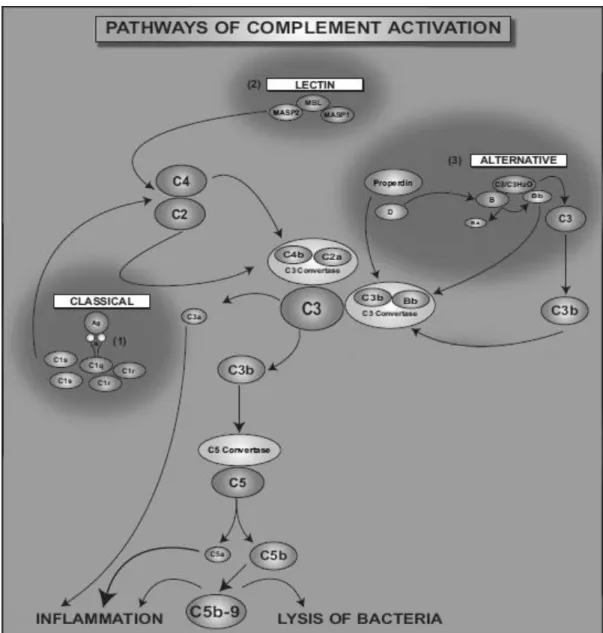
Figure 2. Complement activation though the classical (1), lectin (2), or alternative (3) pathways that converge at the central component of the complement system, C3. C3 and C5 can be cleaved by proteolytic enzymes that are released from leukocytes. (1) The binding of C1 complex to antigen-antibody complexes initiates proteolytic cleavage of the complement components. C1s first cleaves C4, which binds to the bacterial surface, then cleaves C2, leading to the formation of C4b2a complex (the C3 convertase of the classical pathway). (2) The lectin pathway begins by binding of the complex MBL to a bacterial wall, leading to the formation of C3 convertase C4b2a. (3) The alternative way is initiated by spontaneous hydrolysis of C3. The formed complex has the capacity to cleave C3 to C3a and C3b. C3b contributes to the formation of the C5 convertase, which cleaves C5 to C5a and C5b. C3a, C5a and C5b-9 complex are complement effectors that contribute to inflammation. C5ab-9 complex induces bacteria lysis. (Adapted from [23])
The elimination of invading pathogens by the complement system may also damage the host issue due to an inappropriate complement activation or complement deficiency, being potentially destructive to the organisms and causing several diseases. Given the multitude of
10
complement effects, there are mechanisms that prevent its action, limiting the complement activation. This control can be accomplished by inhibition of the C3 convertase activity by soluble or membrane-associated regulatory proteins. These work as cofactors for the proteolysis of the activated proteins C3b and C4b. C4b-binding protein (C4BP) and Factor H (FH) are examples of these proteins [26].
1.6. C4b-binding protein (C4BP)
C4BP is the major soluble inhibitor acting on the C3 convertase, either by increasing the dissociation of the enzyme complex, accelerating the decay of the classical and lectin pathway C3 convertase or by being a cofactor in the proteolytic degradation of C4b. The human plasma protein Factor H binds to the C3b inhibiting his formation and accelerating the decay of the alternative pathway C3 convertase. FH can also act as a cofactor in the proteolytic degradation of C3b, interfering with the complement regulation [25, 27].
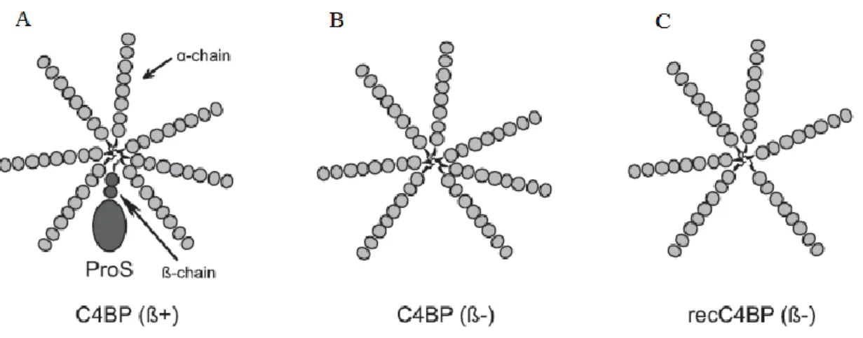
C4b-binding protein is a large oligomeric plasma glycoprotein of 570 kDa that is primarily synthesized in the liver and secreted into the circulation in three main isoforms, which differ in α and β-chains combinations. The major isoform, C4BP (β+), which constitutes 75-80% of C4BP in plasma, is composed of seven identical α-chains of 70 kDa each and a single β-chain of 40 kDa (α7β1). The C-terminal extension of each chain contains two cysteine residues, allowing the different chains to be linked by disulphide bonds, and an amphipathic α-helix region, which is compulsory for intracellular polymerization of the C4BP, resulting in a spider or octopus-like structure, with the seven α-chains forming extended tentacles (Figure 3-A) [26, 27].
The α- and β-chains of C4BP are composed of eight and three domains termed short consensus repeats (SCRs) or complement control protein (CCP), respectively, each one containing 60 amino acid residues [26, 27]. Binding C4b is a prerequisite for the cofactor activity of C4BP, and the CCP1-3 region of the α-chain presents a binding site for C4b, based on ionic-electrostatic interactions between amino acids from both molecules, promoting the cleavage of C4b molecules and the decay of the classical pathway C3-convertase. The β-chain presents a high-affinity binding site for the anticoagulant vitamin K-dependent protein S (ProS) and all the β-chains of C4BP in plasma circulate in complex with protein S (Figure 3-A), which provides C4BP the ability to bind negatively to phospholipids present in cell membranes [27].
11
Two more isoforms of C4BP are known besides the major circulating C4BP (β+) isoform, C4BP α7β1. The minor circulating C4BP (β-) isoform, with seven identical α-chains and without
the β-chain, also recognized as C4BP (β-) or C4BP α
7β0, is inducible under inflammatory
conditions (Figure 3-B). The recombinant C4BP isoform with six α-chains and without the β-chain, identified as recC4BP (β-) or C4BP α
6β0, is produced in vitro (Figure 3-C). However,
during acute-phase conditions, C4BP (β-) becomes the major isoform in plasma, as a consequence of the differential regulation of the genes encoding the α and β chains by proinflammatory cytokines [26].
Figure 3. Schematic structure of the main C4BP isoforms used in this study. (A) The major circulating C4BP (β+) isoform (C4BP α7β1), bound to ProS. (B) The minor circulating C4BP (β-) isoform (C4BP α7β0), inducible under proinflammatory conditions and, (C) a recombinant protein (recC4BP (β-) or C4BP α6β0) produced in vitro from a genetically engineered eukaryotic cell line.
Recently, it was reported that the administration of human C4BP was able to inhibit the development of autoimmune arthritis in mice. This suggests that other mechanisms might be involved in the anti-inflammatory activity of C4BP. It was also demonstrated that the C4BP (β -) isoform is capable of inducing a tolerogenic state in stimulated dendritic cells with phenotypic, morphologic and functional characteristics analogous to immature DC (iDC), and that the CCP6 domain is necessary for the C4BP (β-) tolerogenic activity in these cells. [26].
Major pathologies arising from a deficient complement activation are autoimmune disorders. These occur when a specific adaptive response is directed against self-antigens and, as a result, an immune response develops and leads to chronic inflammation and injury to the host tissues [23]. Systemic Lupus Erythematosus (SLE) is one of these autoimmune disorders, often found associated with the deficiency in C1q of the classical complement activation
12
pathway [21]. Anomalies in the macrophages phenotype and function are also related to autoimmune disease. Some theories affirm that macrophages involved in SLE present a deficient phagocytic function, causing an abnormal accumulation of apoptotic products that gives place to autoimmune disorders [28].
1.7. Systemic Lupus Erythematosus
Systemic Lupus Erythematosus is a multi-organ chronic autoimmune disorder characterized by several clinical manifestations from the skin and mucosal lesions to severe injuries in the central nervous system, kidneys, and heart among other organs. The pathogenesis and precise etiology, such as the mechanisms underlying many other disease manifestations, and the most effective therapeutic options remain unknown. Nevertheless, some genetic and environmental factors, like UV radiation, have been implicated as disease expression contributors [29, 30].
SLE predominantly affects women with ages ranging between 18 to 65 years old, especially in reproductive age. In most studies, 90% or more of patients are women and it is estimated that 200 women per 100 000 express cases of lupus [31], and woman with African American or Hispanic American heritage have 3 to 4 times more risk of developing this disease than Caucasians. SLE is usually associated with periods of illness followed by periods of remission, and the diagnosis of SLE is based on clinical criteria, including fever, skin rashes, photosensitivity, serositis, glomerulonephritis, hematological and neurological disorders [29]. The presence of a high quantity of antibodies against nuclear compounds (antinuclear antibodies), elevated circulating immune complexes and complement consumption, causing damage to cells and tissues, are the main characteristics of the disease [30].
The humoral immune response is being used as a marker of disease activity and the double-stranded (ds) DNA is the main target. Antibody production directed against chromatin components, such as dsDNA, nucleosomes, and histones, are a hallmark of SLE pathogenesis. DNA-histone complexes (nucleosomes) are released in high amounts into circulation in SLE patients, and these nucleosomes are targets of anti-dsDNA autoantibodies [30]. Therefore, despite being unspecific, anti-dsDNA antibody serum levels have been correlated with disease activity and are clinically used as predictive markers. The development of autoantibodies reflects a loss of B- or T-cell tolerance, which possibly result from a combination of genetic
13
predisposition, persistent inflammatory responses, and abnormal handling of apoptotic material and immune complexes, abnormal presentation of self-antigens among other events [29].
Cell death is the most likely phenomenon for supplying autoantigens, and there are two main forms of cells dead, apoptosis and necrosis. When cells die through one of these forms, they must be quickly removed from tissues in order to prevent further inflammation and damage. A highly efficient scavenger system, including macrophages, polymorphonuclear cells, and immature dendritic cells, assure a quick elimination of apoptotic and necrotic cell material. An identification of markers of activity and damage, characterization of key regulatory circuits, identification of suppressors and activators and, in definitive, a deep understanding of the autoimmune pathophysiology, may allow a better understanding, treatment, and prevention of SLE [29].
15 Motivation
Some studies confirm that, annually, around 200 women per 100 000 express lupus manifestations. SLE presents a high incidence and prevalence rates in women, that most often strikes reproductive-age women, being a relevant disease considered a matter of public health. The development of clinical instruments for the assessment of disease activity and disease damage is fundamental in SLE research. Significant advances were made in the past 2 decades in the understanding of the genetic predisposition to disease occurrence and the underlying pathophysiological mechanisms accounting for the inflammatory process and the irreversible organ damage that may result over time.
The minor circulating C4BP (β-) isoform was recently capable of inducing a tolerogenic state in stimulated dendritic cells with phenotypic, morphologic and functional characteristics analogous to immature DC (iDC), evidencing that the C4BP α7β0 isoform could be used as an
anti-inflammatory and immunomodulatory agent in autoimmune diseases. Nevertheless, a complete understanding of C4BP (β-) function and mechanism of action should be fully
elucidated for, in a near future, the C4BP (β-) administration in the correct dosage, be safe and efficient for use in pre-clinical models and patients.
Hypothesis and objectives
Recent studies, performed in vitro, unveil the capacity of the C4BP (β-) isoform or C4BP α7β0, to modulate the behavior of dendritic cells [26]. The plasma-purified and recombinant
C4BP (β-) abolished the proinflammatory phenotype of human monocyte-derived dendritic
cells stimulated with LPS, inducing a semi mature, tolerogenic phenotype on these cells. This resulted in a DC surface expression pattern marked by a decrease of the maturation marker CD83 and reduction of the costimulatory molecules CD80 and CD86. On the other hand, a significant diminution in pro-inflammatory cytokines production, such as IL-12, TNF-α and IFN-γ, and a preferential production of anti-inflammatory cytokines, like IL-10 and TGF-β was evident [26].Therefore, both DC, previously treated with C4BP (β-), and the minor circulating C4BP (β-) isoform on its own could be used as anti-inflammatory and immunomodulatory
16
Although the molecular mechanism explaining how the C4BP induce tolerogenicity in pro-inflammatory stimulated DCs is not yet known, other cellular types, also fundamental in the inflammatory mechanisms such as monocyte-derived macrophages, can present the same tolerogenic characteristics. To verify the therapeutic efficiency of C4BP (β-) in vitro studies analogous to those performed with DCs, and in vivo studies in animal models with inflammatory alterations like NZB/W F1 mice that express spontaneous SLE are essential.
This work is based in in vitro experiments with the objective of understanding if the C4BP isoforms modify the inflammatory activity of monocyte-derived macrophages polarized to M1 and M2. To achieve this goal, phenotype characteristics of macrophages were analyzed. For GM-CSF-differentiated macrophages polarized to M1, the surface markers CD64 and CD80 were analyzed, and for M-CSF-differentiated macrophages polarized to M2. the surface markers CD209 and CD11b were used. The effect of C4BP (β-) on the endocytic and phagocytic activity of M1 and M2 polarized macrophages was also evaluated.
The structural requirements for the functional activities of C4BP are also of interest, because C4BP is the major regulator of the classical pathway of complement, and it may have therapeutic potential. Subsequently, the final goal of this research was to assess the consequences of C4BP (β-)-mediated induction of the phenotype and functional behavior of
pro-inflammatory (M1) or anti-inflammatory, pro-resolving (M2) conditions of monocyte-derived macrophages.
Document Structure
This thesis is organized into five major chapters, each one related to a specific part of the research. The remainder of this section presents a brief theoretical introduction to a better understanding, the main motivations, and objectives of this project.
Chapter 2 starts by presenting each of the most relevant techniques and the respective materials used in the process.
The obtained results are found in Chapter 3 and the respective discussion in Chapter 4. Finally, conclusions and some insight on future work and research are provided in Chapter 5.
17
Chapter 2
Materials and Methods
2.1. Monocyte isolation from peripheral blood
The studies were performed using human monocyte-derived macrophages (MDMs), from PMBCs. These were obtained from buffy coat preparations collected from healthy blood donors from the Blood and Tissues Bank (Barcelona, Spain) and isolated by density gradient centrifugation.
Therefore, 30mL of total blood were placed in 50mL Falcon tubes and centrifuged for 20 min at 1500xg and room temperature (Heareus Multifuge 3 LR, Thermo Scientific, USA). The resulting gradient divided the blood sample into the following phases, according to the cells density: RBCs, WBCs + platelets, and plasma. With a pipette, the leukocytes and platelets were placed in new 50mL Falcon tubes and the volume was increased up to 50mL with PBS (DPBS w/o Calcium and w/o Magnesium, Pan Biotech, Germany). Next, 15mL of Ficoll (Ficoll-Paque, GE Healthcare Bio-Sciences AB, Uppsala, Sweden) were added to 50mL Falcon tubes and 25mL of the previous diluted cells with PBS were added carefully on the top of the tubes with Ficoll and centrifuged for 25 min at 400xg and room temperature. After density gradient separation, a white layer could be easily observed, which corresponds to the PMBCs. These cells were carefully collected with a Pasteur pipette to new 50mL Falcon tubes and washed with PBS to clean the cells. Two additional centrifugations of 10 min at 400xg and room temperature were performed, the supernatant was removed and, finally, the cells were diluted in PBS to a final volume of 10mL.
18 2.2. Cellular count by flow cytometry
The isolated cells were counted by flow cytometry to estimate the necessary volume to be used in the experiment. For functional assays, 40µL of PBS, 20µL of PMBCs and 4µL of monoclonal mouse anti-human CD14 antibody (MACS Miltenyi Biotec, Germany) were placed in a cytometry tube. After 15 min, 120µL of PBS were added to stop the reaction and supplemented with 5µL of 7-Aminoactinomicin D (7-AAD) (7-AAD, Viability Staining Solution, eBioscience) to differentiate alive and dead cells, yielding the percentage of cellular viability, and the tubes were incubated in the darkness during 5 min. After the incubation time, monocytes were purified and counted using 20µL of CD14+ magnetic microbeads (Perfect Count Microspheres, Cytognos SL, Salamanca, Spain). The purity of CD14+ cells was tested by CD14 staining and flow cytometry analysis (Cytometry FACScalibur, BD Biosciences), allowing the assessment of the number of PMBCs and the number of total CD14+(> 90% CD14+ cells).
2.3. Monocyte-derived macrophage differentiation and polarization
Monocytes were cultured at 1×10⁶ cells/mL in a 24 well plate (24-well Tissue Culture Plate, Biofil) in RPMI 1640 medium, supplemented with 100 IU/mL penicillin, 100 µg/mL streptomycin and 2mM L-glutamine without serum (basal medium), and allowed to adhere for 90 min at 37ºC in 5% of CO2. After 90 min, the nonadherent cells were removed by washing
with PBS. Each well was filled with 800µL of RPMI 1640 medium, supplemented with 100 IU/mL penicillin, 100 µg/mL streptomycin, 2mM L-glutamine (all from Invitrogen, Carlsbad, USA) and 10% heat-inactivated FBS (Linus, Cultek, Spain) (complete medium). The wells were also supplemented GM-CSF to those containing monocytes that would subsequently be polarized to M1, or with M-CSF to those containing monocytes that would subsequently be polarized to M2. The factor under study, the C4BP (β-) or C4BP α
7β0 was added to the
corresponding wells with a concentration of 10µg/Ml, and a control of each condition (M0 and M0+β- with GM-CSF and M-CSF) was made (Figure 4). The macrophage differentiation lasts
19
Figure 4. Disposition of a 24 wells plate for the in vitro macrophages differentiation and polarization, and assessment of the C4BP (β-) effect. Monocyte-derived macrophages were polarized to M1 and M2 in the presence of GM-CSF and M-CSF, respectively. The protein under study, the C4BP β- isoform was added to the corresponding wells. M0 and M0+β- corresponds to the controls, also cultivated with GM-CSF or M-CSF. The PBS was added to the surrounding wells to prevent the evaporation of the medium on the cells.
After 6 days, the supernatants were aspired and the cells were resuspended in 500µL of complete medium with GM-CSF and M-CSF, according to the corresponding wells. Uncommitted macrophages (M0) were polarized into M1 using E. coli LPS (40ng/mL) (Sigma, USA) plus rhIFN-γ (40ng/mL) (Invitrogen, USA) and into M2 using rhIL-4 alone (40ng/mL) (Invitrogen, USA) for 2 days (Figure 5). The C4BP (β-) (α7β0) was added to the corresponding
wells at a concentration of 10µg/mL. Macrophages from different donors were polarized in independent experiments.
Figure 5. Human monocytes were differentiated into uncommitted macrophages (M0) by granulocyte macrophage colony-stimulating factor (GM-CSF) or macrophage colony-stimulating factor (M-CSF). After 6 days of incubation at 37ºC in 5% of CO2, macrophages were polarized into classically activated (M1) or alternatively activated (M2) using LPS/IFN-γ or IL-4, respectively, for 2 more days. After 8 days from the beginning of the experiment, the phenotype was analyzed by flow cytometry.
M-CSF GM-CSF Day 8 Cytometry Monocytes IL-4 LPS+IFN-γ Day 6 GM-CSF/ M-CSF Macrophages (M0) M2 M1
20 2.4. Flow cytometry
On day 8, the macrophages were recovered from the wells to 15mL Falcon tubes, centrifuged 10 min at 400xg and room temperature, washed with PBS. The supernatant was preserved at -80ºC on Eppendorf tubes for future immunoassays. The adhered cells were removed by carefully knocking on the plate, pipetting and scrapping. Once the wells were clean, visible on the microscopy, the Falcon tubes were centrifuged for 10 min at 400xg and room temperature. Finally, the supernatant was aspired and the pellet was used for flow cytometry analysis.
2.4.1. Surface marker analysis
To examine the macrophage surface markers, the pellet was resuspended in 20µL of PBS and added to the cytometry tube with 60µL of PBS, 3µL of CD64-APC and 3µL of CD80-PE, in the case of M1 polarized macrophages, or 60µL of PBS, 3µL of CD209-APC and 3µL of CD11b-PE in the case of M2 polarized macrophages. After 15 min of incubation at room temperature, the reaction was stopped with 120µL of PBS and 2,5µL of 7-AAD, and flow cytometry analysis (Cytometry FACSCanto II, Becton Dicknson) was performed to assess the viability and phenotype of the cells.
2.4.2. Endocytosis analysis using OVA and LY
Another cytometry study was performed to investigate the different types of macrophage endocytosis. When macrophages take up and process Ovalbumin (OVA) captured by receptor-mediated endocytosis, and Lucifer Yellow CH (LY) captured by pinocytosis, the cells exhibit a characteristic shift or gain in fluorescence intensity. For this purpose, OVA (Stock 1mg/mL) was diluted in PBS, and 4µL were added to cytometry tubes containing 60µL of RPMI complete medium and 20µL of cells. To demonstrate intracellular uptake, loading was performed at 4°C and at 37°C for 1 h, because intracellular uptake is energy-dependent. After 1 h, the reaction was stopped with 150µL of FACS buffer (PBS + 1% BSA + 0,1% NaN3), 2,5µL of 7-AAD and
flow cytometry analysis (Cytometry FACSCanto II, Becton Dicknson) was performed to assess the viability and endocytosis status of the cells.
21
LY (Stock 10mg/mL) was diluted in H2O and 6µL were added to cytometry tubes
containing 60µL of RPMI complete medium. The same protocol than the OVA uptake was employed, but the incubation time was 2,5 h instead of 1 h. Then, the reaction was stopped with 150µL of FACS buffer and 2 additional washes were performed. The washes consisted of transferring the mixture to Eppendorf tubes, centrifuging them at maximum speed for 3 min, removing supernatant and adding 150µL more of FACS buffer. Finally, the mixture was returned to the cytometry tubes, 2,5µL of 7-AAD were added and the suspension was analyzed by flow cytometry (Cytometry FACSCanto II, Becton Dicknson).
2.4.3. Phagocytosis analysis
Phagocytosis was studied using pHrodo™ Green E. coli BioParticles (Stock 0,909 mg/mL) diluted in 2.2mL of PBS. For each condition, 100µL of PBS and 20µL of the diluted E. coli BioParticles were added, and one of the plates was placed at 4°C and another at 37°C for 1 h. After the incubation time, 130µL of PBS was added making a total of 250µL. The adherent macrophages were removed by scrapping and pipetting. Once removed, the 250µL suspension was collected on a cytometry tube, 2,5µL of 7-AAD were added and flow cytometry analysis was performed (Cytometry FACSCanto II, Becton Dicknson).
2.5. Statistical analysis
All flow cytometry data were analyzed using the FlowJo software (Miltenyi Biotec). Statistical analysis was performed using GraphPad Prism software (GraphPad Prism version xxxx for Windows, GraphPad Software, La Jolla California USA) and data were expressed
23
Chapter 3
Results
3.1. Human monocyte-derived macrophage morphology
Collected from the human peripheral blood, monocytes were differentiated into unstimulated macrophages (M0) by growth factors such as granulocyte-macrophage colony stimulation factor (GM-CSF) and macrophage stimulating factor (M-CSF). M0 macrophages were polarized to M1 and M2 phenotypes.
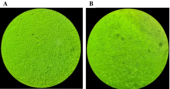
When stimulated in culture with cytokines to induce M1 and M2 polarization, the macrophages displayed markedly different morphologies. Addition of LPS and IFN-γ, which stimulate M1 polarization, caused cells to flatten into a round, pancake-like shape within 48h of stimulation (Figure 6-A). In contrast, the addition of IL-4, which stimulates M2 differentiation, led to cellular elongation (Figure 6-B).
These results prove that phenotypic polarization of macrophages is regulated by the local tissue microenvironment and showing that M2 cells exhibited a significantly higher degree of elongation compared with either M1 or unstimulated M0 cells.
24
Figure 6. Characterization of macrophages morphology after M1 or M2 polarization. (A) After stimulation with LPS/IFN-γ, M1 macrophages showed a rounded morphology. (B) M2 macrophages stimulated with IL4 adopted an elongated cell morphology.
3.2. Human monocyte-derived macrophage treatment with C4BP (β-)
3.2.1. C4BP (β-) affects the polarization phenotype of human macrophages
The expression of a panel of surface molecules on macrophages was analyzed by flow cytometry after 2 days of in vitro polarization of human blood monocytes with IFN-γ/LPS and IL-4, using unpolarized cells (M0 and M0+ β-) as a control.
Macrophages activated with IFN-γ and LPS (M1 phenotype) displayed a robust and specific upregulation of the co-stimulatory molecule CD80 and a downregulation of the CD64 receptor, as compared to the unpolarized cells (Figure 7). The macrophages activated via IL-4 (M2 phenotype) displayed a highly upregulated expression of the myeloid cell markers CD11b and CD209, compared with the control cells (Figure 8).
In summary, these data indicate that human macrophages polarized to M1 with IFN-γ and LPS specifically express CD80 and CD64, and human macrophages polarized to M2 with IL-4 upregulate CD11b and CD209, confirming that the candidate markers were robustly and reproducibly modulated by the polarizing factors and proving their specificity for each type of activated macrophages.
25
When the macrophages were treated with the protein under study, C4BP (β-) or C4BP α7β0 isoform, a decrease of CD64 and CD80 expression was visible in M1 macrophages (Figure
7), and a reduction of CD11b and CD209 expression was observed in M2 macrophages (Figure 8).
Figure 7. Relative mean fluorescent intensity (MFI) for M1 macrophage surface markers and effect of C4BP(β-). Monocyte-derived macrophages polarized with IFN-γ and LPS, presenting a M1 phenotype, express an regulation of CD80 and CD64.When analyzed by flow cytometry for surface marker expression, CD80-PE and CD64-APC antibodies allow the visualization of an increase (CD80) or decrease (CD64) in M1, compared with the control (M0). When exposed to the C4BP(β-) isoform, a significant prevention of CD80 and CD64 overexpression occurs, similar to the control M0 + β-. The results shown are the mean ± SD from five independent experiments. **p < 0.01, ***p < 0.001, ****p < 0.0001, compared to M1. CD64-APC N=5 MF I CD80-PE N=5 MF I
26
3.2.2. C4BP (β-) affects the endocytosis of human macrophages
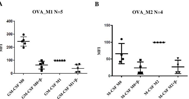
The endocytic capacity on macrophages was analyzed after 2 days of in vitro polarization of human blood monocytes with IFN-γ and LPS for the M1 phenotype, and IL-4 for the M2 phenotype, using unpolarized cells (M0 and M0+ β-) as a control. When macrophages take up Ovalbumin (OVA) captured by receptor-mediated endocytosis (Figure 9), and Lucifer Yellow CH (LY) captured by pinocytosis (Figure 10), exhibit a characteristic shift in fluorescence intensity captured by flow cytometry.
When the macrophages were classically activated (M1 phenotype), a downregulation of their endocytic activity in relation to the control M0 was observed, either in macrophages treated with OVA or with LY (Figures 9-A and 10-A). In contrast, when the macrophages were alternatively activated (M2 phenotype), M2 polarization did not change or slightly increased their endocytic activity, compared to the control M0 (Figures 9-B and 10-B).
CD209-APC N=6 MF I CD11b-PE N=7 MF I
Figure 8. Relative mean fluorescent intensity (MFI) for M2 macrophage surface markers. Monocyte-derived macrophages polarized withIL-4, presenting a M2 phenotype, express an upregulation of CD11b and CD209.When analyzed by flow cytometry for surface marker expression, CD11b-PE and CD209-APC antibodies allow the visualization of an increase in M2, compared with the control (M0). When exposed to the C4BP(β-) isoform, a significant prevention of CD11b and CD209 overexpression occurs, approaching to the control M0 + β-. The results shown are the mean ± SD from six to seven independent experiments. ***p < 0.001, ****p < 0.0001, compared to M2.
27
When the classically and alternatively polarized macrophages were treated with the C4BP (β-) isoform, a decrease of fluorescence occurred, showing a downregulation of the endocytic activity in both M1 and M2 macrophages, and also in non-polarized cells.
MF I OVA_M1 N=5
A
MF I OVA_M2 N=4B
Figure 9. Representation of the endocytic activity of (A) M1 and (B) M2 polarized macrophages when Ovalbumin (OVA) is captured by receptor-mediated endocytosis. (A) When the macrophages are classically polarized (M1), a downregulation of the endocytic activity occurs. (B) An upregulation of the endocytic activity is also visible when macrophages are alternatively activated (M2). In both cases, the C4BP (β-) downregulates endocytosis.
28
3.2.3. C4BP (β-) does not affect the phagocytosis of human macrophages
The phagocytic capacity of macrophages was studied after 2 days of in vitro polarization of human macrophages derived from blood monocytes, with IFN-γ and LPS for the M1 phenotype and IL-4 for the M2 phenotype, using unpolarized cells (M0 and M0+ β-) as a control. When macrophages captured pHrodo™ Green E. coli BioParticles by phagocytosis, a characteristic shift in fluorescence intensity was evident by flow cytometry, allowing quantification of the phagocytic activity.
When macrophages were polarized to the M1 phenotype no difference in the phagocytic activity of macrophages was observed respect to unpolarized macrophages (M0). In contrast, when these cells were polarized into M2 phenotype, an upregulation of the phagocytic activity was detected relative to the control unpolarized cells (M0) (Figure 11).
When the C4BP (β-) was applied on both unpolarized and polarized (M1 and M2)
macrophages, no significant difference in the phagocytic activity was observed respect to untreated cells. LY_M1 N=6 MF I
A
LY_M2 N=5 MF IB
Figure 10. Representation of the endocytic activity of (A) M1 and (B) M2 polarized macrophages when Lucifer Yellow (LY) is captured by pinocytosis. (A) When the macrophages are classically polarized (M1), a downregulation of the endocytic activity occurs. (B) An upregulation of the endocytic activity is also visible when macrophages are alternatively activated (M2). In both phenotypes, the C4BP (β-) downregulates endocytosis.
29
In resume, while M-CSF-differentiated macrophages displayed increased phagocytosis compared with GM-CSF-differentiated macrophages, either M1/M2 polarization or C4BP (β-) treatment did not have any influence in phagocytosis activity (Figure 11).
M
FI
E. coli BioParticles N=6
Figure 11. Representation of the phagocytic activity of M1 and M2 polarized macrophages when pHrodo™ Green E. coli BioParticles is captured by phagocytosis. M2 polarized macrophages displayed an increase of phagocytic activity compared to M1 polarized macrophages. C4BP (β-) treatment did not have any influence in phagocytosis activity.
31
Chapter 4
Discussion
4.1. Human monocyte-derived macrophages morphology
Macrophages are key regulators of the immune response during infection and wound healing, being the first cells sensing pathogens or damage. Their behavior is induced by soluble factors in the local microenvironment, which is critical for regulating the proinflammatory or healing state of these cells.
To fulfill their functionally distinct roles, macrophages are capable of polarizing toward a spectrum of phenotypes, which include classical pro-inflammatory activation (M1), and alternative activation (M2), is responsible for wound healing and tissue repair. In the presence of inflammatory stimuli and danger signals, macrophages polarize to M1 phenotype and release inflammatory cytokines to fight pathogens. In contrast, a wound healing environment promotes polarization toward an M2 phenotype and leads to cellular processes that facilitate tissue repair.
We observed that monocyte-derived macrophages stimulated in culture with cytokines to induce M1 or M2 polarization displayed markedly different cell morphologies (Figure 6). He addition of GM-CSF, LPS and IFN-γ, which stimulate M1 polarization, caused cells to flatten into a round, pancake-like shape within 48h of stimulation (Figure 6-A). On the other hand, the addition of M-CSF and IL-4, which stimulate M2 polarization, led to cellular elongation
(Figure 6-B), suggesting an important role for cell shape involving macrophage function.
4.2. Human monocyte-derived macrophage treatment with C4BP (β-)
The complement system constitutes a key arm of the innate immune response in the fight against pathogen infection. To maintain immune homeostasis, the main fluid-phase complement inhibitor C4BP presents complementary activities in the regulation of excessive