September, 2018
Karina Alexandra Pál
[Nome completo do autor]
[Nome completo do autor]
[Nome completo do autor]
[Nome completo do autor]
[Nome completo do autor]
[Nome completo do autor]
[Nome completo do autor]
Bachelor in Chemical and Biochemical Engineering
[Habilitações Académicas]
[Habilitações Académicas]
[Habilitações Académicas]
[Habilitações Académicas]
[Habilitações Académicas]
[Habilitações Académicas]
[Habilitações Académicas]
Intracellular Stochastic Modelling of
Influenza A Virus and DIP Replication
[Título da Tese]
Dissertation submitted to obtain the degree of Master of Science in
Chemical and Biochemical Engineering
Dissertação para obtenção do Grau de Mestre em
[Engenharia Informática]
Adviser: Daniel Rüdiger, Master of Science,
Max Planck Institute for Dynamics of Complex Technical
Systems
Co-adviser: Rui Manuel Freitas Oliveira, Associated Professor with
Aggregation, Faculty of Science and Technology – NOVA
University of Lisbon
Examination Committee
Chairperson: Prof. Dr. Mário Fernando José Eusébio, Assistant Professor,
NOVA University of Lisbon
Raporteur:
Dr. Rafael Sousa Costa, Postdoctoral Researcher, Instituto
Superior Técnico
Member:
Prof. Dr. Rui Manuel Freitas Oliveira, Associated Professor with
Aggregation, NOVA University of Lisbon
September, 2018
Karina Alexandra Pál
[Nome completo do autor]
[Nome completo do autor]
[Nome completo do autor]
[Nome completo do autor]
[Nome completo do autor]
[Nome completo do autor]
[Nome completo do autor]
Bachelor in Chemical and Biochemical Engineering
[Habilitações Académicas]
[Habilitações Académicas]
[Habilitações Académicas]
[Habilitações Académicas]
[Habilitações Académicas]
[Habilitações Académicas]
[Habilitações Académicas]
Intracellular Stochastic Modelling of
Influenza A Virus and DIP Replication
[Título da Tese]
Dissertation submitted to obtain the degree of Master of Science in
Chemical and Biochemical Engineering
Dissertação para obtenção do Grau de Mestre em
[Engenharia Informática]
Adviser: Daniel Rüdiger, Master of Science,
Max Planck Institute for Dynamics of Complex Technical
Systems
Co-advisers: Rui Manuel Freitas Oliveira, Associated Professor with
Aggregation, Faculty of Science and Technology – NOVA
University of Lisbon
Examination Committee
Chairperson: Prof. Dr. Mário Fernando José Eusébio, Assistant Professor,
NOVA University of Lisbon
Raporteur:
Dr. Rafael Sousa Costa, Postdoctoral Researcher, Instituto
Superior Técnico
Member:
Prof. Dr. Rui Manuel Freitas Oliveira, Associated Professor with
Aggregation, NOVA University of Lisbon
Intracellular Stochastic Modelling of Influenza A Virus and DIP Replication
Copyright © Karina Alexandra Pál e Faculdade de Ciências e Tecnologia – Universidade NOVA de Lisboa.
A Faculdade de Ciências e Tecnologia e a Universidade NOVA de Lisboa têm o direito, perpétuo e sem limites geográficos, de arquivar e publicar esta dissertação através de exemplares impressos reproduzidos em papel ou de forma digital, ou por qualquer outro meio conhecido ou que venha a ser inventado, e de a divulgar através de repositórios científicos e de admitir a sua cópia e distribuição com objetivos educacionais ou de investigação, não comerciais, desde que seja dado crédito ao autor e editor.
v
The past, like the future, is indefinite and exists only as a spectrum of possibilities. Stephen Hawking
To lead of my acknowledgments, I would like to thank firstly to my adviser Daniel Rüdiger for giving me the opportunity to work in a field that I was very excited to learn more about and guiding me in the right direction through my research. He showed an amazing availability whenever I had any question or run into dead ends. I am gratefully for his valuable comments and corrections on this thesis, I learned so much with him and he did everything in his power to help me achieve my goal. In second, I am sure that professor Rui Oliveira contributions were also very important in providing me all the support before and during my Erasmus mobility, always showing a great concern about my progress.
I believe that Tanja, Sacha and my office partner Lukas, gave me an important feedback and valuable advises which helped me improving my thesis, also professor Udo Reichl and the Bioprocess Engineering Group were very helpful in giving me the opportunity to work at the MPI for Dynamics of Complex Technical Systems and providing me all the resources needed to pursue my research.
I also feel immensely grateful towards my parents, Aurelia and Ferenc, for supporting my decisions in life and providing me all the means to pursue my goals. A big share of gratitude to my grandmother, Măriuța, for all her words and advises that helped me not to give up and made me go chase my dreams. I would also like to thank my boyfriend, Gustavo, for this amazing 4 years and for all the patience during the more stressful times. We learned a lot together and I am very proud for what we achieved together, so far.
I would also like to praise my partners in crime, Cláudia and Suzana, for supporting me through this amazing journey in Magdeburg. Even during hard times, we were always there to ensure that each of us were laughing and that we could overcome the obstacles together. I am sure we will keep in touch and our friendship bound is for life. I would also want to thank to José, Nino, Lars, Clair and Weiwei for all the evenings spent drinking good German beer which helped me to take my mind off work and relax. I feel also appreciate for meeting Juliana and João, for having the possibility to talk in Portuguese and making me feel at home. I am so grateful for this experience, for allowing me to meet such amazing people and all the moments that we spent together that surly, I will keep them forever in my heart.
I also believe that my friends Filipa, David, Tai, Daniela, Daniel, Nídia, Sara and Inês deserve recognition for all the nights spent at the university while working on Project I and II. Their companionship helped me so much during the hardest times of the course and I do not know if it would be possible to accomplish everything without them. A big thank you to professor Mário Eusébio for the advises and support during this period.
Finally, I want to thank my life long friends Samantha, Gabriela, Beatriz and Diogo for always being there for me and accompanying me closely for such a long time.
vii
Abstract
Defective interfering particles (DIPs) are mutated versions of viruses that are characterized by carrying internal deletions in their genome. These deletions are introduced randomly during virus replication and the truncated genomes interfere with the propagation of their standard virus (STV) leading to reduced infectious virus titers. Therefore, DIPs were recently proposed to be used for antiviral therapy which increased the demand for a reliable production process and a better understanding of the interference mechanism. The infection dynamics were analysed by a deterministic modelling approach, however, the impact of stochastic effects introduced by cell-to-cell variability, different coinfection scenarios and an independent genome segment replication remain largely elusive. Hence, we developed a stochastic model of influenza A virus and DIP replication which considers the influence of these random effects on STV and DIP release. We found that the viral nucleoprotein (NP), which is essential for encapsidation of the naked viral RNA (vRNA), is strongly affected by fluctuations and three distinct sub-populations emerged in our model. Furthermore, simulations performed with one DIP and one STV infecting the cell resulted in mostly non-productive simulations, mainly caused by failures during the endocytosis of particles and by the random degradation of vRNAs. Moreover, the optimal DIP production was achieved when STV enters nucleus first and the DIP entry is delayed between 1.5 and 3 hours. Lastly, we demonstrate that the implementation of a two-step packaging process, which separates the formation of genome complexes and the assembly of all required proteins for release, is crucial to achieve a substantial DIP advantage over STV production. Overall, our simulations suggest that a combination of various random effects influences the replication of STVs and DIPs inducing a broad distribution of progeny particle release. The stochastic model developed in this thesis provides an ideal basis for the analysis of these effects and their impact on DIP interference and production.
Keywords: Defective interfering particle, influenza A virus, interference mechanism, stochastic mathematical model, stochastic effects
ix
Resumo
Partículas defeituosas interferentes (DIPs) são versões mutagénicas do vírus, sendo caracterizadas por apresentarem secções internas do seu genoma excluídas. Estas secções são geradas aleatoriamente durante a replicação do vírus e o genoma incompleto interfere com a propagação do seu vírus standard (STV), advindo numa redução da concentração de vírus infeciosos. Consequentemente, o uso de DIPs foi recentemente proposto para terapia antiviral, o que resultou num aumento da procura de um processo de produção viável e de um conhecimento mais aprofundado do mecanismo de interferência. A dinâmica de infeção foi analisada através de uma abordagem de modelação determinística, no entanto, o impacto de efeitos estocásticos causados pela variabilidade celular, diferentes cenários de coinfecção e replicação independente dos diferentes segmentos do genoma permanecem assim largamente elusivos. Portanto, desenvolveu-se um modelo estocástico da replicação do vírus influenza A e DIPs que considera o impacto de efeitos aleatórios na produção de STVs e DIPs. Foi descoberto que a nucleoproteina viral (NP), que é essencial para a encapsidação do RNA viral (vRNA), é fortemente afetado por flutuações e três subpopulações distintas advêm do nosso modelo. Além disso, simulações efetuadas em que um DIP e um STV infetam a célula, resultam maioritariamente em simulações não produtivas causadas geralmente por uma falha durante a endocitose de partículas e pela degradação aleatória de vRNAs. Mais adiante, a produção de DIP é otimizada quando o STV entra no núcleo primeiro e a entrada da DIP é atrasada entre 1.5 e 3 horas. Finalmente, demonstrou-se que a implementação de um processo de empacotamento com duas etapas, separando a formação de complexos genómicos e a montagem de todas as proteínas necessárias para a libertação de partículas, é crucial para conferir uma vantagem substancial à DIP em relação à produção de STV. Em geral, as simulações sugerem que as combinações de vários efeitos aleatórios influenciam a replicação de STVs e DIPs, resultando numa distribuição de partículas produzidas muito alargada. O modelo estocástico desenvolvido nesta tese consiste uma base ideal para a análise destes efeitos e do seu impacto na produção e interferência das DIPs.
Termos-chave: Partículas defeituosas interferentes, vírus influenza A, mecanismo de interferência, modelo matemático estocástico, efeitos estocásticos
xi
Contents
Abstract ...vii
Resumo ... ix
List of Figures ... xiii
List of Tables ... xv
List of Equations ... xvii
List of Abbreviations ...xix
List of Symbols ...xxi
1 Introduction ... 1
2 Theoretical Background ... 3
2.1 Discovery of defective interfering particles ... 3
2.2 Influenza A virus structure and DIP de novo generation ... 3
2.3 Influenza A virus intracellular life cycle ... 5
2.4 DIP interference ... 9
2.5 Therapeutic potential of DIPs ... 10
2.6 Intracellular deterministic model of influenza A virus and DIP replication ... 10
2.7 Intracellular stochastic model of influenza A virus replication ... 11
2.8 Stochastic simulation algorithms ... 12
3 Models and Methods ... 15
3.1 Implementation of biochemical reactions ... 15
3.2 Model variations ... 24
3.3 Computation and simulation assumptions ... 27
xii
4 Results ... 31
4.1 DIP interference and comparison with the deterministic model ... 31
4.1.1 High MOI and MODIP ... 31
4.1.2 Low MOI and MODIP ... 37
4.1.3 Impact of MOI and MODIP ... 42
4.2 Non-productive simulations ... 45
4.2.1 Effects of MOI and MODIP ... 45
4.2.2 Filtering non-productive simulations... 50
4.3 Timing investigation ... 51
4.3.1 Randomly generated delay ... 51
4.3.2 Induced delay ... 54
4.4 Model variations ... 59
4.4.1 Minimum packaging model ... 59
4.4.2 Replication model ... 63
5 Discussion ... 67
6 Conclusion ... 73
Bibliography ... 75
xiii
List of Figures
Figure 2.1 | Influenza A virus particle, genome structure and encoding proteins. ... 4
Figure 2.2 | Structure of a defective interfering particle (DIP) and de novo generation mechanism of defective RNA. ... 5
Figure 2.3 | Diagram of the influenza A virus life cycle. ... 6
Figure 2.4 | Different hypothesis for the transition mechanism from transcription to replication. ... 7
Figure 3.1| DIP advantage over STV replication... 25
Figure 3.2 | Average converges with increased number of simulations. ... 28
Figure 3.3 | Averaged stochastic dynamics and distribution. ... 29
Figure 4.1 | Distribution of standard virus (STV) and DIP production at MOI/MODIP 10/10. ... 31
Figure 4.2 | Comparison of the stochastic and deterministic simulation. ... 32
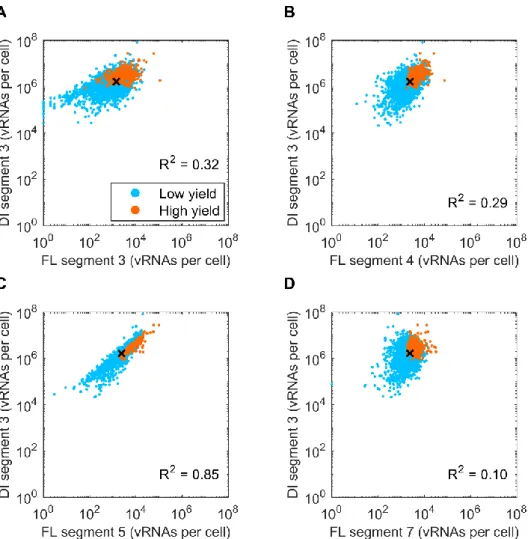
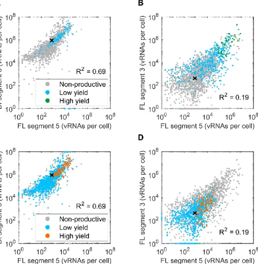
Figure 4.3 | Correlation of the defective interfering vRNA and various vRNA segments and DIP production... 33
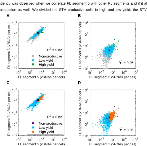
Figure 4.4 | Correlation of vRNA segment 5 with defective and functional segment 3 and its influence in STV and DIP production... 34
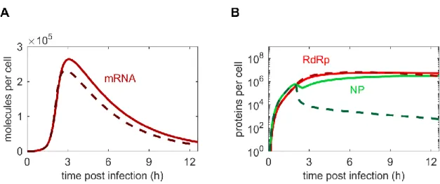
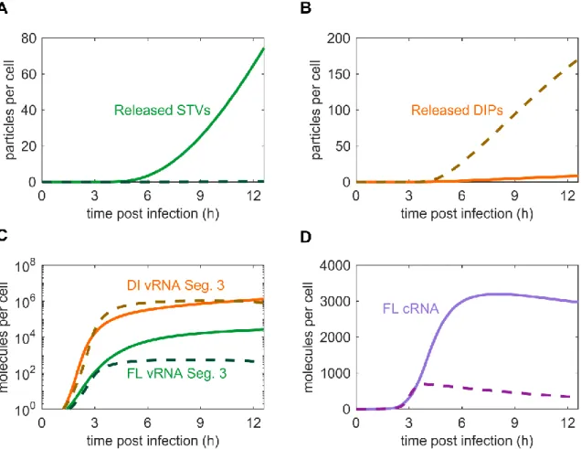
Figure 4.5 | Comparison of mRNA and protein dynamics for stochastic and deterministic model simulations at MOI/MODIP 10/10. ... 35
Figure 4.6 | Levels and distributions of HA and NP related to STV and DIP yield. ... 36
Figure 4.7 | Distribution of STV and DIP production at MOI/MODIP 1/1. ... 37
Figure 4.8 | Higher differences between the stochastic and deterministic simulation at MOI/MODIP 1/1. ... 38
Figure 4.9 | Correlation of vRNA segment 5 with defective and functional segment 3 and its influence in STV and DIP production... 39
Figure 4.10 | Comparison of mRNA and protein dynamics for stochastic and deterministic model simulations at MOI/MODIP 1/1. ... 40
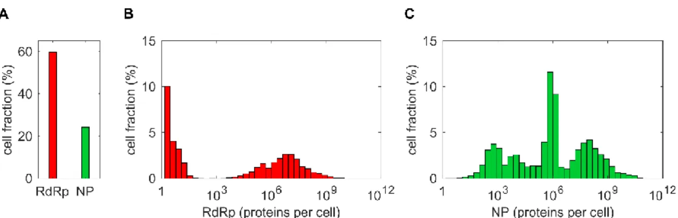
Figure 4.11 | Distributions of RdRp and NP in MOI/MODIP 1/1 scenario... 41
Figure 4.12 | Levels of RdRp and NP related to STV and DIP yield. ... 42
xiv
Figure 4.14 | Average cumulative DIP released and increased MODIP. ... 44
Figure 4.15 | vRNP ratios and impact on STV and DIP release. ... 45
Figure 4.16 | Factors which develop STV non-productive cells. ... 46
Figure 4.17 | Factors preventing DIP production. ... 47
Figure 4.18 | Detailed factors which prevent DIP production at different MOIs and a fixed MODIP of 1. ... 48
Figure 4.19 | Percentage of segment loss for each vRNA segment at MODIP 1 and different MOI. ... 48
Figure 4.20 | Detailed factors which prevent DIP production at different MODIPs and a fixed MOI of 1. ... 49
Figure 4.21 | Percentage of segment loss for each vRNA segment MOI 1 and different MODIP. ... 49
Figure 4.22 | Filtered results and comparison with the deterministic model at MOI/MODIP 1/1. ... 50
Figure 4.23 | Standard deviation and filtered results during infection at MOI/MODIP 1/1. ... 51
Figure 4.24 | Nucleus entry delay influence STV and DIP production. ... 52
Figure 4.25 | STV non-productive and 1 STV release simulations and nucleus entry delay. ... 53
Figure 4.26 | DIP release and non-productive simulations with nucleus entry delay. ... 54
Figure 4.27 | STV release and non-productive simulations with an induced delay. ... 55
Figure 4.28 | Distribution of DIP release with induced delay. ... 56
Figure 4.29 | DIP release results with compiled random and induced delays. ... 57
Figure 4.30 | DIP release when DIP entry is delayed for DI derived from segments 3 and 4. ... 58
Figure 4.31 | DIP release when STV entry is delayed for DI derived from segments 3 and 4. ... 59
Figure 4.32 | Distribution of fused particles at MOI/MODIP 10/10. ... 60
Figure 4.33 | STV and DIP release with different implementations of particle packaging. ... 61
Figure 4.34 | Release dynamics using different packaging approaches. ... 62
Figure 4.35 | Complex formation and release rates using different packaging approaches. ... 63
Figure 4.36 | STV and DIP release in the Replication Model. ... 64
Figure 4.37 | Effect of the defective cRNA replication advantage on particle release and genome segment levels. ... 65
xv
List of Tables
Table 3.1 | Example of the implementation of DIP binding and detachment reactions in the stochastic
model. ... 28
Table 4.1 | Number of simulations which showed STV or DIP production despite losing (at least) one genome segment ... 50
Table A.1.1 | List of implemented parameters of the model ... 83
Table A.1.2 | List of additional implemented parameters in the Replication Model ... 85
Table A.2.1 | Number of simulations performed for different initial infection conditions. ... 86
Table A.2.2 | Number of simulations obtained for each random generated delay. ... 86
Table A.2.3 | Number of simulations performed for each induced delay and a DIP carrying a DI segment 3. ... 86
Table A.2.4 | Number of simulations performed for each induced delay and a DIP carrying a DI segment 4. ... 87
xvii
List of Equations
Equation 3.1 | Extracellular STV attachment and detachment to free binding sites ... 15
Equation 3.2 | Extracellular DIP attachment and detachment to free binding sites ... 15
Equation 3.3 | STV endocytosis ... 16
Equation 3.4 | DIP endocytosis ... 16
Equation 3.5 | STV fusion ... 16
Equation 3.6 | DIP fusion ... 16
Equation 3.7 | STV degradation in lysosomes ... 16
Equation 3.8 | DIP degradation in lysosomes ... 16
Equation 3.9 | STV nuclear import of vRNPs ... 17
Equation 3.10 | DIP nuclear import of vRNPs ... 17
Equation 3.11 | Synthesis of FL cRNAs ... 17
Equation 3.12 | Synthesis of DI cRNAs ... 17
Equation 3.13 | RdRp binding to FL cRNAs ... 18
Equation 3.14 | RdRp binding to DI cRNAs ... 18
Equation 3.15 | NP attachment to FL RdRp-cRNA complexes ... 18
Equation 3.16 | NP attachment to DI RdRp-cRNA complexes ... 18
Equation 3.17 | Synthesis of FL vRNAs ... 18
Equation 3.18 | Synthesis of DI vRNAs ... 18
Equation 3.19 | RdRp binding to FL vRNAs ... 18
Equation 3.20 | RdRp binding to DI vRNAs ... 18
Equation 3.21 | NP attachment to FL RdRp-vRNA complexes ... 18
Equation 3.22 | NP attachment to DI RdRp-vRNA complexes ... 18
Equation 3.23 | M1 binding to FL vRNPs ... 19
Equation 3.24 | M1 binding to DI vRNPs ... 19
Equation 3.25 | NEP attachment and nuclear export of FL vRNPs ... 19
Equation 3.26 | NEP attachment and nuclear export of DI vRNPs ... 19
Equation 3.27 | Transcription of mRNAs ... 20
Equation 3.28 | Formation of RdRp ... 20
Equation 3.29 | Synthesis of HA ... 20
xviii
Equation 3.31 | Synthesis of NA ... 20
Equation 3.32 | Synthesis of M1 ... 21
Equation 3.33 | Synthesis of M2 ... 21
Equation 3.34 | Synthesis of NEP ... 21
Equation 3.35 | Formation of vRNP-complexes containing eight FL vRNPs ... 21
Equation 3.36 | Formation of vRNP-complexes containing seven FL and one DI vRNPs ... 21
Equation 3.37 | Release of progeny STVs ... 22
Equation 3.38 | Release of progeny DIPs ... 22
Equation 3.39 | STV release rate ... 22
Equation 3.40 | DIP release rate ... 22
Equation 3.41 | Degradation of all molecules containing RNAs ... 23
Equation 3.42 | Degradation rates assuming the stabilization hypothesis ... 23
Equation 3.43 | Total number of FL vRNAs of each segment ... 23
Equation 3.44 | Total number of FL vRNA of segment 3 ... 24
Equation 3.45 | Total number of DI vRNA ... 24
Equation 3.46 | DIP replication advantage on the DI cRNA synthesis ... 24
Equation 3.47 | STV complex formation rate ... 25
Equation 3.48 | DIP complex formation rate ... 25
Equation 3.49 | Modified STV complex formation rate... 25
Equation 3.50 | Modified DIP complex formation rate... 25
Equation 3.51 | Release of progeny STVs considering a single-step packaging reaction ... 26
Equation 3.52 | Release of progeny DIPs considering a single-step packaging reaction ... 26
Equation 3.53 | Modified STV release rate ... 27
Equation 3.54 | Modified DIP release rate ... 27
Equation 4.1 | Average of all FL vRNPs divided by the FL segment 3 ratio and DI vRNP segment 3 over the complementary FL segment ratio ... 44
xix
List of Abbreviations
cRNA complementary RNA DI defective interfering DI RNA defective interfering RNA DIP defective interfering particle
FL full-length
HA hemagglutinin
hpi hours post infection IAV influenza A virus
M1 matrix protein 1
M2 matrix protein 2
MOI multiplicity of infection
MODIP multiplicity of defective interfering particles mRNA messenger viral RNA
NA neuraminidase
NEP nuclear export protein
NP nucleoprotein
NS1 non-structural protein 1 ODE ordinary differential equation
ORF open reading frame
PA polymerase acidic protein PB1 polymerase basic protein 1 PB2 polymerase basic protein 2 RdRp RNA-dependent RNA polymerase
RNA ribonucleic acid
RT-qPCR quantitative reverse transcription polymerase chain reaction SSA stochastic simulation algorithm
STV standard virus
vRNA viral genomic RNA vRNP viral ribonucleoprotein VSV vesicular stomatitis virus
xxi
List of Symbols
Symbol Description Unit
𝑎𝑗 propensity function cells∙h-1
𝐵𝐻𝑖 number of free high-affinity binding sites sites
𝐵𝐿𝑜 number of free low-affinity binding sites sites
𝐵𝐻𝑖𝑇𝑜𝑡 total number of high-affinity binding sites sites
𝐵𝐿𝑜𝑇𝑜𝑡 total number of low-affinity binding sites sites
𝐶𝑝𝐷𝐼 number of nuclear DI cRNPs molecules∙cell-1
𝐶𝑝𝑖 number of nuclear cRNPs of segment 𝑖 molecules∙cell-1
𝐷𝑅𝑖𝑏 distance between two adjacent ribosomes on an mRNA nucleotides
𝐷𝐻𝑖𝐴𝑡𝑡 number of DIPs attached to high-affinity binding sites virions∙cell-1
𝐷𝐿𝑜𝐴𝑡𝑡 number of DIPs attached to low-affinity binding sites virions∙cell-1
𝐷𝐶𝑦𝑡 defective complex of parental vRNPs in the cytoplasm molecules∙cell-1
𝐷𝐶𝑝𝑙𝑥𝐶𝑦𝑡 defective complex of progeny vRNPs in the cytoplasm molecules∙cell-1
𝐷𝐸𝑛 number of DIPs in endosomes virions∙cell-1
𝐷𝐸𝑥 number of DIPs in the extracellular medium virions∙cell-1
𝐷𝑅𝑒𝑙 number of progeny DIPs virions∙cell-1
𝐹𝐴𝑑𝑣 replication advantage of DI RNA -
𝐹𝐹𝑢𝑠 fraction of fusion-competent virions -
𝐹𝑆𝑝𝑙7 fraction of M2-encoding mRNAs -
𝐹𝑆𝑝𝑙8 fraction of NEP-encoding mRNAs -
𝐾𝑉𝑅𝑒𝑙 influence of protein concentration on virus release virions
𝑘𝐻𝑖𝐴𝑡𝑡 attachment rate to high-affinity binding sites site-1∙h-1
𝑘𝐿𝑜𝐴𝑡𝑡 attachment rate to low-affinity binding sites site-1∙h-1
𝑘𝑀1𝐵𝑖𝑛𝑑 binding rate of M1 to nuclear vRNPs molecule-1∙h-1
xxii
𝑘𝑅𝑑𝑅𝑝𝐵𝑖𝑛𝑑 binding rate of RdRp-complexes to vRNA/cRNA molecule-1∙h-1
𝑘𝐶𝑝𝑙𝑥 formation constant of complexes containing eight vRNPs molecule-7∙h-1
𝑘𝐸𝑛𝐷𝑒𝑔 degradation rate of virions in lysosomes h-1
𝑘𝑀 𝐷𝑒𝑔
degradation rate of mRNAs h-1
𝑘𝑅𝐷𝑒𝑔 degradation rate of naked cRNA/vRNA h-1
𝑘𝑅𝑛𝑝𝐷𝑒𝑔 degradation rate of RNPs h-1
𝑘𝑅𝑅𝑑𝑅𝑝𝐷𝑒𝑔 degradation rate of RdRp-RNA complexes h-1
𝑘𝐻𝑖𝐷𝑖𝑠 detachment rate from high-affinity binding sites h-1
𝑘𝐿𝑜𝐷𝑖𝑠 detachment rate from high-affinity binding sites h-1
𝑘𝐸𝑛 endocytosis rate h-1
𝑘𝐻𝑖𝐸𝑞 equilibrium constant of high-affinity binding sites site-1
𝑘𝐿𝑜𝐸𝑞 equilibrium constant of low-affinity binding sites site-1
𝑘𝐸𝑥𝑝 rate of NEP binding and nuclear export molecule-1∙h-1
𝑘𝐹𝑢𝑠 fusion with endosomes rate h-1
𝑘𝐼𝑚𝑝 nuclear import rate h-1
𝑘𝑅𝑒𝑙 virus release constant virions∙molecule-1∙h-1
𝑘𝐶 𝑆𝑦𝑛
cRNA synthesis rate h-1
𝑘𝑀𝑆𝑦𝑛 mRNA synthesis rate nucleotides∙h-1
𝑘𝑃 𝑆𝑦𝑛
protein synthesis rate nucleotides∙h-1
𝑘𝑉𝑆𝑦𝑛 vRNA synthesis rate h-1
𝐿𝑀𝑖 length of the mRNA of segment 𝑖 nucleotides
𝐿𝑉𝐷𝐼 length of the vRNA and cRNA of DI segment nucleotides
𝐿𝑖𝑉 length of the vRNA and cRNA of segment 𝑖 nucleotides
𝑁𝑃𝑗
number of proteins of type 𝑗 =
{𝑅𝑑𝑅𝑝, 𝐻𝐴, 𝑁𝐴, 𝑁𝑃, 𝑀1, 𝑀2, 𝑁𝐸𝑃} in a virus particle molecules∙virion-1 𝑁𝑀1𝑁𝑢𝑐 number of nucleotides bound by one M1 molecule nucleotides
𝑁𝑁𝑃𝑁𝑢𝑐 number of nucleotides bound by one NP molecule nucleotides
𝑃𝑗 number of proteins of type 𝑗 ={𝑅𝑑𝑅𝑝, 𝐻𝐴, 𝑁𝐴, 𝑁𝑃, 𝑀1, 𝑀2, 𝑁𝐸𝑃} molecules∙cell-1
𝑅𝑗 chemical reaction channel 𝑗 -
𝑅𝐷𝐼𝐶 number of naked DI cRNAs molecules∙cell-1
𝑅𝑖𝐶 number of naked cRNAs of segment 𝑖 molecules∙cell-1
𝑅𝑅𝑑𝑅𝑝,𝐷𝐼𝐶 number of DI RdRp-cRNA complexes molecules∙cell-1
𝑅𝑅𝑑𝑅𝑝,𝑖𝐶 number of RdRp-cRNA complexes of segment 𝑖 molecules∙cell-1
xxiii
𝑟𝐷𝐼𝑃𝐶𝑝𝑙𝑥 DIP complex formation rate molecules∙cell-1∙h-1
𝑅𝑖𝑀 number of mRNA of segment 𝑖 molecules∙cell-1
𝑟𝐷𝑅𝑒𝑙 DIP release rate virions∙cell-1∙h-1
𝑟𝑉𝑅𝑒𝑙 virus release rate virions∙cell-1∙h-1
𝑟𝑅𝑑𝑅𝑝 formation rate of polymerase complexes molecules∙cell-1∙h-1
𝑅𝐷𝐼𝑉 number of naked DI vRNAs molecules∙cell-1
𝑅𝑖𝑉 number of naked vRNAs of segment 𝑖 molecules∙cell-1
𝑅𝑅𝑑𝑅𝑝,𝐷𝐼𝑉 number of DI RdRp-vRNA complexes molecules∙cell-1
𝑅𝑅𝑑𝑅𝑝,𝑖𝑉 number of RdRp-vRNA complexes of segment 𝑖 molecules∙cell-1
𝑅𝑇𝑜𝑡,𝐷𝐼𝑉 total number of DI vRNAs in a cell molecules∙cell-1
𝑅𝑇𝑜𝑡,𝑖𝑉 total number of vRNAs of segment 𝑖 in a cell molecules∙cell-1
𝑆𝑖 chemical specie 𝑖 -
𝑡 time h
𝜏 time step during which multiple reactions can occur
simultaneously h
𝜈𝑖𝑗 state-change matrix -
𝑉𝐻𝑖𝐴𝑡𝑡 number of virions attached to high-affinity binding sites virions∙cell-1
𝑉𝐿𝑜𝐴𝑡𝑡 number of virions attached to low-affinity binding sites virions∙cell-1
𝑉𝐶𝑦𝑡 complex of eight parental vRNPs in the cytoplasm molecules∙cell-1
𝑉𝐶𝑝𝑙𝑥𝐶𝑦𝑡 complex of eight progeny vRNPs in the cytoplasm molecules∙cell-1
𝑉𝐸𝑛 number of virions in endosomes virions∙cell-1
𝑉𝐸𝑥 number of virions in the extracellular medium virions∙cell-1
𝑉𝑅𝑒𝑙 number of progeny virions virions∙cell-1
𝑉𝑝𝑀1,𝐷𝐼 𝐶𝑦𝑡
cytoplasmatic DI M1-NEP-vRNP complexes molecules∙cell-1
𝑉𝑝𝑀1,𝑖𝐶𝑦𝑡 cytoplasmatic M1-NEP-vRNP complexes of segment 𝑖 molecules∙cell-1
𝑉𝑝𝐷𝐼𝑁𝑢𝑐 number of nuclear DI vRNPs molecules∙cell-1
𝑉𝑝𝑖𝑁𝑢𝑐 number of nuclear vRNPs of segment 𝑖 molecules∙cell-1
𝑉𝑝𝑀1,𝐷𝐼𝑁𝑢𝑐 number of nuclear DI M1-vRNP complexes molecules∙cell-1
𝑉𝑝𝑀1,𝑖𝑁𝑢𝑐 number of nuclear M1-vRNP complexes of segment 𝑖 molecules∙cell-1
𝑥 realization of the state vector -
1
1 Introduction
Influenza A viruses (IAVs) are intracellular pathogens that infect cells and take over the biosynthetic machinery and cell resources to spread the infection by producing progeny viruses. They infect an extensive number of species, e.g. poultry, wild birds, pigs, horses, dogs, sea mammals and humans [1]. An influenza infection in humans causes the flu which is a contamination of the epithelial cells of the upper respiratory tract characterized by symptoms as high fever, dry caught, headache and rhinitis [2]. The seasonal infection in healthy individuals is usually not severe and patients recover after one or two weeks of treatment. However, the elderly, the young and individuals with compromised immune system are most susceptible to this contagious disease which leads to an increased mortality among these risk groups [2]. The World Health Organization estimates that IAV annually causes up to 5 million cases of severe illness and 250 000 to 500 000 deaths [1]. Due to its segmented genome comprising eight single-stranded RNAs, antigenic drift and shift can easily occur during replication resulting in new and more potent IAV strains [3]. Besides the seasonal epidemics, specific mutations which may include a combination of viruses from different host species are responsible for global pandemics which occur every 50-60 years [4]. The most disastrous outburst was the “Spanish flu” in 1918 with an estimated number of 50 million deaths [5]. Moreover, during the most recent pandemic of 2009 22 million cases were reported worldwide and the estimated number of casualties rose up to 203 000 [6].
These dramatic events increased the demand for research in the field of IAV infection with the aim of developing treatment and prevention strategies. Currently various antiviral drugs are available to treat IAV infections, however, the most effective method to prevent severe illness and propagation is vaccination. Usually, researchers are focused on infectious influenza virus particles, i.e. virus particles which are responsible for infecting cells, producing progeny virions and spreading the disease. However, it has been found that in both IAV infection and vaccine production, the majority of progeny particles are non-infectious [7,8]. There are different types of non-infectious particles, but we focused on the study of defective interfering particles (DIPs) which are characterized by carrying an internal deletion in at least one of their genome segments. Since their coding sequence is lacking a part of the genetic information, they are unable to produce all the proteins required for their propagation. Consequently, their replication depends on the coinfection with a complete functional virus that will provide the missing protein(s) [9]. Several experiments show that DIPs can impair the replication and
2
production of IAV [7,10]. Moreover, it has been suggested that these defective particles can also impact other properties of the virus, e.g. its evolution and pathogenicity [11,12]. Since DIPs can interfere with the virus replication and considerably reduce the production of infectious virions, they have been proposed as a potential antiviral agent [13,14]. Recent studies showed that the administration of DIPs in mice and ferrets protects them from severe illness and death [15,16]. Furthermore, it has been suggested that DIP production can potentially overcome the limitations of conventional vaccination methods [13].
In order to find novel and efficient antiviral strategies, an extensive understanding of the complex steps of the viral life cycle is crucial. However, the inherent biological mechanism of DIP interference on IAV replication is not completely understood. Systems biology approaches, which comprise the computational and mathematical modelling of complex biochemical processes, can support the elucidation of the intracellular interactions during DIP replication. These approaches have a special importance for virology since the resources and reactants used in viral experiments can be very expensive. Mathematical models can overcome this burden by reducing costs associated with this research and provide a prediction of the system dynamics in different initial infection conditions. However, is important to notice that the model needs to be supported by experimental data to achieve a reliable description. Therefore, it is essential to validate the model predictions with laboratorial experiments. In the last decades, theoretical studies of intracellular IAV replication have been used for process optimization of vaccine production and the developing new treatment methods [17]. Frequently, these mathematical models are developed assuming that the system dynamics can be described with a deterministic approach. However, stochastic effects have a major impact in systems with a low number of molecules [18]. Since a single virus can infect a cell and replicate, this process is highly susceptible to stochastic fluctuations which are caused by the random nature of biochemical reactions [19]. Such random effects impact virus replication resulting in a wide-spread distribution of virus yields and a large cell-to-cell heterogeneity. These random scenarios can be simulated and analysed using a stochastic modelling approach [20].
3
2 Theoretical Background
2.1 Discovery of defective interfering particles
Cells infected by IAV release infectious and non-infectious particles. Moreover, the majority of progeny particles are non-infectious and were observed for the first time in 1944 by Friedewald and Pickels during sedimentation experiments using high speed centrifugation [21]. Then, 10 years later von Magnus suggested that these particles might impair influenza virus replication since he observed a reduction in the ratio of infectious over non-infectious virus particles after successive passages in embryonated chicken eggs at high multiplicity of infection (MOI) [21]. In 1970, Huang and Baltimore coined the term “defective interfering particles” (DIPs) to describe these non-infectious virus particles. DIPs do not encode for all viral proteins due to an internal deletion in at least one of their genome segments (“defective”). Therefore, they depend on a coinfection with a completely functional standard virus (STV) that provides the missing resources for replication [22]. Additionally, DIPs impair STV propagation, as shown by von Magnus, by interfering with the regular virus replication (“interfering”). The exact mechanisms of this interference are not fully understood, however, an advantage of the DIP at the replication and/or packaging level was suggested by previous studies [10,23].
2.2 Influenza A virus structure and DIP de novo generation
IAV is a member of the Orthomyxoviridae family and contains a segmented genome which consists of eight single-stranded viral RNAs (vRNAs) of negative polarity (Figure 2.1A). The genome segments are present inside the spherical virus particle as viral ribonucleoprotein complexes (vRNPs). These complexes include the vRNA which is associated to the polymerase complex (RdRp) and multiple copies of the nucleoprotein (NP) (Figure 2.1C) [24]. Each segment has a double-helical structure and the polymerase is attached to both 5’ and 3’ ends of the vRNA [25]. The NP encapsidation stabilizes the vRNA which prevents degradation processes in the host cell nucleus [26]. The eight genome vRNPs form a “7+1” configuration inside the virus particle: a central segment is surrounded by the other seven vRNPs [27].
Each genome segment encodes for at least one protein which is essential to virus propagation (Figure 2.1B) [28]. The vRNP segments 1 to 3 encode for three protein sub-units which form the RdRp
4
complex: polymerase basic proteins 1 and 2 (PB1 and PB2) and the polymerase acidic protein (PA). NP, which is responsible for stabilizing the vRNA, is derived from segment 5. The virus envelope surface contains the glycoproteins hemagglutinin (HA) and neuraminidase (NA) which are encoded by segments 4 and 6, respectively. Furthermore, the matrix protein 2 (M2) is also located on the lipid membrane surface. M1 forms a layer underneath the virus envelope and is also associated to vRNPs. Both proteins are transcribed from spliced viral mRNA of segment 7. The shortest genome segment 8 encodes for the nuclear export protein (NEP) and the non-structural protein 1 (NS1). Other viral proteins were observed in the IAV particles, however, they are not essential for virus replication as they were only expressed under laboratory experiments and in a few virus strains [29].
DIPs carry at least one defective interfering (DI) segment which contains internal deletions in the coding sequence of its genome (Figure 2.2A). These deletions can vary in size and affect different segments [8,12]. However, it has been found that deletions on segments 1 to 3, which encode for the three RdRp subunits, are most common [30,31]. The average size of deletions is between 300 to 500 nucleotides (nt), but DI vRNAs can lack more than 80% of their original functional segment [12,30]. Consequently, DIPs are incapable to replicate on their own and require a coinfection with a STV which provides the missing protein(s). Since the DI segment has the 3’ and 5’ promoters which enable the polymerase attachment, DI vRNA replication is possible (Figure 2.2B) [32]. Furthermore, transcription of DI mRNA can occur and truncated versions of proteins, which usually lost their function, can be synthesized. Due to the double-helical structure of vRNPs, it has been suggested that DIPs might be generated by an erroneous translocation of the viral polymerase during replication [8,12]. This can be Figure 2.1 | Influenza A virus particle, genome structure and encoding proteins. (A) Structure and proteins of
an influenza A virus particle. The represented proteins are: PB – polymerase basic protein, PA – polymerase acidic protein, HA – hemagglutinin, NP – nucleoprotein, NA – neuraminidase, M – matrix protein, NEP – nuclear export protein. (B) Diagram of different genome segments encoding viral proteins. The boxes represent the encoded proteins and the lines at the end of each box are the non-coding regions. The V-shapes indicate the introns of the spliced mRNAs of segment 7 and 8. NS1 – non-structural protein 1. (C) Structure of an influenza A virus ribonucleoprotein (vRNP). Figure taken from the PhD dissertation of Heldt [1].
5
caused by the premature dissociation of the RdRp from the template and reattachment further on the sequence space (Figure 2.2C). However, the molecular mechanism of the DIP de novo generation is still largely elusive. Concerning this matter, our research group is currently modelling and developing different hypothesis to further understand how DIPs are generated.
2.3 Influenza A virus intracellular life cycle
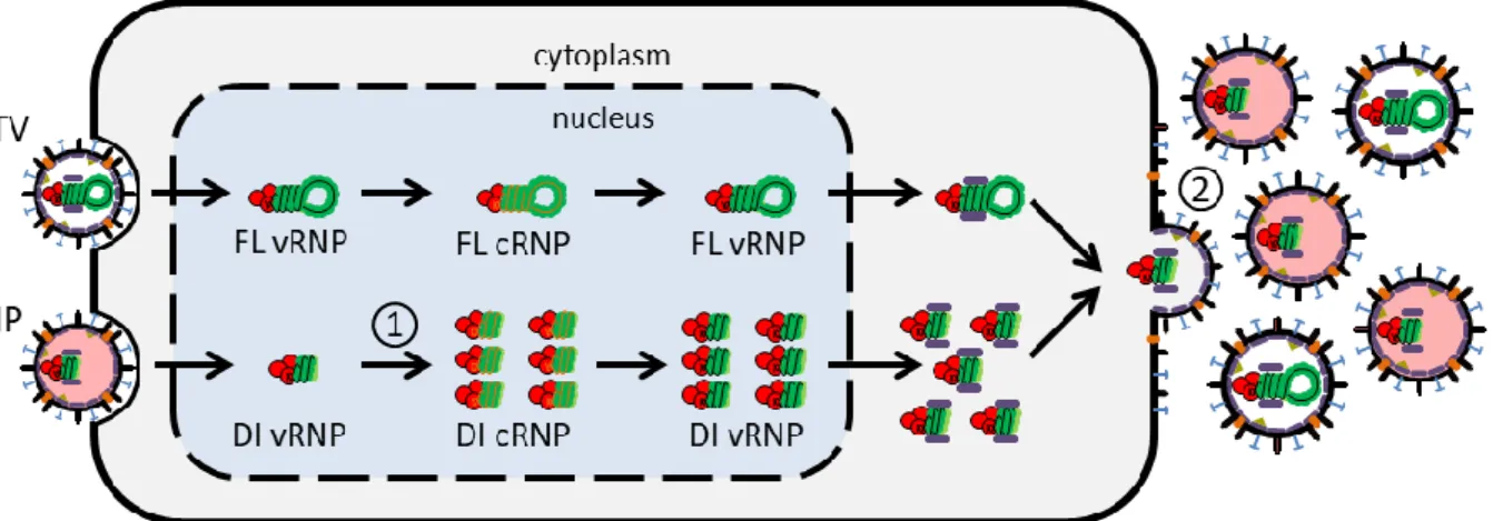
The deterministic intracellular model of IAV replication was developed by Heldt et al. and describes the IAV life cycle in a single mammalian cell using ordinary differential equations (ODEs) [33]. The detailed description of this model is provided by Frank Stefan Heldt as part of his PhD dissertation [1]. The IAV life cycle comprises the virus entry into the host cell, the vRNA replication and synthesis of all viral proteins and the release of progeny virions into the extracellular space. We assumed in our model that the core mechanisms for STV and DIP replication are very similar. In this section we explain the essential steps of the viral life cycle implemented in the original model (Figure 2.3) and in the next section we will highlight the adjustments for DIP replication.
Virus entry
The IAV entry into the host cell is initiated by the interaction of specific viral envelope proteins with the receptor binding sites of the cell: HA protein attaches to neuraminic acids (sialic acids) on the host Figure 2.2 | Structure of a defective interfering particle (DIP) and de novo generation mechanism of defective RNA. (A) Morphology of a DIP with a defective segment 3 (B) Structure of a full-length (FL) and defective
interfering (DI) RNA. The functional RNA has an open reading frame (ORF) represented by the straight lines. The defective RNA carries an internal deletion in its segment which is indicated by the V-shape line. The red boxes represent the terminal promoter sequences. (C) Potential mechanism for DI RNA de novo generation. The polymerase normal pathway along the template is represented by the sequential numbers and black arrows. The translocation of the polymerase with dissociation and reattachment at number 2 and 4, respectively deletes the internal coding sequence and originate DIPs. This path is indicated by the red dashed line. Figure adapted from the PhD dissertation of Heldt [1].
6
cell surface which promotes virus entry [34]. An endosome is formed on the cell surface which induces virus entry through a receptor-mediated endocytosis. The virus goes through endocytosis until the acidification in late endosomes which enables the virus envelope to fuse with the endosomal membrane [34,35]. This reaction results in viral uncoating, i.e. protons enter the virus particle through M2 ion channels causing the vRNPs to detach from M1 proteins which promotes the release of vRNPs into the host cell cytoplasm. During endocytosis, the virus can either successfully fuse with the viral envelope and release its vRNPs or fail to fuse and the virus particle will eventually be degraded. The fusion process in late endosomes is the only step during cell entry in which the virus can be degraded in the model, which can prevent further replication. Note that other biological processes can cause virus degradation, e.g. fail to attach the cell surface or cytoplasmatic transport, however such mechanisms were neglected in the model.
Nuclear import of vRNPs
After the viral fusion the vRNPs travel through the cytoplasm. Since the virus does not possess all the enzymes and resources necessary for its replication, it requires to take control over the biosynthesis machinery in a host cell’s nucleus to promote its propagation. Experimental data suggest that the eight vRNPs are transported together across the cytoplasm and only separate when they reach the karyoplasm [36]. The model does not directly consider cell compartments, however, it contains an inactive cytoplasmic state of vRNPs which is converted into separated nuclear vRNP segments when Figure 2.3 | Diagram of the influenza A virus life cycle. To simplify the figure, only one of the eight vRNPs is
represented and non-structural proteins were omitted. Solid arrows indicate synthesis or protein attachment and dashed arrows represent transport processes. The different life cycle steps are indicated by the numbers: 1 – attachment, 2 – endocytosis, 3 – fusion in late endosomes, 4 – nuclear import, 5 – transcription, 6 – replication (cRNA synthesis), 7 – protein translation, 8 – cRNA encapsidation, 9 – replication (vRNA synthesis), 10 – vRNA encapsidation, 11 – M1 and NEP binding, 12 – nuclear export, 13 – virus assembly and budding. Figure taken from the PhD dissertation of Heldt [1].
7
the genomes reach the nucleus. The nuclear import of vRNPs occurs via an active transport mechanism mediated by transport receptors located on the nucleus membrane [37,38]. Inside the nucleus each vRNP segment behaves as an independent functional structure [36].
Viral replication
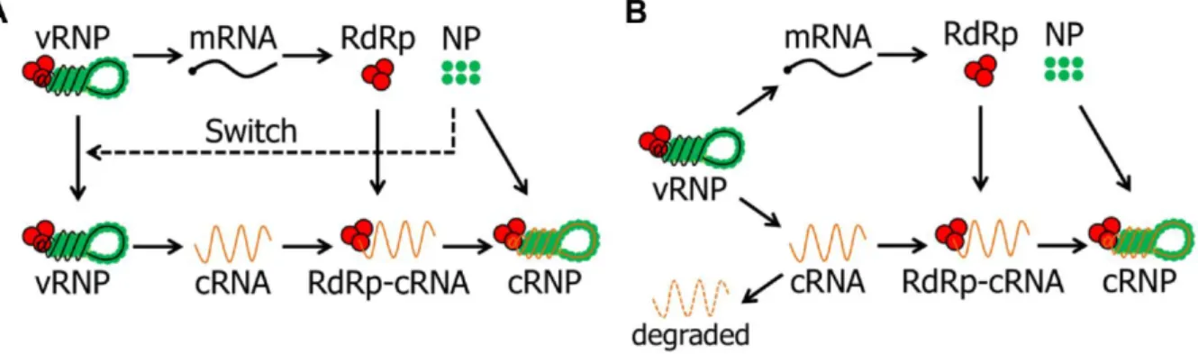
Once inside the nucleus, the eight vRNP segments are used as templates to produce viral mRNA and the complementary RNA (cRNA). However, the mechanism which coordinates the synthesis of both molecules is still under debate and different hypothesis were proposed in the past decades [25]. The most accepted theories are the switching and the stabilization hypothesis.
The switching hypothesis suggests that before cRNA synthesis, an initial round of viral protein translation occurs which promotes the accumulation of soluble NP, i.e. free NP that is not attached to vRNAs. This hypothesis proposed that NP switches the vRNP activity from transcription to replication (Figure 2.4A) [39]. The theory is supported by laboratory experiments which show that the synthesis of cRNA depends on NP [40] and a study with temperature-sensitive NP mutants which revealed a reduced cRNA synthesis and normal viral mRNA production [41]. However, other experimental data showed that over expressed NP did not increase cRNA replication [42]. By contrast, the stabilization hypothesis suggests that both viral mRNA and cRNA are synthesised early on after vRNPs nuclear import. This theory describes that due to the attachment of RdRp and NP the nascent cRNA forms cRNP which prevents its degradation by cellular nucleases, i.e. cRNA does not accumulate unless these proteins stabilize it (Figure 2.4B) [43]. This hypothesis is supported by in vitro experiments which showed that both transcription and replication occur early on and in the absence of free NP [44]. The vRNA synthesis proceeds from the positive-stranded cRNP which was formed previously. In a similar way, the vRNA needs to be stabilized by the viral polymerase and NP such that vRNPs can be formed in the host nucleus [45]. Is important to notice that in the model all intermediate molecules (cRNA, vRNA, cRNP and vRNP) can be degraded by nucleases during replication, however, the stabilized RNPs are degraded significantly slower.
Figure 2.4 | Different hypothesis for the transition mechanism from transcription to replication.
(A) Switching hypothesis – the transcription of mRNA by the polymerase occurs early in infection. Accumulation of NP switches the polymerase toward replication. (B) Stabilization hypothesis – vRNPs engage both transcription and replication early in infection. cRNA requires stabilization of viral polymerase and NP to prevent its degradation by cellular nucleases. Figure taken from the PhD dissertation of Heldt [1].
8
Transcription and protein synthesis
Experimental data showed that in infections with IAV the number of viral mRNA is negatively correlated to its length, i.e. shorter mRNAs are in higher abundance [46]. The transcribed viral mRNAs are then transported to the cytoplasm where the protein translation will take place. The viral mRNA needs to compete for transcripts and resources of the cell. To achieve this, several mechanisms are triggered that enables the viral mRNA to have preferential access to these resources and take over the cell translation machinery [47]. The synthesised proteins can engage two different pathways: they can either enter the cell nucleus or be transported to the plasma membrane [37,38]. RdRp and NP enter the nucleus to stabilize the newly produced vRNA and cRNA. Furthermore, a fraction of M1 and NEP takes the first path, and the remaining fraction takes the second route since they are involved in the nuclear export of vRNPs as well as in the process of virus particle release. Finally, the surface proteins HA, NA and M2 are processed in the endoplasmic reticulum and Golgi complex and transported to the plasma membrane.
Nuclear export of vRNPs
After replication, the newly synthesised vRNPs can either be used as template for viral mRNA and cRNA production or be exported from the nucleus to be assembled into progeny virions. It has been suggested that the nuclear vRNPs can be divided into two types: the active vRNPs which participate in viral mRNA and cRNA synthesis and the inactive ones that leave the nucleus and produce new viral particles [48]. Laboratory experiments showed that overexpression of M1 protein inhibit the viral mRNA transcription suggesting that this protein is a the mediator for vRNP inactivation [49,50]. There is further evidence that M1 binds to NP [49] and also to RNA [51] to promote the nuclear export of vRNPs. Additional studies illustrate the importance of M1: experiments with a fluorescence marker for NP, which can be used to localize vRNPs, showed that cells that lack M1 or were treated with antibodies which retain M1 in the cytoplasm, lead to an accumulation of vRNPs inside the nucleus [52]. Furthermore, another study, in which an M1 production inhibitor was applied, resulted in the nuclear retention of vRNPs [53]. It was also suggested that NEP participates in the export process: experimental work revealed that no vRNP was exported from the nucleus after injection of anti-NEP antibodies [54]. However, it has also been observed that even in complete absence of NEP nuclear export still occurs [55]. This results indicates that NEP is not a determinant factor for vRNP nuclear export or at least suggests that this protein is not required in high quantities [39].
Viral packaging and release
After replication and protein synthesis, the intracellular IAV continues its life cycle with the processes of virus packaging and finally the release from the host cell. To form a functional virus particle, one copy of each vRNP genome segment is required. However, whether the eight vRNP segments are assembled by a random mechanism or in a segment-specific packaging has been
9
debated for several years [56]. Experimental studies showed that the genome segments form a “7+1” configuration where seven segments display a ring around a central vRNP [27,57]. Further experiments indicated that this complex configuration might be mediated by RNA-RNA interactions between the IAV genome segments [58,59]. In addition, it has been observed that DIPs compete for their full-length (FL) segments in a segment-specific way during packaging [7,10]. These experimental results provide strong evidence that the viral genome assembly is controlled by a specific packaging mechanism. The virus assembly includes the aggregation of vRNP segments and the packaging of the structural proteins. It has been suggested that the M1 protein has a very important role during the virus assembly since observations showed it attaches to vRNPs, forms a layer underneath the virus envelope and interacts with HA and NA facilitating an association with the cell membrane [60]. The virus particle release is initiated by the formation of a bud which is mediated by the interaction of HA, NA and M2 with the cell membrane. Then, the bud is extended and the vRNPs segments are incorporated into the progeny particle. In the final steps M2 promotes the membrane curvature and the separation of the budding particle [61]. Once in the cell exterior, NA removes the sialic acids from the viral envelope which prevents HA from binding to other progeny particles.
2.4 DIP interference
Since influenza virus DIPs carry an internal deletion in at least one of their genome segments, they do not possess the complete coding sequence to synthesise all proteins. Consequently, they depend on coinfections with STVs that provide the missing protein(s) to form progeny particles. During these coinfections, DI RNA interferes with the STV replication which results in a reduction of the infectious virus titer and mainly progeny DIPs are produced [11,13]. However, the molecular mechanism inducing this interference is still not completely understood [12].
Several experiments showed a preferential amplification of the DI RNAs over the FL genome segments [7,62] which indicates that the interference can emerge during RNA replication. cRNA synthesis has been suggested to be the step comprising the replication advantage [63]. In this regard, it has been proposed that the reduced length of the DI vRNA induces this advantage, because synthesis processes could occur faster [12,13]. Since the viral polymerase synthesizes a constant number of nucleotides per unit time, shorter RNAs can be generated in greater abundance if the necessary resources are available. This theory is in agreement with experimental data from a dual luciferase reporter assay for RNA replication [64]. In this experiment, the shorter of the two luciferase-encoding influenza virus-like RNAs showed a higher interference potential on luciferase expression when compared to the longer reporter segment. Although shorter DI segments are assumed to be synthesised faster, large deletions can also disrupt terminal packaging signals [13]. This indicates that there may exist an optimal length of DI RNA at which DIPs achieve the maximum replication advantage and are still efficiently packaged into progeny particles. However, it has been observed that some DI RNAs do not accumulate to high levels in coinfected cells [7] which indicates that RNA length is not the only determinant factor of interference.
10
Other interference mechanism has been suggested to be the competition for limited viral and/or cellular resources [13]. Experimental studies support this assumption, because the competition between DI RNA and FL segments for viral polymerases has been observed in vesicular stomatitis virus (VSV) and influenza virus. Furthermore, different DI segments showed a more efficient packaging which results in an increased production of DIPs to the detriment of STV release [10,23]. Besides the competition for resources, it has also been hypothesised that the proteins synthesised from the DI RNA could contribute to the interference mechanism. Since the DI segment has the 3’ and 5’ promoters, the polymerase can transcribe DI mRNAs and synthesise truncated versions of proteins [62]. However, these proteins are shorter than the functional ones, are therefore very likely to lose their function and do not show an increased interference potential [12,62].
2.5 Therapeutic potential of DIPs
In recent years, DIPs have been suggested to be used in antiviral therapies due to their interference with STV replication which efficiently reduces the infectious virus yield [13,14]. Experimental results revealed that the administration of DIPs prevented mice and ferrets from developing lethal infections caused by different IAV strains [15,16]. More precisely, it reduced fever, weight loss, respiratory symptoms and the infectious load. Additionally, it was shown that the administration of DIPs between 24 and 48h post virus infection completely prevented clinical disease and death in mice [15]. Furthermore, it was observed that coinfections with STV and DIPs activate the immune response inducing anybody production [16]. Hence, research results suggest that DIPs have a major potential to be used for the prevention of IAV infections and as antiviral medication, especially when the infectious strain is unknown or resistant to other antiviral drugs [15].
2.6 Intracellular deterministic model of influenza A virus and DIP
replication
A deterministic intracellular model of DIP replication was developed by Laske et al. [9] which constitutes an extension from the previously implemented deterministic model of the IAV life cycle [33]. This model provided novel insights about the factors which influence DIP production focussing in the replication advantage, coinfection timing and DI RNA originated from different genome segments. The model comprises a DIP carrying a DI vRNA which has a replication advantage at the cRNA synthesis level due to its reduced length.
Using the deterministic model of DIP replication, Laske et al. evaluated the effect of the length dependent replication advantage on STV and DIP production. The obtained results suggested that there is an optimal DI RNA synthesis rate which results in a maximum DIP production. Model simulations showed that when the replication advantage increases above its optimum, the production of DI segments consumes an increasing amount of proteins which leads to the depletion of NP.
11
Consequently, the DIP release is reduced due to protein limitation. The authors concluded that at the optimum replication advantage, a balance between the FL and DI RNA synthesis is established such that the pool of viral resources would not be depleted.
Additionally, the authors evaluated the impact of different time delays of successive confections with STV and DIP. The results showed that DIP produced progeny particles only if DIP infection was delayed no longer than 3 hours after STV infection. It has been suggested that when the STV replication already progressed too far, DIP replication does not occur. In this case, the high levels of M1 and NEP accumulated in the cytoplasm promote the attachment of these proteins to the DI vRNP which become inactive. To compile these findings, simulations with different DI segments were performed. The model results showed that a defect in segment 3 provides an advantage in DIP replication compared to a DIPs carrying a defective segment 4 when STV coinfection is delayed more than 3 hours. Furthermore, simulations with DI segments 5 showed to be less productive than a DI segment 3 for all tested delays. The authors concluded that defects in segments which encode for proteins of the polymerase complex can provide an advantage in DIP replication. Hence, these DIPs can overcome DIPs with other defective segments after several passages and emerge as the dominant species, explaining why most DI RNAs of IAV are originated from segments 1 to 3.
2.7 Intracellular stochastic model of influenza A virus replication
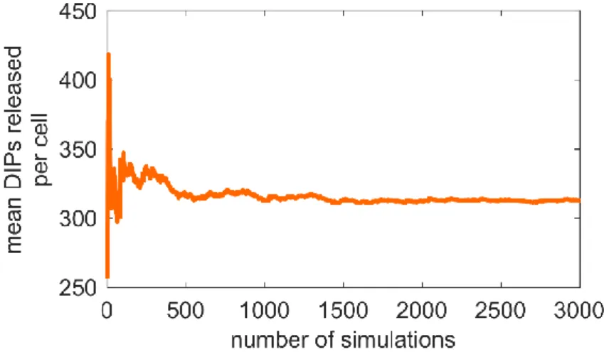
Cell-to-cell variability is a phenomenon usually observed in nature and most of these fluctuations are caused by the random nature of biochemical reactions [19]. By contrast to the deterministic model, which considers that the reactants in the system are continuous variables, the stochastic approach defines them as discrete numbers of molecules that can be changed by random events. Stochastic models can provide valuable information about how the system dynamics respond to the random effects inherent to biological processes [65]. Furthermore, for the same initial conditions the deterministic model provides one definitive result, however, stochastic model simulations result in a distribution that reflects the variance introduced by the randomness of biochemical reactions [3]. The stochastic effects are more pronounced in systems with a low number of molecules [18]. This is the case in virus infections since one single viral particle can be sufficient to infect an entire organism. However, stochastic simulations are usually more computationally demanding since the credibility of the stochastic results is related to the number of simulations performed [66].
A stochastic intracellular model of IAV replication was implemented by Heldt & Dorl [3,67] which is a new approach to the previous deterministic model of the IAV life cycle [33]. Model simulation results showed that the randomness of biochemical reactions, i.e. stochastic effects, have a significant impact on IAV replication at the cell level. The experiments performed in this study include the single-cell analysis of viral replication dynamics using real-time RT-qPCR and the observed results were complemented with the stochastic intracellular model. The authors focussed on different sources of noise affecting virus replication, genome segmentation, infections at low MOI and stochastic effects during virus entry.
12
The stochastic model of IAV replication showed that one of the major sources of variability in IAV infection is the segmented genome since each segment is affected by stochastic effects independently. Furthermore, simulations indicated that the viral replication process, which is assumed to be autocatalytic, amplifies heterogeneity in the levels of viral RNA. The authors suggested that the genome segmentation combined with the autocatalytic synthesis of vRNA from cRNA and vice versa, are the cause for the large fluctuations detected in the model for RNA and proteins levels which affect virus production.
The model predicted that nearly 93% of simulations performed at MOI 1 did not release any progeny virion. In half of these simulation runs the virus failed to fuse its envelope with the endosomal membrane. Another reason for this high percentage of non-productive infections is segment loss which is caused by the degradation of vRNA in the nucleus by cellular nucleases. If the vRNA of one or more genome segments is completely degraded before replication, the virus lacks part of its genome and, consequently, is unable to produce any viral particle. Due to these observations, the authors concluded that fusion failure and segment loss events are the major steps in virus infection that induce the cell-to-cell heterogeneity observed in IAV production.
2.8 Stochastic simulation algorithms
The stochastic chemical kinetics considers that in a well-stirred system
𝑁
chemical species{𝑆
1, 𝑆
2, … , 𝑆
𝑁}
interact through𝑀
chemical reactions{𝑅
1, 𝑅
2, … , 𝑅
𝑀}. 𝑋
𝑖(𝑡)
denotes the number of molecules of species𝑆
𝑖 in the system at time𝑡
. The goal of a stochastic simulation is to estimate the state vector𝑋(𝑡) ≡ (𝑋
1(𝑡), 𝑋
2(𝑡), … , 𝑋
𝑁(𝑡))
knowing that the system was in state𝑋(𝑡
0) = 𝑥
0 at some initial time𝑡
0[20]. The changes in the species populations are caused by the chemical reactions. In this context, stochastic simulations use state-change vectors and propensity functions to characterize each reaction channel𝑅
𝑗. The change in the system is not given by ODEs as in the deterministic model, but rather using a state change vector which is represented as:𝜈
𝑗≡ (𝜈
1𝑗, 𝜈
2𝑗, … , 𝜈
2𝑗)
, where𝜈
𝑖𝑗 is the change in the molecular population of𝑆
𝑖 induced by a single𝑅
𝑗reaction. If the system is in state𝑥
and one𝑅
𝑗reaction occurs, the system will jump to state𝑥 + 𝜈
𝑗. Furthermore, the reaction constants are described in the form of propensity functions rather than reaction rates as in the deterministic approach. The propensity function is represented as:𝑎
𝑗(𝑥) 𝑑𝑡
, which is the probability, given𝑋(𝑡) = 𝑥,
that one𝑅
𝑗reaction will occur in the next infinitesimal time interval[𝑡, 𝑡 + 𝑑𝑡]
[20]. The time to the next reaction occurring is an exponentially distributed random variable with average1 𝑎
⁄
0(𝑥)
and𝑎
0(𝑥) = ∑ 𝑎
𝑗(𝑥),
which represents the sum of all propensity functions.These concepts are included in the chemical master equation (CME) which determines the probability that each species will have a specific molecular population at a given future time point [68]. However, an analytical solution for the CME is usually quite complicated to obtain even for simple systems. To approximate a solution, Gillespie proposed a Monte Carlo procedure to simulate time
13
trajectories of the molecular populations described by the CME. This procedure was named stochastic simulation algorithm (SSA) and has been used as the basis for stochastic simulations until today [69,70]. The SSA is usually preferred over other iteration methods due to its accurate results and simple coding [71]. However, it simulates every single reaction separately which makes it very inefficient for systems with a high number of molecules, as is the case for IAV infection. During later stages of infection, this problem becomes even more challenging as the amount of interacting molecules increases rapidly from a low number to millions of molecules. As the population of some species increases, the values of the propensity functions increase substantially [3]. Consequently,
𝑎
0(𝑥)
achieves higher values and the time until the next reaction is on average reduced which results in more simulations per time unit. To improve computational speed, Gillespie proposed an extension to the SSA named Tau-leaping in 2001 [72]. This algorithm approximates the result of the SSA by advancing the system by a time step𝜏
during which multiple reaction events can occur simultaneously. Hence, computational efficiency can be increased by this method as it allows to aggregate a high number of individual reactions in a single computational step. However, the leap condition has to be considered:𝜏
has to be small enough that none of the states 𝑋𝑖 in the system change by an amount above a certainthreshold, which is normally defined as 3%.
As mentioned before, the stochastic algorithm to simulate influenza virus infection was previously developed by Heldt & Dorl and we opted to use this algorithm as a basis for our model [3]. The SSA implementation followed the steps described in the original work of Gillespie [70] and the Tau-leaping procedure was based on the work of Cao et al. [73], which has been indicated as the most efficient method for explicit tau-leaping [20]. The step-by-step explanation of the algorithm procedure can be consulted in the Bachelor thesis of Sebastian Dorl [3].
15
3 Models and Methods
3.1 Implementation of biochemical reactions
Our stochastic model of DIP replication is based on a previous stochastic model of the intracellular life cycle of IAV developed by Heldt & Dorl [3,67]. We extended this model by implementing the mechanism of DIP interference described in the deterministic model of DIP replication implemented by Laske et al. [9]. For most of our simulations, we established a DI RNA derived from segment 3, which encodes for the polymerase acidic protein (PA). This specific segment was chosen, because it is one of the polymerase-encoding segments which are the most abundant DI segments found in IAV production [30,31]. Furthermore, the previous deterministic DIP model also established a DI RNA for segment 3 and to enable the comparison between both models we opted to apply the same modification in our implementation. In the following, we provide a complete list of the biochemical equations established in our model. For a comparison between different DI segments, we also applied a DI RNA derived from segment 4 which encodes for HA.
Virus entry
The extracellular STVs