UNIVERSIDADE DE LISBOA
FACULDADE DE CIÊNCIAS
DEPARTAMENTO DE BIOLOGIA ANIMAL
The role of the extracellular matrix in
the patterning and morphogenesis of
the sclerotome
Patrícia Gomes de Almeida
Mestrado em Biologia Evolutiva e do Desenvolvimento
2011
UNIVERSIDADE DE LISBOA
FACULDADE DE CIÊNCIAS
DEPARTAMENTO DE BIOLOGIA ANIMAL
The role of the extracellular matrix in
the patterning and morphogenesis of
the sclerotome
Patrícia Gomes de Almeida
Dissertação orientada por:
Professora Doutora Sólveig Thorsteinsdóttir
Mestrado em Biologia Evolutiva e do Desenvolvimento
2011
ACKNOWLEDGEMENTS
Muitas são as pessoas que de alguma forma contribuíram para que este ano fosse fantástico, e a quem quero agradecer do fundo do coração.
Em primeiro lugar e em especial, à Professora Sólveig, a quem tenho tanto a agradecer! Primeiramente por aceitar ser minha orientadora, mas também por toda a ajuda e paciência, por se mostrar sempre preocupada e entusiasmada com o trabalho, pela confiança que depositou em mim a tantos níveis, por nunca se chatear com os meus erros e vibrar com os resultados que fui tendo, e por estar sempre disponível para responder às minhas dúvidas ou questões, independentemente do quão inconveniente e tonta pudesse ser. No entanto, agradeço principalmente a compreensão e apoio que me deu quando mais precisei. A sua preocupação, mais do que com o trabalho, com o meu bem-estar, é algo que não tem preço e pelo qual estou profundamente grata. Sinto sinceramente que não podia ter melhor orientadora!
Tenho ainda a sorte de estar inserida num grupo fantástico, e ser uma BD deixa-me orgulhosa. Agradeço a todos vocês pelo bom ambiente e por tornarem o meu dia-a-dia tão agradável num ano de stress e corrida contra o tempo. Em particular, agradeço imenso ao Pedro, por se mostrar sempre disponível para me ajudar quando me surgiram dúvidas ou problemas, ou asneiras…, por tudo o que me ensinou e por me fazer acreditar que consigo cortar notocordas ao meio com os olhos fechados. À Raquel Vaz, por toda a ajuda, confiança e desabafos mútuos, pelas conversas e pela companhia, por se esforçar por manter o pessoal na linha e por me ensinar tanto! Marianne, thank you so much for your wisdom, friendship and great advices, for the conversations and the laughs! Ao Luís, por ser um geek especial e por me fazer rir com gosto mesmo naqueles dias em que é a última coisa que apetece fazer. Ao P’sor Gabriel, por conseguir sempre arranjar um bocadinho para mim e para me ajudar no que pudesse, e também à Prof. Gabriela, pela constante simpatia e boa disposição. E, claro, aos meus companheiros mestrandos! À Raquel, pelos infindáveis favores, companheirismo, risos, desabafos, ajudas e devaneios; ao André, por levar sempre tudo na brincadeira e dar música ao laboratório, unplugged; à Joana, pelas gargalhadas, confiança, partilha e por juntar-se a mim em cantorias de bandas sonoras embaraçosas. Não posso deixar de agradecer também à Josefina, Raquel, Carla, Eduardo e a todo o grupo de Evolução Experimental pelos momentos de descontracção, desabafos e insanidade. Em especial um muito obrigado à Raquel, pela companhia, por aturar o meu mau humor e desatinos matinais e pelos minutos extra de sono que me permitiu ter em tantos dias de boleia!
Agradeço também à Sara Sousa, à Dra. Raquel Andrade e em especial à (Dra.) Isabel Palmeirim, pela disponibilidade em ajudar a resolver os problemas técnicos estranhos que me surgiram, pela simpatia e pelas sugestões preciosas. Agradeço ainda à D. Branca, porque as coisas não aparecem prontas a usar e esterilizadas sozinhas, e por dar sempre uma mão amiga para ajudar quando se precisa!
Às minhas tontas, por serem as maiores e pelas tardes e noites bem passadas, não só este ano, mas desde o início do curso. Em especial, um gigante obrigado à Andreia, por ser a melhor amiga que alguém pode desejar, não tenho palavras para te
agradecer, nunca seria o suficiente; à Margarida, por ser uma querida mesmo não admitindo, por estar sempre lá e por ser um apoio e companhia constantes durante este ano, não sei como me aturas; à Catarina, por toda a confiança, compreensão, companhia e momentos de insanidade conjuntos mas, mais do que isso, por todo o apoio que me deste quando mais precisei. À Inês, pelo apoio incondicional e por estar sempre lá para os bons e os menos bons momentos, mesmo quando nem temos tempo para respirar; à Sofia pelas gargalhadas e boa disposição e à Joana, pela amizade e confiança. Sem vocês nada disto seria possível, vocês fazem qualquer noite de menos horas dormidas valer a pena. Ao Nelson, por todo o apoio, compreensão e motivação. À Xana e à Joana, por uma vida inteira de amizade. À Vanessa, por largar tudo quando preciso de uma palavra de conforto. A todos os amigos que tenho a sorte de ter e que de alguma forma me apoiaram neste tempo de loucura, lembrando-me que a vida não é só trabalho, muito obrigado!
Agradeço também a toda a minha família, em particular aos meus pais, por todos os sacrifícios, por me terem possibilitado chegar até aqui, e por não torcerem o nariz quando decidi seguir Biologia, apoiando-me sempre na decisão. Aos meus avós, por serem uma inspiração. Esta tese é para vocês.
I
ABSTRACT
Extracellular matrices have several important functions during vertebrate development. They are responsible for polarizing cells, supporting cell migration and maintaining tissue boundaries, and also regulate cell shape, proliferation, growth and differentiation. Fibronectin is a major component of embryonic extracellular matrices and is involved in numerous processes during embryogenesis. It has recently been implicated in the formation of epithelial somites, transient structures responsible for the development of the axial skeleton, muscle and dermis. Significantly, when the assembly of the fibronectin matrix is impaired, somite formation is severely affected; moreover, somites formed before this impairment have defective morphologies.
With this work, we addressed whether the blocking of fibronectin matrix assembly would have implications in the development of older somites and their derivatives, in particular the sclerotome. We found that when chick embryo whole or bisected posterior explants, including all tissues caudal to somite X, are cultured for 6 hours with a 70kDa fibronectin fragment, thus abrogating further fibronectin matrix assembly, not only is somitogenesis halted, but the morphology of all the somites is affected, suggesting a delay in somite development. Furthermore, somite patterning is also altered, as seen by a delay in the expression of Pax1, a sclerotome marker. This delay in the activation of the sclerotome identity is accompanied by a failure of its full de-epithelialization at the correct time. Moreover, the myogenic program, as visualized by MyoD expression, also fails to be activated and maintained. Together these results demonstrate that an intact fibronectin matrix is essential for both sclerotome and myotome development. Given that both of these somite compartments are patterned by the morphogen Sonic hedgehog (Shh), we investigated whether inhibiting fibronectin matrix assembly affects Shh signaling. We found that although the notochord expresses Shh normally, the expression of Patched genes, a readout of Shh signaling, is perturbed in the somites. All these results highlight the complexity of paraxial mesoderm development, and demonstrate the importance of the extracellular matrix not only as a supportive scaffold, but as an active player in somite patterning and morphogenesis.
Key words: Extracellular matrix, fibronectin, sclerotome morphogenesis and
II .
III
RESUMO
Nos Vertebrados, a mesoderme paraxial forma-se de cada lado do tubo neural como consequência da gastrulação. Este tecido é composto por células mesenquimatosas que constituem a mesoderme presomítica, cuja parte rostral sofre periodicamente uma transição mesênquima-epitélio, formando um par de sómitos epiteliais. Estes são estruturas transientes, precursores da musculatura, esqueleto axial e derme e são responsáveis pelo arranjo segmentar de várias estruturas. No embrião de galinha (Gallus gallus), um novo par de sómitos é formado a cada 90 minutos, resultando num gradiente de maturação rostro-caudal em que os sómitos anteriores são mais desenvolvidos do que os sómitos posteriores, mais recentes.
A maturação do sómito resulta na formação de vários derivados. Pouco tempo após a sua formação, a parte ventral do sómito sofre uma transição epitélio-mesênquima para formar o esclerótomo, responsável pela formação das vértebras e costelas. A região dorsal do sómito mantém-se epitelial, designando-se o dermamiótomo. Na região mais mediana do sómito inicia-se a formação do miótomo, precursor do músculo esquelético axial. A formação destes derivados somíticos é dependente dos tecidos envolventes, que produzem morfogénios responsáveis pela indução dos vários tipos celulares específicos de cada derivado. Um destes morfogénios é o Sonic hedgehog (Shh), importante para inúmeros processos durante o desenvolvimento embrionário, nos quais se inclui a formação dos sómitos no intervalo de tempo correcto. O Shh é produzido pelas estruturas axiais, a notocorda e o floor plate do tubo neural, sinalizando ao sómito ventral de forma a induzir a formação do esclerótomo. É ainda um dos responsáveis pela indução do miótomo, sendo que concentrações intermédias de Shh, em conjunto com concentrações intermédias de Wnt produzido pela ectoderme, induzem a activação do programa miogénico na parte mediana do sómito.
A matriz extracelular que ocupa os espaços intercelulares nos tecidos é outro componente de extrema importância no desenvolvimento embrionário. A matriz extracelular têm inúmeros papéis durante a embriogénese – além de providenciarem suporte físico às células, fornecem sinais bioquímicos e biomecânicos cruciais para a morfogénese e diferenciação dos tecidos, polarizam as células, servem de substrato à sua migração e mantêm as fronteiras entre tecidos. Variações na sua deposição, composição, rigidez e elasticidade são ainda responsáveis por regular profundamente o comportamento e a fisiologia celulares, tendo importante impacto no crescimento, sobrevivência, forma e diferenciação das células.
Um dos componentes mais ubíquos das matrizes extracelulares embrionárias é a fibronectina. Esta matriz tem inúmeros papéis durante o desenvolvimento embrionário, desde a polarização das células, à separação de diferentes tecidos e ao desenvolvimento correcto da mesoderme, incluindo a mesoderme paraxial. De facto, nos modelos de ratinho, peixe-zebra e Xenopus, a ausência ou deficiência de fibronectina leva à não formação de sómitos e o mesmo é observado em embriões de galinha. Estudos anteriores no nosso laboratório demonstraram que quando explantes posteriores de embrião de galinha são cultivados na presença de um fragmento de fibronectina de 70kDa, que se liga aos domínios de ligação fibronectina-fibronectina e impede a sua fibrilogénese, o processo de somitogénese é atrasado e mais tarde parado. Para além disto, a morfologia dos sómitos formados antes do período de cultura é afectada, ficando mais desorganizada. A fibronectina é assim importante não só na polarização das células da mesoderme presomítica, de forma a formarem o sómito epitelial, como é também importante na manutenção desta estrutura ao longo do seu desenvolvimento.
Para além deste efeito do bloqueio da construção da matriz de fibronectina na somitogénese e morfologia dos sómitos formados antes da cultura, foi também observado que estes sómitos não chegam a formar o esclerótomo no tempo devido. Assim, o objectivo deste projecto foi analisar a morfogénese e a padronização do
IV esclerótomo quando a construção da matriz de fibronectina é impedida, analisando possíveis efeitos na morfologia, indução e padronização pelo Shh, e expressão de genes marcadores de diferenciação característicos do esclerótomo e do miótomo. Para o efeito, explantes posteriores de embriões de galinha de estádio HH11 a HH14, englobando os 10 sómitos mais posteriores (sómitos SI a SX) e a PSM, foram cultivados com o fragmento de 70kDa de fibronectina durante 6 horas. Para determinar se a morfologia do esclerótomo é alterada quando a construção da matriz de fibronectina é bloqueada, explantes cultivados foram processados para imunohistoquímica para N-caderina. Verificou-se que a morfologia de todos os sómitos sofre alteração, incluindo nos sómitos formados antes da cultura, sugerindo um atraso no seu desenvolvimento. A principal alteração observada foi a não de-epitelização do sómito ventral para dar formação ao esclerótomo no tempo correcto, confirmando os resultados preliminares dos estudos anteriores.
Processando explantes cultivados para hibridação in situ utilizando sondas para o mRNA de marcadores específicos dos derivados somíticos, foi possível constatar que os sómitos mais anteriores mantêm a identidade de esclerótomo, confirmada pela análise da expressão do marcador Pax1. No entanto, tal como verificado para a morfologia, a activação deste gene é atrasada na ausência de uma matriz de fibronectina intacta. Assim, a matriz de fibronectina é importante não só para a activação do programa de diferenciação do esclerótomo no tempo certo, mas também para a sua correcta morfogénese mais tarde na sua maturação.
Além dos efeitos da inibição da construção da matriz de fibronectina na morfogénese do esclerótomo, foi também identificada uma severa alteração no programa de miogénese. Quando cultivados com o fragmento de 70kDa de fibronectina, não só a expressão de MyoD não é activada no tempo correcto, como sómitos formados antes da cultura perdem a forte expressão de MyoD que apresentavam anteriormente. Assim, os nossos resultados sugerem que a matriz de fibronectina tem um papel determinante não só na activação mas também na manutenção do programa miogénico.
Foi ainda identificado, através de hibridação in situ para os receptores e genes-alvo de Shh Patched1 e Patched2, que a sinalização Shh é afectada quando a fibrilogénese de fibronectina é impedida. Em explantes cultivados com o fragmento de 70kDa, a expressão de Patched1 nos sómitos diminui, e não chega a ser restrita à parte mediana do sómito, como acontece em explantes controlo e no embrião. Por outro lado, a alteração na expressão de Patched2 é ainda mais severa, desaparecendo da mesoderme presomítica anterior e dos sómitos, incluindo sómitos formados antes da cultura, sendo apenas mantida nos sómitos mais rostrais. Assim, embora o Shh seja produzido correctamente pelas estruturais axiais, como comprovado por hibridação in situ para Shh, o seu sinal não é recebido correctamente pelo sómito na ausência de uma matriz de fibronectina intacta. Deste modo, a sinalização de Shh parece depender directa ou indirectamente da matriz de fibronectina de modo a induzir correctamente a diferenciação e morfogénese dos sómitos. É possível que a alteração na matriz de fibronectina tenha um papel na difusão, restrição ou apresentação da molécula de Shh aos tecidos alvo. Por outro lado, a alteração na sua construção pode levar a alterações noutros componentes da matriz extracelular que por sua vez interajam com o Shh como, por exemplo, a laminina ou proteoglicanos.
Os resultados deste projecto demonstram assim a importância da matriz extracelular não só como estrutura de suporte, mas também como tendo um papel directo e crucial na padronização e morfogénese dos sómitos e seus derivados.
Palavras-chave: Matriz extracelular, fibronectina, morfogénese e padronização do
VI
TABLE OF CONTENTS
ABSTRACT ... I RESUMO ... III
I. INTRODUCTION ... 1
I.1. Paraxial mesoderm development ... 1
I.1.1. Somitogenesis ... 1
I.1.2. Somite maturation and patterning ... 1
I.1.3. Epithelial to mesenchymal transitions (EMTs) ... 3
I.2. The morphogen Sonic hedgehog ... 3
I.2.1. The pathway ... 3
I.2.2. Shh pathway components in the vertebrate embryo ... 5
I.2.3. Shh role in the dorso-ventral patterning of the paraxial mesoderm ... 5
I.3. The extracellular matrix ... 6
I.3.1. Extracellular matrices ... 6
I.3.2. Integrins ... 6
I.3.3. Laminins ... 7
I.3.4. Fibronectin ... 7
I.3.5. Fibronectin in embryonic development ... 8
I.3.6. Fibronectin in the somitogenesis ... 8
I.4. Objectives ... 10
II. MATERIALS AND METHODS ... 12
II.1. Embryo collection ... 12
II.2. Cultures ... 12
II.2.1. Embryo collection and surgical procedures ... 12
II.2.2. Explant culture ... 12
II.2.3. Formation of somites in explant cultures and somite terminology ... 12
II.3. In situ probe production ... 15
II.3.1. Total RNA isolation and cDNA production ... 15
II.3.2. Primer designing and gene amplification ... 15
II.3.3. PCR product ligation and plasmids used ... 15
II.3.4. Bacteria transformation ... 15
VII
II.3.6. Linearization of the vector ... 16
II.3.7. Transcription of the genes of interest with Dioxygenin ... 16
II.4. In situ hybridization ... 16
II.5. Embedding and cryossectioning ... 17
II.6. Immunohistochemistry ... 17
II.7. Image acquisition and treatment ... 18
III. RESULTS ... 20
III.1. The role of the fibronectin matrix in the morphogenesis of the sclerotome ... 20
III.1.1. Impairing fibronectin matrix assembly affects sclerotome morphology ... 20
III.2. The role of the fibronectin matrix in the patterning of the sclerotome ... 22
III.2.1. Shh is produced normally when fibronectin fibrillogenesis is impaired ... 23
III.2.2. Shh is not signaling properly to the somites ... 24
III.2.3 Sclerotome development is delayed when fibronectin assembly is impaired ... 28
III.2.4. The sclerotome of rostral somites successfully downregulates Pax3 when fibronectin assembly is impaired ... 28
III.3. The role of the fibronectin matrix in myogenesis ... 30
III.3.1. Inhibiting fibronectin assembly into a matrix leads to severely impaired myogenesis ... 31
III.4. The role of fibronectin matrix in regulating the expression of ECM components ... 32
III.4.1. Fibronectin expression in unaffected when fibronectin fibrillogenesis is impaired ... 32
III.4.2. Fibronectin regulates Lama5 mRNA expression in the lateral somite ... 33
IV. DISCUSSION ... 36
IV.1. The fibronectin matrix is important for the morphology of all the somites ... 36
IV.2. The morphological defects of the paraxial mesoderm of experimental explants are accompanied by changes in gene expression ... 37
IV.3. Affecting the fibronectin matrix interferes with Shh signaling ... 38
IV.4. Future prospects ... 39
V. REFERENCES ... 41 SUPPLEMENTARY MATERIAL ... A
1
I. INTRODUCTION
I.1. Paraxial mesoderm development
I.1.1. SomitogenesisDuring vertebrate gastrulation, two bands of loosely arranged mesoderm (the presomitic mesoderm, PSM) are formed on each side of the neural tube and notochord
(Stockdale et al., 2000; Christ et al., 2007). While new cells are continuously added to the caudal end of the PSM, the rostral end undergoes a mesenchyme to epithelial transition (MET), forming epithelial somites (Duband et al. 1987; Stockdale et al., 2000). Somites, the precursors of dermis, skeletal muscle and axial skeleton, are transient structures composed of epitheloid cells arranged in an aster-like organization, surrounding a mesenchymal core, the somitocoel (Christ et al., 2007).
The PSM is characterized by the cyclic expression of specific genes, such as hairy1 and lunatic fringe which reveal the existence of a molecular clock defining the time interval at which the somites form. This interval is precise and species-specific. In the chick embryo, it takes 90 min for a somite pair to form (Andrade et al., 2007; Palmeirim et al., 2008).
Because somites are progressively formed in the anterior end of the PSM and they mature in a caudal to rostral direction, this generates a posterior to anterior differentiation gradient in which the caudal-most somites represent early maturation phases, while rostral-most somites represent the oldest stages. Somites not only constitute a metameric pattern within the embryo, giving a segmental arrangement to their derivatives, the vertebral column, ribs, muscles, tendons, ligaments, but also impose a segmented pattern on neighboring structures such as dorsal root ganglia, peripheral nerves, and blood vessels. Therefore, the processes of somitogenesis and somite maturation must be tightly regulated for the embryo to develop normally (Christ and Ordahl, 1995; Stockdale et al., 2000; Christ et al., 2007).
I.1.2. Somite maturation and patterning
During somite maturation, the ventral somite undergoes an epithelium to mesenchyme transition (EMT), where cells lose their cell-cell adhesions and become motile, originating the mesenchymal sclerotome, precursor of the axial skeleton (Fig. 1;
Christ et al., 2007). The sclerotome is characterized by the expression of Pax1 (Fig.1) and Pax9 (Balling et al., 1996). Pax1 is an early marker of sclerotome fate and begins to be expressed in the somitocoele and ventral two-thirds of somite IV (according to Christ and Ordahl, 1995), while Pax9 is only expressed when the sclerotome fully de-epithelializes (Ebensperger et al., 1995; Borycki et al., 1997; Monsoro-burq, 2005). Pax1 is not necessary for the dissociation of the ventral somite into the sclerotome but it is required for the development of ventral vertebral structures (Wallin et al., 1994).
The dorsal half of the somite, the dermomyotome, remains epithelial (Fig. 1; Stockdale et al., 2000; Christ et al., 2007). Its apical side faces the underlying sclerotome, and its borders curve downwards to form the dermomyotomal lips (Stockdale et al., 2000; Christ et al., 2007; Thorsteinsdóttir et al., 2011). The dermomyotome is the precursor of the dermis and all the trunk and limb muscles,
2
expressing the transcription factors Pax3 and Pax7 (Scaal and Christ, 2004; Buckingham, 2006). Pax3 expression first occurs throughout the whole somite, but becomes restricted to the dorsal compartment when Pax1 expression is activated in the ventral domain (Cairns et al., 2008).
Myogenesis starts in the dorsomedial lip (DML) of the dermomyotome, but eventually all lips contribute towards the morphogenesis of the myotome, the first skeletal muscle of the embryo. Committed myogenic precursors express specific transcription factors, the myogenic regulatory factors (MRFs) MyoD and Myf5 (Buckingham, 2006).
Fig. 1. Somite differentiation. A. Somites are transient structures composed of an epithelioid
wall of cells surrounding a mesenchymal core, the somitocoel. Somites are surrounded by a discontinuous basement membrane, which in turn is in close contact with a more exterior fibronectin-rich extracellular matrix. B. Signals from the surrounding tissues are responsible for the differentiation of the cells from different domains of the somite, which acquire specific fates and gene-expression profiles. Shh produced by the notochord and the floor plate of the neural tube signals to the ventral somite, which de-epithelializes forming the mesenchymal sclerotome, composed of Pax1-positive cells. The dorsal region of the somite, the dermomyotome, remains epithelial and expresses Pax3. The DML of the dermomyotome originates the first cells committed to myogenesis, which express MyoD and then enter the myotome. Extracellular matrices are always in close contact with these structures, with a basement membrane surrounding the basal (dorsal) side of the dermomyotome and separating the myotome from the sclerotome, while a fibronectin matrix is present surrounding the dermomyotome and the sclerotomal cells. NT – neural tube. Not – notochord DML – dorso-medial lip. Adapted from
3
I.1.3. Epithelial to mesenchymal transitions (EMTs)
EMT is a process by which epithelial cells disrupt their cell-cell attachments and enhance their motile behavior, becoming mesenchymal (Fig. 2; Guarino et al., 2007; Baum et al., 2008). Several factors can induce EMT in a given context, namely growth factors and cell surface receptors, extracellular matrix-related molecules and signal transduction pathways (Guarino et al., 2007; Baum et al., 2008). EMT involves several drastic changes in cell morphology, behavior, physiology and expression of genes involved in cell adhesion, polarity, motility and viability. Cell-cell junctions are disassembled and the extracellular matrix is degraded, allowing delamination and migration of the now mesenchymal cells (Guarino et al., 2007; Baum et al., 2008).
Regulation of EMT is crucial for normal tissue homeostasis even in the adult. An uncontrolled EMT event may contribute to pathologies such as fibrosis and cancer (Guarino et al., 2007; Baum et al., 2008). Thus, it is of extreme importance to study these tissue rearrangements and transitions. Since both EMT and MET occurs several times and in distinct contexts during development, and cancer is characterized by the reactivation of embryonic programs, the study of the embryonic MET and EMT, such as those that occur during paraxial mesoderm development, provide an excellent study model (Guarino et al., 2007; Baum et al., 2008; Acloque et al., 2009).
Fig. 2. The processes of epithelium-to-mesenchyme (EMT) transitions. During EMT,
epithelial cells lose their attachment to their neighbors by disassembling their adherens junctions. These cells acquire mesenchymal characteristics and behavior, becoming motile and abandon the epithelium. This process occurs in a variety of embryonic contexts and is thought to underlie sclerotome formation. From Acloque et al., 2011.
I.2. The morphogen Sonic hedgehog
I.2.1. The pathwayThe hedgehog (Hh) signaling pathway has been extensively studied since the first Hh molecule was discovered in Drosophila melanogaster (Nusslein-Volhard and Wieschaus, 1980) and found to be essential for the normal patterning of the larval cuticle (Hidalgo and Ingham, 1990). There are three hedgehog orthologs common to all vertebrates, Sonic (Shh), Indian (Ihh) and Desert (Dhh) hedgehog, each with different and specialized functions in development (Ehlen et al., 2006; Ingham and Placzek, 2006). Shh, a morphogen and regulator of cell survival and proliferation, is crucial for numerous processes during vertebrate development, including the regulation of left-right asymmetry, limb and digit patterning, and neural tube and brain development (Ehlen et al., 2006; Ingham and Placzek, 2006).
4 Hh signaling involves multiple downstream effectors and a negative feedback mechanism (Fig. 3). The core components and transduction of this signaling pathway are highly conserved in both Drosophila and vertebrates. Considering what happens in Drosophila, in the absence of the Hh signaling molecule in the target cell, the transmembrane Hh receptor Patched represses the activity of Smoothened. This repression leads to the proteolytic cleavage of the transcription factor Cubitus interruptus (Ci), resulting in a truncated form of the protein that translocates to the nucleus and represses the transcription of Hh target genes. When Hh binds to the receptor Patched on the target cell, the repression of Smoothened is released, preventing the cleavage of Ci, which enters the nucleus and activates Hh target gene transcription. Patched itself is a target of Hh signalling, and because its upregulation leads to an increase in Smoothened inhibition and cleavage of Ci, a negative feedback loop is created (Ehlen et al., 2006; Ingham and Placzek, 2006).
There are two Patched genes in Vertebrates, Patched1 and Patched2. Cubitus interruptus has three vertebrate orthologs, the Glioma-associated oncogene homologues Gli1, Gli2, and Gli3. However, only Gli2 and Gli3 are processed to repressors, whereas Gli1 cannot be phosphorylated and processed, functioning only as a transcription activator (Ehlen et al., 2006).
Fig. 3. Hh signaling pathway. A. In the absence of Hh molecule, its receptor Patched
represses Smoothened, leading to the proteolylic cleavage of Cubitus interuptus (CI), whose truncated form enters the nucleus and functions as a repressor of Hh target genes. B. When Hh arrives to target cells and binds to Patched, the inhibition of Smoothened is relieved, which in turn prevents the cleavage of CI. The full-length CI protein can then enter the nucleus to activate target gene transcription. From Ingham and Placzek, 2006.
5
I.2.2. Shh pathway components in the vertebrate embryo
In the early vertebrate embryo, Shh is produced by the notochord and the floor plate of the neural tube, the zone of polarizing activity in the limb and by the zona limitans intrathalamica in the midbrain (Marti et al., 1995). Cells expressing low levels of Patched are capable of receiving and transducing the Shh signal, while high levels of Patched transcription is a readout of active Shh signaling, marking cells directly responding to Shh (Hidalgo and Ingham 1990). In the 48h chick embryo, Patched1 is expressed in the ventral neural tube, the notochord and splanchnic mesoderm (Marigo and Tabin, 1996). Posterior somites show Patched1 expression in the ventral domain, whereas anterior somites express Patched1 in the dorsomedial domain, becoming progressively more restricted to the myotome-forming region and sclerotome (Marigo and Tabin, 1996; Borycki et al., 1998). Patched2 expression in the neural tube and paraxial mesoderm is similar to that of Patched1. However, differential expression of these Shh receptors in the midline of the early embryo and the limb bud suggests a different regulatory mechanism for these two genes.
It is nevertheless clear that both Patched1 and Patched2 are dependent on Shh signaling for their activation. When Shh signaling is blocked, Patched1 and Patched2 expression are dramatically downregulated. Conversely, expression of ectopic Shh leads to a broader expression of Patched1 and especially Patched2, confirming that these are target genes of the Shh pathway (Pearse et al., 2001).
I.2.3. Shh role in the dorso-ventral patterning of the paraxial mesoderm
The dorso-ventral (DV) patterning of the somite is dependent on signals emitted by the surrounding tissues that both antagonize and synergize depending on the context (Dietrich et al., 1997). A gradient of Shh protein produced by the notochord is one of the major morphogens responsible for the DV patterning of the somite. When the notochord is surgically separated from the paraxial mesoderm or Shh signaling is blocked, both Pax1 and MyoD expression are absent. Conversely, the presence of exogenous Shh recovers their expression, implicating Shh produced by the axial structures as necessary for the induction and maintenance of both the sclerotome and myotome (Borycki et al., 1998; Marcelle et al., 1999).
Different levels of Shh elicit different responses from the somitic cells. The Shh gradient is opposed by Wnt6 and BMP4 produced by the overlying ectoderm, and Wnt1/Wnt3a from the dorsal neural tube. In this way, low levels of Shh in combination with high concentration of Wnts promote Pax3 expression and dermomyotome development, while intermediate levels of both Shh and Wnt signaling induce the activation of myogenesis, with the expression of MyoD. In the ventral somites, high levels of Shh signaling result in Pax1 expression and the development of the sclerotome (Dietrich et al., 1997). Thus, Shh secreted by the notochord and floor plate of the neural tube, and Wnt signaling from the ectoderm and dorsal neural tube, function synergistically to regulate the expression of somite-specific genes and pattern somitic cells to adopt different cell fates (Borycki et al., 2000; Cairns et al., 2008).
6
I.3. The extracellular matrix
I.3.1. Extracellular matricesThe extracellular matrix (ECM) occupies the noncellular space within tissues, serving not only as a physical scaffold, but also being responsible for providing biochemical and biomechanical cues crucial for tissue morphogenesis, differentiation and homeostasis (Frantz et al., 2010). ECM components include glycoproteins, such as collagens, fibronectin, laminins and tenascins, and polysaccharide-rich molecules, mostly glycosaminoglycans and proteoglycans. Once secreted by the producing cells, these molecules are organized into intricate, tissue-specific networks (Frantz et al., 2010; Thorsteinsdóttir et al., 2011). There are two main types of ECMs. Certain collagens, fibronectin and other ECM components constitute the interstitial matrix that surrounds mesenchymal cells and characterizes connective tissue. On the other hand, basement membranes, which are mainly composed of laminins and type IV collagen, form a sheet-like structure close to the basal side of epithelial and endothelial cells (Frantz et al., 2010; Thorsteinsdóttir et al., 2011). The ECM is a complex and highly dynamic entity which is constantly modified, degraded and reassembled in concert with the events occurring throughout development (Daley et al., 2008; Frantz et al., 2010).
The ECM has various functions during development. It polarizes cells, supports cell migration, creates space and maintains tissue boundaries, which in turn is important for the maintenance of tissue identity and integrity (Rozario and DeSimone, 2010). Moreover, differences in ECM concentration gradient, stiffness, viscosity, elasticity, deposition and composition profoundly regulate cell behavior and physiology (Ingber, 2006; Larsen et al., 2006; Daley et al., 2008; Frantz et al., 2010; Rozario and DeSimone, 2010; Thorsteinsdóttir et al., 2011). In addition, the ECM is also capable of determining cell fates. For example, mouse and human neural stem cell precursors differentiate into neurons on laminin, but not fibronectin (Flanagan et al., 2006).
I.3.2. Integrins
Cells attach and communicate with the ECM through integrins (Danen and Sonnenberg 2003). These are transmembrane glycoproteins composed of an α and a β subunit, whose intracellular domain is attached to the actin cytoskeleton through adaptor proteins, with the extracellular domain contacting with the ECM. A total of 24 different integrins are known in mammals, formed by different combinations of 18 α subunits and 8 β subunits, resulting in heterodimeric receptors for one or more ECM molecule (Barczyk et al., 2010).
Integrins function as mechanotransducers, transforming the mechanical forces created by the ECM or the cytoskeleton into chemical signals. Changes in the ECM result in integrin intracellular signaling with effects on gene expression. In addition, integrins are also able to control nuclear lamins through the cytoskeleton, also leading to changes in gene transcription and mRNA processing (Rozario and DeSimone, 2010). Furthermore, crosstalk between integrin signaling and growth factor signaling pathways is possible. Thus, through binding to its receptors, the ECM not only provides mechanical support to cells, but also alters their gene expression (Ingber, 2006; Larsen et al., 2006; Rozario and DeSimone, 2010; Thorsteinsdottir et al., 2011).
7
I.3.3. Laminins
Laminins are glycoproteins composed of one α, one β and one γ chain, and are the principal constituents of basement membranes (Colognato & Yurchenco 2000). In the mouse, 5 α chains, 3 β chains and 3 γ chains were described, combining to form at least 16 different laminins (Colognato and Yurchenco, 2000; Durbeej, 2010). If basement membrane assembly is impaired or misregulated, embryonic development is arrested, suggesting an essential role of laminins in embryo viability (Colognato and Yurchenco 2000; Thorsteinsdóttir at al., 2011). The earliest laminin to be expressed during embryogenesis is Laminin111 (α1β1γ1), which is present in the blastocyst. Embryogenesis is eventually abrogated in the absence of each of these laminin chains, suggesting a great importance of laminin in early development (Miner and Yurchenco, 2004).
In the mouse, a basement membrane is assembled around the epithelial somites as soon as they are formed, and since Lama1 is already transcribed in the PSM, Laminin111 is probably the first component of this matrix (Duband et al., 1987; Bajanca et al., 2004; Anderson et al., 2009). In the mouse, laminin has been found to repress myogenesis in the dermomyotome (Bajanca et al., 2006). Moreover, the laminin matrix separating the myotome from the sclerotome confines the myotomal cells to the myotomal space (Tajbakhsh et al., 1996; Bajanca et al., 2006).
Interestingly, Shh regulates the expression of Lama1 in the epithelial somites and the sclerotome, and is also necessary for Myf5 activation in the DML, which in turn controls their interaction with laminin (Bajanca et al., 2006, Anderson et al., 2009). Shh also regulates basement membrane assembly in the zebrafish myotome (Henry et al., 2005). Finally, Shh-laminin interactions are important in the maintenance of undifferentiated cells in the developing cerebellum (Blaess et al., 2004).
I.3.4. Fibronectin
Fibronectin is a 230-270 kDa homodimeric protein common in the interstitial and pericellular ECMs. Each subunit comprises three types of repeating modules, types I, II and III, which enclose binding domains for cells, other ECM components, and fibronectin itself. Once outside the producing cell, fibronectin is incorporated into a fibrillar matrix in a cell-dependent process, normally involving the α5β1 integrin (Mao and Schwarzbauer, 2005). When α5β1 interacts with the Arg-Gly-Asp (RGD) motif and modulatory synergy site of the globular secreted fibronectin, α5β1 conformation changes and renders it capable of extending the fibronectin molecule. This exposes fibronectin-fibronectin binding sites, leading to the formation of fibronectin fibrils (Fig. 4; Mao and Schwarzbauer 2005; Singh et al., 2010).
Fibronectin matrix is very dynamic. Fibrils are stretched, contracted and deformed with cellular movements, while fibronectin is continuously produced and assembled in order to maintain its normal function, since fibronectin matrix rapidly disappears when assembly is inhibited or fibronectin is removed (Mao and Schwarzbauer, 2005).
8
Fig. 4. Fibronectin matrix assembly. A. α5β1 binds to and extends the fibronectin molecule,
exposing fibronectin-fibronectin binding sites, leading to the formation of fibronectin fibrils. B. The 70kDa fibronectin fragment binds to and saturates fibronectin-fibronectin binding sites, which cannot bind to other fibronectin molecules. This results in an impairment of fibronectin fibrillogenesis and a net loss of this matrix. Adapted from Mao and Schwarzbauer, 2005.
I.3.5. Fibronectin in embryonic development
Fibronectin has several important roles during embryonic development. When fibronectin is knocked down in Xenopus, epiboly and gastrulation movements fail to occur properly, blastopore closure is delayed, and the tadpoles show extensive defects, including a short AP axis and lack of heart and blood vessels (Marsden and DeSimone, 2001). Zebrafish mutant for fibronectin have defects in epithelial organization and migration of myocardial precursor cells, resulting in a phenotype of cardia bifida, also observed in Fn-null mice (George et al., 1993; Trinh and Stainer, 2004). Fibronectin is also important for somite and myotome development in zebrafish
(Snow et al., 2008).Mice null for Fn1 have a shortened AP axis and a general deficit in mesoderm (George et al., 1993). Importantly, these mice lack both somites and notochord, and Shh expression in the midline is disrupted. When the α5 integrin (Itga5) subunit is ablated, similar phenotypes are observed, albeit less severe. However, variable Pax1 and altered Pax3 expressions are observed (Goh et al., 1997).
I.3.6. Fibronectin in the somitogenesis
The fact that mouse, zebrafish and Xenopus embryos with no fibronectin fail to form somites highlights the importance of this molecule in somitogenesis (George et al., 1993; Koshida et al., 2005: Kragtorp and Miller, 2007; Snow et al., 2008). The rostral PSM and the epithelial somites of the avian embryo are surrounded by a fibronectin matrix. The bulk of fibronectin that surrounds the paraxial mesoderm is produced by the overlying ectoderm and α5β1 integrin in the PSM mediates its assembly (Rifes et al., 2007; Martins et al., 2009). The building of this matrix is essential for supporting somite formation. When chick PSM explants are separated from their surrounding fibronectin matrix and cultured for 6h, somitogenesis does not
9 occur. If the fibronectin matrix is maintained, 2.5 somites form, while in the presence of the ectoderm, the expected 3.5-4 somites form normally. Moreover, addition of exogenous fibronectin to fibronectin-stripped PSM explants partially recovers their ability to form somites, even in the absence of the ectoderm (Rifes et al., 2007).
The addition of an N-terminal 70kDa fibronectin fragment to the culture medium of whole posterior embryo explants, which maintain their original fibronectin matrix and all the surrounding tissues, results in an impaired ability of these explants to form somites. Somitogenesis is first delayed and then effectively halted, resulting in the formation of only 1-2 somites during the 6h culture period (Martins et al., 2009). This 70 kDa fragment binds to the fibronectin-fibronectin binding domain necessary for fibronectin assembly, inhibiting further binding to other molecules (Fig. 4). Since embryonic fibronectin matrices are in constant turnover and this fragment abrogates further fibronectin matrix construction, this matrix is progressively disrupted and gradually lost (McKeown-Longo and Mosher, 1985; Daley et al., 2008; Martins et al., 2009). Thus, an intact fibronectin matrix is absolutely essential for somitogenesis to occur (Rifes et. al., 2007; Martins et al., 2009).
Rostral PSM and somites formed after the addition of the 70 kDa fibronectin fragment are disorganized and misshapen compared to axially equivalent somites of control embryos, showing a clear defect in epithelialization. Furthermore, somites formed before the culture period also have a defective morphology, meaning that a fibronectin matrix is also crucial for the maintenance of the epithelial organization in older somites. Indeed, the rostral PSM of 70 kDa fibronectin-fragment treated embryos never polarizes N-cadherin to the apical side and the maintenance of N-cadherin in the adherens junctions of somites formed before the culture is defective (Martins et al., 2009).
In addition to their role in somite formation, fibronectin matrices are likely to be important in the myotome. Elongated myocytes are in a position to interact with the intersegmental fibronectin matrix in all vertebrate model embryos, suggesting a role in myocyte elongation and/or stability (Thorsteinsdóttir et al., 2011). In agreement with this hypothesis, the knockdown of both fibronectin genes in zebrafish (Fn1+Fn3) results in disruption of myotome boundaries, with defects in the organization, alignment and size of myofibers (Snow et al., 2008).
The ECM may also be important for the morphogenesis of the sclerotome. Its extracellular matrix is mainly composed of the glycosaminoglycan hyaluronic acid, which has a crucial role in the expansion of the sclerotome towards the notochord. Its great capacity for hydration leads to the expansion of the intracellular spaces, increasing the volume of the sclerotome and expanding its mass (Solursh et al., 1979). Although fibronectin has also been found surrounding sclerotomal cells (Duband et. al., 1987), little is known about its role in the morphogenesis of the sclerotome.
10
I.4. Objectives
As mentioned previously, fibronectin has been found to be necessary for the cells of the anterior PSM to polarize and epithelialize correctly into a somite, and for the maintenance of the somite epithelium (Martins et al., 2009). In addition to these defects in the somites, blocking fibronectin assembly in explants appeared to result in a delay in sclerotome formation (Rifes, unpublished). Therefore, the objective of this project is to analyze the morphogenesis of the sclerotome when fibronectin matrix assembly is blocked and determine if any potential defects in sclerotome formation are due to a delay or inhibition in its development and/or a patterning defect. Thus, the main aims are to determine if there are differences in the morphology of the sclerotome, whether Shh patterns the sclerotome and whether sclerotomal differentiation markers are expressed.
12
II. MATERIALS AND METHODS
II.1. Embryo collection
Embryos were collected from fertilized chicken (Gallus gallus) eggs, purchased from Quinta da Freiria and stored at 13ºC until further use. To obtain the desired developmental stages, eggs were incubated at 37.5ºC in a humidified atmosphere for 48h to reach stages HH11 to HH14 according to Hamburger and Hamilton (1951).
II.2. Cultures
II.2.1. Embryo collection and surgical procedures
Chicken embryos from stages HH11 to HH14 were collected in sterile complete
Phosphate Buffered Saline (PBS, with Ca++ and Mg++; for recipe of all underlined
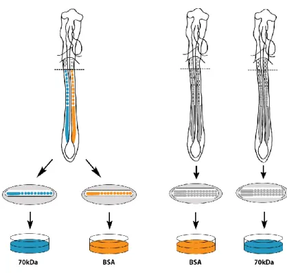
solutions see Annex S-I). A transverse cut was done immediately rostral to somite X (according to Christ and Ordahl, 1995) and prepared as shown in Fig. 5.
II.2.2. Explant culture
Posterior explants were placed in a drop of M199 culture medium supplemented with 10% chicken serum (Invitrogen), 5% fetal bovine serum (Invitrogen) and 1% penicillin/streptomycin (Invitrogen) on top of 0,8μm Millipore filters (IsoporeTM), floating
on 200-250µl of the same culture medium in 4-well plates (VWR). The N-terminal 70kDa fragment of fibronectin (Sigma-Aldrich) was added giving a final concentration of 100µg/ml for experimental explants, whereas bovine serum albumen (final concentration 100µg/ml; Sigma-Aldrich) was used for of control explants (Rifes et al., 2007; Martins et al., 2009) (Fig. 5). Explants were then incubated at 37ºC, in 5% CO2 humidified atmosphere for 6 hours.
II.2.3. Formation of somites in explant cultures and somite terminology
A developing chick embryo normally forms 4 pairs of somites in 6h, 1 for every 90 min, and so do control explants in culture (Palmeirim et al., 1997). However, when the 70kDa fibronectin fragment is added to the culture medium, the explants form only 1-2 more somite pairs (Rifes et al., 2007; Martins et al., 2009). In our hands, whole posterior explants cultured for 6h in control medium formed on average 3,9 somites (n=28) and bisected posterior explants formed on average 4,0 somites (n=106). In contrast, whole posterior explants cultured with the 70 kDa fragment formed an average of 1,5 somites (n=47) whereas bisected posterior explants formed 1,7 somites (n=104). These quantifications are graphically represented in Figure 6.
13
Fig. 5. Schematic representation of the explant culture. A. HH11-HH14 embryos were
transversally sectioned at the level of sX and then bisected along the midline, making sure to leave approximately the same amount of the axial structures (neural tube and notochord) on both contra-lateral sides. The posterior explants were then separated and placed on top of a filter in a drop of culture medium, with one being cultured with the 70kDa FN fragment (blue) and the contra-lateral side cultured with BSA (control; orange). B. HH11-HH14 embryos were transversally sectioned at the level of the sX. In this case, these posterior explants were left intact, and the whole explants were placed on top of filters. Different explants from different embryos were then cultured either with the 70kDa FN fragment or BSA.
Fig. 6. Average number of somites formed during the culture period. Control explants formed consistently more somites than experimental explants, regardless of being whole or bisected posterior explants. Bars represent the standard deviation.
14
Fig. 7. Graphic representation of somites
formed before and during a 6h culture period of posterior HH11-14 chick explants. Before culture, all explants start with 10 somites and the PSM (grey). After 6h of culture, control explants form the expected 4 somites, with normal morphology (orange). In contrast, experimental explants form 1-2 somites, here represented as 2 somites (blue). Somite staging is according to Christ and Ordahl (1995). Because previous studies have demonstrated that the somites of embryos cultured with the 70kDa fragment have a defective morphology, including those formed before the culture period (Martins et al., 2009), we marked the somite stage with quotation marks in order to stress the differences (both in terms of axial level and in morphology) between normal somites and those of 70kDa cultured explants.
Asterisks mark somites formed during culture. Dashed lines between control and experimental explants represent axial equivalents in terms of tissue ageing. Dashed-outlined somites in the PSM of experimental explant represent the somites that would have formed under normal conditions.
There are two ways of evaluating the development of explants cultured with the 70 kDa fragment:
1. The first way is to compare control with experimental explants using the
axial level as an indicator (dashed lines in Figure 7) even if the two tissues
compared have different morphologies. It has previously been shown that isolated PSM cultured in the absence of all surrounding tissues proceed in their developmental programme, as evaluated by delta mRNA expression, even if they do not form morphological somites (Palmeirim et al., 1998). Thus comparing explants according to axial level seems to be an appropriate method to compare tissue maturity in terms of gene expression. 2. The second way to compare control and experimental explants is to compare the somite morphology. According to this comparison, the first epithelial somite, sI, in the explants cultured in BSA is compared to the first epithelial somite in the experimental explants, “sI” (Figure 7) and so on, even if they are not at the same axial level. This comparison places and emphasis on morphology as a measure of maturity.
15
II.3. In situ probe production
II.3.1. Total RNA isolation and cDNA production
Total RNA isolation from 48h chicken embryos was performed using the RNeasy Mini Kit (Qiagen). The RNA obtained was diluted in RNase free water (Sigma-Aldrich), the concentration and purity was measured in a Nanodrop device and the RNA then stored at -80ºC. cDNA production followed Protocol A in Annex S-II, and the resulting cDNA was stored at -20ºC.
II.3.2. Primer designing and gene amplification
To produce the desired RNA probes, primers for the amplification of the genes of interest were designed. Lama1, Lama5 and Lamc1 transcript RNA sequences were obtained from the NCBI database (http://www.ncbi.nlm.nih.gov/nuccore). Forward and reverse primers for these genes (Annex S-III) were designed in Primer3 (http://biotools.umassmed.edu/bioapps/primer3_www.cgi) and their homology with other sequences confirmed in BLAST (http://blast.ncbi.nlm.nih.gov/Blast.cgi) to guarantee that the specificity of the final RNA probe.
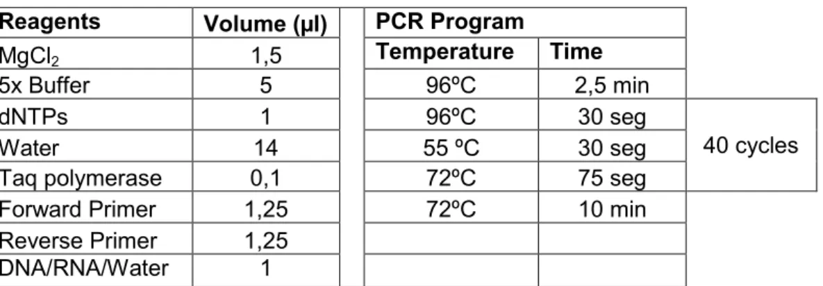
These genes were amplified in a polymerase chain reaction (PCR), the details of which are present in Table S-1 (Annex S-IV). RNA and H2O were used as negative
controls. The PCR product size was then analyzed with DNA electrophoresis in a 1%
agarose gel. Gel green (VWR) was added to the agarose gel to allow the detection of
the nucleic acids through the incidence UV light, and the gel was subjected to a 100V voltage for 30-45min.
II.3.3. PCR product ligation and plasmids used
The fresh PCR products were used to insert the amplified fragments in the TOPO TA Cloning pCR®II vector (Invitrogen). The product of this reaction was used to transform bacteria for amplification of the plasmids. In addition to these plasmids, a number of plasmids already constructed with inserts of interest were also used to transform bacteria (see Table S-2, Annex S-IV).
II.3.4. Bacteria transformation
1µl of the plasmid was added to 50µl of DH5α E. coli, which were incubated on ice for 30min. Next, a heat-shock was applied to the bacteria by placing them for 20s at 42ºC and transferring them immediately to ice for 2min. 950µl of pre-warmed LB
medium was added to the bacteria, which were allowed to grow for 1h at 37ºC and
225rpm and then plated in selective plates previously plated with 50µl of X-gal (VWR) to permit discrimination between transformant and non-transformant bacteria. The plates were inverted and incubated overnight at 37ºC.
16
II.3.5. Plasmid amplification and extraction
2-4 white bacteria colonies were placed in 4ml of selective medium for 8h at 37ºC and 300rpm. 1ml of these primary cultures was used to perform a Mini Prep (Protocol B in Annex S-II), to check which colony grew better and to confirm the transformation of the bacteria. Then 50µl of the primary culture of the best bacteria colony were added to 25ml of selective medium and incubated for 16h at 37ºC and 300rpm. The plasmid was then extracted using the JETSTAR 2.0 Midi Prep (Invitrogen) and then diluted in 10µl of TE and stored at -20ºC.
II.3.6. Linearization of the vector
To prepare the in situ hybridization probes, 10µg of the plasmid was digested for 3h30 hours at 37ºC with 5U of the appropriate restriction enzyme (see Table S-2), Complete linearization of the plasmid was confirmed by analyzing in a 1% agarose
gel. 100µl of ultrapure water and a solution of 25:24:1 of phenol:chloroform:isoamylic
alcohol (Sigma-Aldrich) were added to the linearized plasmid, followed by a 30s vortex and 5min centrifugation at 13000rpm. The aqueous phase was collected, and 200µl of ultra pure water was added to the organic phase, followed by new vortex, centrifugation and aqueous phase collection. 400µl of chloroform (Sigma-Aldrich) was added to the aqueous phase, and new vortex, centrifugation and acqueous phase collection were performed. 10% of 3M NaAc and 2.5x of 100% EtOH (Merck) of the volume was added, and the DNA left at -20ºC for 1h to precipitate, followed by a 13000rpm centrifugation at 4ºC. The pellet was washed in 200µl of 70% EtOH in ultra pure water, centrifuged for 15min at 4ºC and dried. The DNA pellet was then re-suspended in 20µl of ultra pure water, the concentration measured in the Nanodrop, followed by storage at -20ºC.
II.3.7. Transcription of the genes of interest with Dioxygenin
1 µg of linearized DNA was incubated for 4h at 37ºC with 2µl of Transcription Buffer (Roche), 2µl of DIG labeling mix, 1µl of DTT (Promega), 0,5µl of Rnasin (Promega) and 1µl of the appropriate RNA polymerase (see Table S-2), in a total of 19,5µl of ultra-pure water. After this incubation period, 1µl of 0,5M EDTA, pH8, 2,5µl of
4M LiCl and 75µl of 100% EtOH were added to precipitate the RNA, which was left at
-20ºC overnight. After a 45min centrifugation at 4ºC, the pellet was washed with 200µl of 70% EtOH in ultra pure water, centrifuged for 15min at 4ºC and left to dry. The pellet was then re-suspended in 50µl of ultra pure water and measured in Nanodrop for concentration and purity. Part of the probe was diluted in Hybmix, at a concentration of 1µg/ml and stored at -20ºC, while the remaining probe was stored at -80ºC for future use.
II.4. In situ hybridization
HH11 to HH14 chicken embryos and cultured explants were fixed overnight at 4ºC with WISH fixative. The embryos and explants were then dehydrated in a series of increasing concentrations of methanol (VWR) in PBT and stored in 100% methanol at -20ºC for at least 24h.
17 Day 1
Embryos and explants were re-hydrated with 75%, 50%, 25% of methanol-PBT and digested in a solution of Proteinase K (Roche) in PBT, with the time of digestion corresponding to the developmental stage of the embryo (e.g., HH12 embryos were digested for 12min). The explants were digested in Proteinase K for 12-14min. Afterwards, they were fixed in 4% formaldehyde/0,4% glutaraldehyde
post-fixative solution, washed with PBT, pre-warmed 1:1 PBT-Hybmix and placed in
Hybmix for 1h at 70ºC. Finally, the embryos and explants were incubated in the probe solution at 70ºC overnight.
Day 2
Embryos and explants were washed with and placed in Hybmix (pre-warmed at 70ºC) and left for 30min at this temperature. They were then washed with 1:1 MABT-Hybmix for 10min, 100% MABT for 15min and left for 1h in a solution of MABT+BL+SS, all at RT. Embryos/explants were then placed in the anti-Digoxygenin antibody conjugated with alkaline phosphatase (Roche) diluted in MABT+BL+SS at 1:2000 and incubated overnight at 4ºC.
Day 3
Embryos/explants were washed 3x with MABT, and then 3x1h with MABT again, with agitation, all at RT. After 2x10min washes with NTMT, the hybridized mRNA probe was detected by incubating the embryos in BM Purple (Roche) or a solution of BCIP/NBT (Roche) at RT for hours to days, depending on the probe. The reaction was stopped with PBT, and embryos stored at 4ºC in PBT-azide or 80% glycerol in PBT-azide.
II.5. Embedding and cryossectioning
Embryos and explants processed for in situ hybridization were embedded for cryosectioning by passing through 0.12M phosphate buffer with increasing concentrations of sucrose, finally being placed in a solution of phosphate buffer with 15% sucrose and 7.5% gelatin, and frozen on isopentane cooled with dry ice. Embryos were then stored at -80ºC until sectioning. 16µm sections were obtained with a Bright Clinicut 60 Cryostat, placed on SuperFrost Ultra Plus microscope slides (Menzel-Gläser) and mounted in Aquatex (VWR).
II.6. Immunohistochemistry
Explants cultured for 6h were fixed overnight in 4% PFA at 4ºC. After washing with PBS, they were incubated in a solution of 1% Triton and 1% BSA in PBS (ID) for 2-4h at room temperature. Ncad primary antibody (BD) was diluted in this solution and the explants incubated overnight at 4ºC. After washing, the explants were incubated overnight at 4ºC with the secondary antibodies, 1:800 of ToPro3 and 1:100 of RNase, also diluted in ID, and post-fixed with 4% PFA for 1-4h. The explants were then washed in PBS, slowly dehydrated in increasing concentrations of methanol in PBS and stored in 100% methanol at -20ºC. At the time of mounting, methylsalicylate was slowly added in order to transparentize the explants, which were then mounted in methylsalicylate on coverslips sealed with paraffin (Martins et al., 2009).
18
II.7. Image acquisition and treatment
Whole mount embryos and explants processed for in situ hybridization were photographed using a Wild M8 stereoscope coupled to an Olympus C-4040Z digital camera, while cryossections obtained from these embryos and explants were photographed with an Olympus BX60 microscope coupled to an Olympus DP50 digital camera. Explants processed for immunohystochemistry were analyzed in a Leica SPE Confocal System using 20x 0.7NA dry and 40x 1.3NA oil immersion lenses. Z-stack images were aquired using LAS software. The images were analyzed and treated using ImageJ, Amira v4.2 and Imaris v5.7.2 softwares
20
III. RESULTS
III.1. The role of the fibronectin matrix in the morphogenesis of the
sclerotome
III.1.1. Impairing fibronectin matrix assembly affects sclerotome morphology
To determine whether sclerotome morphology is altered in the absence of de novo fibronectin matrix assembly, control and experimental explants were processed for in toto immunohistochemistry for N-cadherin (Ncad) and analyzed with confocal microscopy. One of a total of 3 controls was chosen as representative in terms of somite morphology, to serve as a reference for comparing with the axially equivalent somites of a total of 5 experimental explants (Fig. 8). The experimental explants shown in Figure 8 represent the mildest and strongest effects observed in the presence of the 70kDa fragment.
Comparing the somites of the same axial levels, differences between control and experimental somites are evident. For clarity, when describing these differences we use the somite stage before culture as reference (for somite stages after culture, see Fig. 8). In somite III before culture (Fig. 8H,P,Z), there is a clear difference in Ncad organization in control and experimental explants. While the control shows a clear polarization of Ncad to the apical side of the somite cells, in both experimental explants, Ncad is much less polarized (Fig. 8H,P,Z, arrows). Considering somite V before culture, while the control somite starts downregulating Ncad in the ventral region, the same is not verified for the experimental explant with the strongest effect of the 70kDa fragment (compare Fig. 8F and V, arrows). A clear difference is also visible in the de-epithelialization of the lateral side of somite VIII, which is often defective compared to that of the control (Fig. 8, compare C and S, arrows). As for the experimental explant with mildest effect of the 70kDa fragment, it is clear that there are also differences in the de-epithelialization of the ventral somite comparing to the control, as evidenced by the comparison of somite X before culture, showing a defective de-epithelialization of the medial and ventral part of the somite (compare Fig. 8A and I, arrows).
In an attempt to quantify these differences, a staging score was created for the morphology of the somites of control embryos, to which the somite morphology of experimental explants was then compared, at each corresponding axial level (Table 1). Comparing the maturation score of control and experimental explants it is evident that somites of experimental explants are delayed in their maturation when compared to control somites at the same axial level. It is also clear from the average differences between control and experimental explants that this delay is more severe in the more rostral somites when compared to the caudal somites of experimental explants. Thus progressively more rostral somites are progressively more delayed in their morphological maturation when compared to the control.
21
Fig. 8. Somite morphology in control and experimental explants. Transverse
reconstructions of explants submitted for in toto immunohistochemistry for Ncad, at the levels of somites III to X before culture. Representative control explants (A-H). Experimental explant with the mildest (I-P) and strongest (Q-Z) effect observed. Scale bar=100µm.
Thus, impairing fibronectin matrix assembly effectively delays the morphogenesis of the sclerotome, which fails to fully de-epithelialize and expand at the correct developmental time, and that this delay is more accentuated in the rostral somites of the explants when compared to the caudal ones.
22
Somite stage
before culture III IV V VI VII VIII IX X Somite stage
after culture VII VIII IX X XI XII XIII XIV
Control M1 M2 M3 M4 M5 M6 M7 M8
Somite stage
after culture “V” “VI” “VII” “VIII” “IX” “X” “XI” “XII”
70kDa 1 M1 M2 M3 M4 M4 M4 M5 M6
70kDa 2 M1 M1 M2 M2 M3 M3,5 M4 M5
70kDa 3 M1 M1 M2 M3 M3 M4 M4 -
70kDa 4 M1 M2 M3 M4 M4 M5 M5 M6
70kDa 5 M1 M1 M1 M2 M3 M4 M4 M5
Average 70 kDa stage M1 M1,4 M2,2 M3,0 M3,4 M4,1 M4,4 M5,5 Average differences
BSA-70kDa 0 0,6 0,8 1 1,6 1,9 2,6 2,5
Table 1 – Staging score for the morphology of somites of cultured experimental explants (n=5)
compared to the same axial level of a reference control explant (Fig. 8A-H). Somites III to X before culture of the control reference explant were arbitrarily staged M1 to M8, representing progressively more mature somite stages. Experimental explants were compared to the reference control explant, and assigned to the stage with the most morphological similarities. The morphology was assessed by the visual analysis of the degree of de-epithelialization of the ventral, lateral and medial somite or the full de-epithelialized state of the sclerotome. it is important to stress that this scoring is only visual, and its sole objective is to get an idea of the differences in the morphology of somites of the same axial level of control vs. experimental explantsThe average M stage of the experimental explants was calculated and differences between the control M value and this average was calculated.
III.2. The role of the fibronectin matrix in the patterning of the sclerotome
Since impairing fibronectin matrix fibrillogenesis results in alteration of sclerotome morphology in somites of equivalent axial levels, we next asked whether this effect is merely morphological or whether it correlates with a patterning defect. Posterior explants of HH11-14 chick embryos were cultured with the 70kDa fragment and processed for in situ hybridization for a variety of differentiation markers. In a first approach, these posterior explants were sectioned along the midline, making sure that approximately the same amount of notochord and neural tube tissue was present in the two contra-lateral halves of the explant. This is extremely important, since the patterning of the sclerotome depends on Shh coming from the notochord and floor plate. One side of the explant was then cultured in control medium, while the contra-lateral side was cultured with the 70kDa fibronectin fragment.
Despite the efforts to divide the notochord along the midline, it is technically difficult to make sure that there is exactly the same amount of notochord tissue along the full anterior-posterior (AP) axis of the explant. When these explants were processed for in situ hybridization with a probe for Patched1, a Shh receptor whose