Contents lists available atScienceDirect
Redox Biology
journal homepage:www.elsevier.com/locate/redox
Research Paper
Regulation of the mechanism of Type-II NADH: Quinone oxidoreductase
from S. aureus
Filipa V. Sena
a, Filipe M. Sousa
a, A. So
fia F. Oliveira
b, Cláudio M. Soares
a, Teresa Catarino
c,
Manuela M. Pereira
a,d,⁎aInstituto de Tecnologia Química e Biológica– António Xavier, Universidade Nova de Lisboa, Av. da Republica EAN, 2780-157 Oeiras, Portugal bSchool of Biochemistry, University of Bristol, University Walk, Bristol BS8 1TD, UK
cDepartamento de Química, Faculdade de Ciências e Tecnologia, Universidade Nova de Lisboa, 2829-516 Caparica, Portugal
dUniversity of Lisbon, Faculty of Sciences, BioISI - Biosystems & Integrative Sciences Institute, Campo Grande, C8, 1749-016 Lisboa, Portugal
A R T I C L E I N F O
Keywords: FAD Respiratory chain Flavoprotein Electron transfer Bacterial respiration KineticsA B S T R A C T
Type-II NADH:quinone oxidoreductases (NDH-2s) are membrane proteins involved in respiratory chains and the only enzymes with NADH:quinone oxidoreductase activity expressed in Staphylococcus aureus (S. aureus), one of the most common causes of clinical infections. NDH-2s are members of the two-Dinucleotide Binding Domains Flavoprotein (tDBDF) superfamily, having aflavin adenine dinucleotide, FAD, as prosthetic group and NAD(P)H as substrate. The establishment of a Charge-Transfer Complex (CTC) between the isoalloxazine ring of the re-ducedflavin and the nicotinamide ring of NAD+ in NDH-2 was described, and in this work we explored its role in the kinetic mechanism using different electron donors and electron acceptors. We observed that CTC slows down the rate of the second half reaction (quinone reduction) and determines the effect of HQNO, an inhibitor. Also, protonation equilibrium simulations clearly indicate that the protonation probability of an important re-sidue for proton transfer to the active site (D302) is influenced by the presence of the CTC. We propose that CTC is critical for the overall mechanism of NDH-2 and possibly relevant to keep a low quinol/quinone ratio and avoid excessive ROS production in vivo.
1. Introduction
Flavoproteins account for ~1% of proteins and are highly versatile enzymes, which perform many diverse reactions[1,2]. In many cases a charge-transfer complex (CTC) is formed whose role remains elusive [3–10]. In this way, we used type II NADH: quinone oxidoreductases (NDH-2s) to investigate how the flavin reactivity is controlled and modulated by the presence of the CTC.
NDH-2s are membrane proteins involved in respiratory chains and in the metabolic regeneration of NAD+. They are recognized as suitable targets for novel antimicrobial therapies considering they are the only enzymes with NADH:quinone oxidoreductase activity present in many pathogenic organisms, both bacteria and protozoa[11]. These enzymes belong to the two-Dinucleotide Binding Domains Flavoprotein (tDBDF) superfamily, which share two structurally related Rossmann fold do-mains for the binding of dinucleotides (FAD and NADH)[12]. Struc-tural studies on NDH-2 have long contributed to the understanding of this enzyme, solving several controversies including the number and location of the substrate binding sites[5,13–15]. Also the possible role
of NDH-2 as a drug target as been thoroughly explored [16–18]. Nevertheless, functional information is still not fully congruent and the reaction mechanism of NDH-2 remains unknown.
Previous pre-steady state kinetic studies of NDH-2 from S. aureus, reported the presence of a CTC formed between NAD+and the reduced flavin, which is dissociated upon oxidation by the quinone (2,3-Dimethyl-1,4-naphthoquinone, DMN)[5], indicating the involvement of a ternary complex in the catalytic mechanism of NDH-2. These stu-dies also showed that FAD reduction by NADH was an order of mag-nitude faster than FADH2oxidation by the quinone (180 ± 30 s−1vs 5.0 ± 0.5 s−1, respectively) and 2-n-Heptyl-4-hydroxyquinoline N-oxide (HQNO) inhibition only affected the kinetically limiting second half-reaction of the enzymatic turnover (0.08 ± 0.02 s). Intriguingly, NDH-2 from Caldalkalibacillus thermarum was also reported to establish a CTC upon reduction with NADH, but with similar FAD reduction and oxidation half-reaction rates (200 s−1) [4]. In this case a different quinone analogue, menadione, was used, which has one less methyl group and a higher reduction potential then DMN (0 vs −90 mV) [19–21].
https://doi.org/10.1016/j.redox.2018.02.004
Received 23 January 2018; Received in revised form 8 February 2018; Accepted 10 February 2018
⁎Corresponding author at: Instituto de Tecnologia Química e Biológica– António Xavier, Universidade Nova de Lisboa, Av. da Republica EAN, 2780-157 Oeiras, Portugal.
E-mail address:mpereira@itqb.unl.pt(M.M. Pereira).
Available online 17 February 2018
2213-2317/ © 2018 The Authors. Published by Elsevier B.V. This is an open access article under the CC BY-NC-ND license (http://creativecommons.org/licenses/BY-NC-ND/4.0/).
Marreiros et al. [22], recently proposed the first catalytic me-chanism for NDH-2 family. In this study, which contemplates CTC formation coincidentally with NADH binding, aspartate 302 (D302), present in the vicinity of FAD, was observed to be totally conserved (100%), even among other members of the tDBDF superfamily. The authors suggested the involvement of D302 in the catalytic mechanism of NDH-2 as a putative proton donor in FAD reduction or proton ac-ceptor in FADH2oxidation. The authors have also observed the pre-sence of a conserved glutamate residue (E172) in the proximity of the NADH binding site, and proposed it as part of a proton conductive pathway. This residue was later proved to be further involved in the stabilization of the NADH binding and CTC establishment[23].
In the present work, we aimed at unraveling the role of the CTC in the reaction mechanism of NDH-2 and understanding its influence on flavin reactivity, in general.
2. Materials and methods
2.1. Enzyme expression and purification
E. coli Rosetta 2 (DE3) pLysS cells were transformed with the plasmid pET-28a(+) (Novagen) carrying the gene SAOUHSC_00878, coding for NDH-2 from S. aureus NCTC8325. Protein expression and purification were conducted as described previously [24]. Protein concentration was determined by the Pierce™ BCA protein assay kit or using the FAD extinction coefficient of 11.3 M−1cm−1[25].
2.2. Kinetic studies
Pre-steady state kinetic experiments were performed in a TgK Scientific stopped-flow instrument placed inside an anaerobic chamber and equipped with a diode array detector, capable of acquiring spectra in the wavelength range of 350–700 nm at a rate of 1 spectrum per 1.5 ms. The temperature of the drive syringes and mixing chamber was maintained at 15 °C and the pH was controlled with 100 mM potassium phosphate buffer pH 7.0, 250 mM NaCl. All solutions were prepared inside the anaerobic chamber with degassed water. The reductive re-action was monitored after mixing oxidized NDH-2 with NADH or so-dium dithionite, Na2S2O4(1:1 ratio). Reduced NDH-2 was mixed with quinone or O2(1:2 ratio) to study the oxidative reaction. Complete turnovers were monitored using two equivalents of NADH/ sodium dithionite and quinone. Similar assays were performed in the presence of the inhibitor, HQNO.
2.3. Simulation of equilibrium protonation states
pH titration curves were calculated for all ionizable amino acid residues, including D302, of NDH-2 from S. aureus (PDB: 5NA1), based on the thermodynamics of proton binding as described before in detail [26,27]. These methodologies use a combination of Poisson-Boltzmann (PB) calculations, performed with the program MEAD (version 2.2.9) [28–30], and Metropolis Monte Carlo (MC) simulations, using the program PETIT (version 1.5)[31]. Here, two systems were simulated: the fully reduced state FADH2similar to that obtained with dithionite, and the fully reduced protein with NAD+forming the charge transfer complex in the catalytic site, FADH2-NAD+. The position of the NAD+ was inferred upon fitting to the structure of the S. cerevisiae enzyme (PDB entry: 4G73).
3. Results and discussion
3.1. The presence of a Charge-transfer Complex
In the oxidized form, NDH-2 presents an absorption spectrum with maxima at 375 and 450 nm and a shoulder close to 475 nm, typical of flavoproteins (Fig. 1). Upon reduction with NADH, the absorption at
375–450 nm decreases and a broad absorption band at the 650–800 nm region appears. This band is considered to be originated by the estab-lishment of a CTC resulting from aπ-π interaction between the parallel stacked isoalloxazine and nicotinamide rings of reduced FAD and NAD+ respectively. The enzyme was also reduced with sodium di-thionite and in this case the decrease in absorption at 375–450 nm was also observed, but the band resulting from the establishment of the CTC was not detected (Fig. 1). A CTC could also be observed by adding NAD+to the dithionite-reduced NDH-2[5], meaning that the CTC was formed only in the presence of the NAD+/FADH
2, independently of the reducing agent.
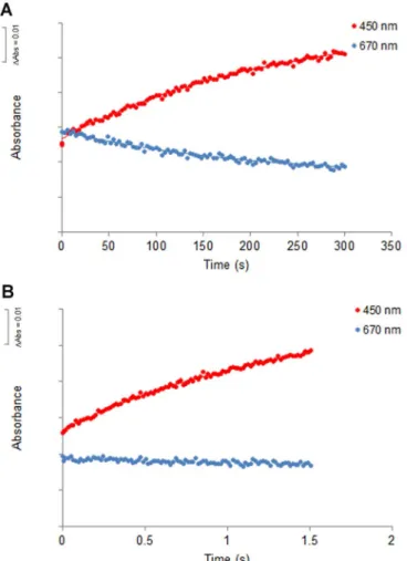
3.2. The Charge-Transfer Complex is a limiting factor in protein oxidation In order to investigate the role of the charge-transfer complex on the catalytic mechanism of NDH-2, we compared the oxidation of NDH-2 when reduced by NADH, which establishes a CTC, to that of the enzyme reduced by dithionite, which does not establish a CTC. Upon addition of quinone (in a 1:1 ratio) to NDH-2 previously reduced with NADH, spectral changes showed an increase in the absorbance peak at 450 nm and the disappearance of the broad band at 650–800 nm. The rate constant obtained for this second half-reaction was equal to 5.0 ± 0.5 s−1(Fig. 2A)[5]. The dithionite-reduced NDH-2 was mixed with DMN in a 1:1 ratio (Fig. 2B) and a rate constant equal to 196 ± 15 s−1 was measured. This reaction was approximately 40 times faster than the oxidation of the NADH-reduced NDH-2 (Fig. 2A) [5]and interestingly similar to the rate constant for the reduction of the protein by NADH. In order to confirm whether the difference observed in the oxidation reaction of FAD was due to the presence of the CTC we incubated the dithionite-reduced enzyme with NAD+and monitored its reaction with DMN (all the components were in a 1:1 ratio) (Fig. 2C). The rate constant obtained was 11.4 ± 2.2 s−1, which is comparable to that observed for the oxidation of the NADH-reduced enzyme by the quinone. These data indicate the presence of the CTC is a limiting factor in protein oxidation.
3.3. The Charge-Transfer Complex decreases the oxidation rate independently of the oxidant
We also investigated whether the observed effect of the CTC on the oxidation reaction was dependent on the quinone, i.e., of the oxidant.
0.00 0.02 0.04 0.06 0.08 0.10 325 425 525 625 725 825 Absorbance Wavelength (nm) oxidized, as purified NADH reduced
Sodium dithionite reduced
Fig. 1. UV–visible absorption spectra of NDH-2 as purified (black solid line), reduced with NADH (blue solid line) or reduced with sodium dithionite (blue dashed line). The insertedfigure expands absorption spectra in the 600–800 region (For interpretation of the references to color in thisfigure legend, the reader is referred to the web version of this article.).
For this we used oxygen, O2, as the electron acceptor. In thefirst ex-periment 10 µM NDH-2 was reduced with a stoichiometric amount of NADH and then the reduced NDH-2 was mixed with buffer containing 24 µM O2in approximately 1:2 protein:O2ratio (Fig. 3A).
The oxidation of the protein by oxygen was very slow, with a rate constant of 0.0044 ± 0.0007 s−1. In this experiment we could also observe a decrease in absorbance at 650–800 nm due to the dissociation of the CTC (Fig. 3A). In a second experiment, we reduced the protein with sodium dithionite (1:1 ratio) and monitored its reaction with O2 (1:2 ratio). The rate constant obtained was 0.76 ± 0.2 s−1higher than that measured for the oxidation of the NADH-reduced NDH-2. Also, as expected, the absorbance in the 650–800 nm region did not change, as a
CTC was not present in the reduced enzyme to begin with. These results reinforce the idea that the presence of the CTC determines the oxidation rate of the protein.
3.4. The presence of the CTC determines the inhibitory effect of HQNO We previously showed that HQNO, a quinone derivative, acts as an inhibitor of S. aureus´NDH-2 activity with an apparent Ki value of 6.8 ± 0.4 µM. It was also demonstrated that the oxidation of the en-zyme by DMN is strongly impaired in the presence of HQNO (unlike the first half reaction which is not affected) and that the binding sites for HQNO and the quinone are superimposed or very close[5].
Here we tested whether the presence of the CTC, which also in flu-ences the second half reaction, would alter the effect of the inhibitor. For this we compared the effect of HQNO (10 µM) on the rate of protein oxidation by DMN (10 µM), in the case of NADH or dithionite reduced protein (10 µM). The rate of oxidation of dithionite-reduced protein by DMN in the presence of HQNO was 179 ± 12 s−1, a value similar to that obtained in the absence of the inhibitor, which suggests that in the absence of CTC the HQNO has no effect (Fig. 4B). This is completely different from the result obtained previously, where the oxidation of NDH-2 by DMN, after its reduction by NADH, was slowed-down by a factor of 50 (to 0.08 ± 0.02 s−1) in the presence of HQNO (Fig. 4A) [5]. Again, in order to confirm whether the difference observed in the effect of HQNO was due to the presence of the CTC we incubated the dithionite-reduced enzyme with NAD+and monitored its reaction with DMN in the presence of the inhibitor. In this situation we observed the Fig. 2. Time-resolved oxidation by DMN of (A) NADH-reduced NDH-2 (1:1, protein:DMN
ratio), (B) sodium dithionite-reduced NDH-2 (1:1, protein:DMN ratio) and (C) sodium dithionite-reduced NDH-2 in the presence of NAD+(1:1:1, protein: DMN:NAD+ratio),
monitored at 450 nm (red dots) and at 670 nm (blue dots) (For interpretation of the re-ferences to color in thisfigure legend, the reader is referred to the web version of this article.).
Fig. 3. Time-resolved oxidation by O2of (A) NADH-reduced NDH-2 (1:2, NDH-2: O2
ratio) and (B) dithionite-reduced NDH-2 (1:2, NDH-2: O2ratio) monitored at 450 nm (red
dots) and at 670 nm (blue dots) (For interpretation of the references to color in thisfigure legend, the reader is referred to the web version of this article.).
protein oxidation was also very slow, with a rate constant of 0.1 ± 0.0006 s−1, a value that is similar to that obtained when the enzyme was reduced with NADH and then oxidized by DMN in the presence of HQNO (Fig. 4C).
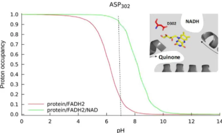
3.5. The presence of the CTC influences the protonation probability of D302 We performed protonation equilibrium simulations for all the io-nizable amino acid residues of NDH-2 from S. aureus. Two different systems were simulated: the fully reduced state (FADH2), similar to that obtained with dithionite, and the fully reduced protein with NAD+
(FADH2-NAD+) in which the CTC is present.
Our protonation equilibrium simulations for NDH-2 from S. aureus indicate that the proton occupancy probability of the majority of the ionizable amino acid residues was not significantly altered by the pre-sence of the CTC. D302 was the exception, for which our simulations clearly showed that its proton occupancy probability in the reduced enzyme is influenced by the presence of the CTC (Fig. 5).
According to the proposed catalytic mechanism of Marreiros et al. [22], upon binding of NADH, FAD is reduced through hydride transfer to the N5 of the FAD isoalloxazine ring. The second proton needed for the full protonation of the reduced FAD is suggested to be provided by rearrangement of the hydrogen bond network around N1 in which a strictly conserved D302 is present. The high conservation of D302 is extended to several families of the tDBDF superfamily, further sug-gesting its importance in the oxidation/reduction processes of FAD. Upon quinone binding, FADH2transfers two electrons to the quinone, which is provided of two protons by the previously observed putative proton pathways[22]. After FADH2oxidation, theflavin returns to its original conformation, leading to the release of the protons at N5 and N1. This proton at N1 undergoes the reverse process that restores the initial hydrogen bonding network around D302.
We observed that the proton occupancy probability curve of D302 in the case of FADH2(in the absence of CTC) is different from that obtained in the case of FADH2-NAD+(in the presence of the CTC). The curve in the presence of the CTC is shifted to higher pH values com-paring to that obtained in its absence. This means that at pH 7 it is more difficult to oxidize FADH2-NAD+, because in this state D302 is almost 100% protonated and therefore unable to receive protons. In the ab-sence of NAD+at pH 7, D302 is expected to have a much lower proton occupancy, allowing proton transfer from the active site and faster oxidation of the FADH2.
Therefore, the difference observed for the proton occupancy prob-ability of D302 of the reduced protein in the presence and absence of the CTC can contribute to explain the different oxidation rates observed by pre-steady state kinetics.
4. Conclusions
The establishment of a CTC betweenflavoproteins and NADH is well documented, and has been observed for many enzymes catalyzing dif-ferent reactions [32]. Likewise, in previous work we described the formation of a CTC in the reduction of NDH-2 with NADH, which dis-appears upon oxidation by the quinone. In this work, we describe, for thefirst time, that the presence of the CTC controls the rate of the second half reaction and influences the effect of the inhibitor HQNO.
We show that the oxidation reaction is limited by the presence of the CTC because the dithionite-reduced NDH-2 oxidation rate is 40 Fig. 4. Time-resolved oxidation by DMN of (A) NADH- reduced NDH-2 in the presence of
HQNO (1:1:5, NDH-2:DMN:HQNO ratio), (B) dithionite- reduced NDH-2 in the presence of HQNO (1:1:5, NDH-2:DMN:HQNO ratio) and (C) dithionite- reduced NDH-2 in the presence of HQNO and NAD+(1:1:5:1, NDH-2:DMN:HQNO:NAD+ratio).
Fig. 5. Proton equilibrium occupancy probability curves for D302 in two reduced con-ditions: FADH2(in red) and FADH2-NAD+(in green) (For interpretation of the references
times faster than the oxidation rate of the NADH-reduced NDH-2. The effect of the presence of the CTC is also observed when oxygen is the electron acceptor, revealing that the presence of the CTC decreases the rate of the second half reaction, independently of the oxidant. This set of results, summarized inTable 1, can be partially explained by the protonation equilibrium simulations which revealed that the protona-tion of D302 in the reduced enzyme is strongly influenced by the pre-sence of NAD+: protonated D302 populations at pH 7, which decreases from almost 100% in the presence of NAD+to only 20% in its absence. When testing HQNO, we noticed it decreases the rate of oxidation of the enzyme when the CTC is observed, having no effect when the en-zyme is reduced with dithionite. In this case one could hypothesize that DMN could accept electrons from the unoccupied site of NADH. However we can rule out this hypothesis based on our previous data obtained by STD-NMR and Fluorescence spectroscopies, which showed that NADH and DMN interact with the protein at two different sites, without interfering with each other. We also observed that HQNO only affects the binding of DMN[5] discarding the possibility of HQNO binding to the NADH site when the nucleotide is absent. Thus our present result indicates that the presence of the CTC produces con-formational changes at the HQNO and quinone binding pocket(s), which may be favorable for the binding of HQNO and disadvantageous for the binding of the quinone. In this way, HQNO acted as a probe reflecting conformational changes at the quinone binding site. The hypothesis that the establishment of the CTC is disadvantageous for the binding of the quinone can also contribute to explain the observed difference in the rates of the second half reaction in the presence or absence of the CTC, in addition to its influence on the protonation probability of D302.
We can conclude that the presence of the CTC slows down the second half reaction. The physiological relevance of this effect is un-known, however we hypothesize it may contribute to keep a low quinol/quinone ratio and thus avoid the production of radical oxygen species by unspecific oxidation of quinol. We believe that our hypoth-esis for the physiological relevance of the molecular control of the en-zymatic mechanism will foster further discussion and pave the way for in vivo studies.
Acknowledgments
FVS and FMS are recipients of fellowships by Fundação para a Ciência e a Tecnologia (PD/BD/113985/2015, PD/BD/128213/2016, respectively (within the scope of the PhD program Molecular Biosciences PD/00133/2012)). The work was funded by Fundação para a Ciência e a Tecnologia (IF/01507/2015 to MMP). This work was also supported by Project LISBOA-01-0145-FEDER-007660 (Microbiologia Molecular, Estrutural e Celular) funded by FEDER funds through COMPETE2020 - Programa Operacional Competitividade e Internacionalização (POCI) and by national funds through FCT -Fundação para a Ciência e a Tecnologia and by national funds through FCT - Fundação para a Ciência e a Tecnologia and by UID/MULTI/ 04046/2013 centre grant from FCT, Portugal (to BioISI).
References
[1] V. Piano, B.A. Palfey, A. Mattevi, Flavins as covalent catalysts: new mechanisms
emerge, Trends Biochem. Sci. 42 (2017) 457–469,http://dx.doi.org/10.1016/j. tibs.2017.02.005.
[2] W.P. Dijkman, G. De Gonzalo, A. Mattevi, M.W. Fraaije, Flavoprotein oxidases: classification and applications, Appl. Microbiol. Biotechnol. 97 (2013) 5177–5188,
http://dx.doi.org/10.1007/s00253-013-4925-7.
[3] A.P. Landry, D.P. Ballou, R. Banerjee, H2S oxidation by nanodisc-embedded human sulfide quinone oxidoreductase, J. Biol. Chem. 292 (2017) 11641–11649,http://dx. doi.org/10.1074/jbc.M117.788547.
[4] J.N. Blaza, H.R. Bridges, D. Aragão, E.A. Dunn, A. Heikal, G.M. Cook, Y. Nakatani, J. Hirst, The mechanism of catalysis by type-II NADH:quinone oxidoreductases, Sci. Rep. 7 (2017),http://dx.doi.org/10.1038/srep40165.
[5] F.V. Sena, A.P. Batista, T. Catarino, J.A. Brito, M. Archer, M. Viertler, T. Madl, E.J. Cabrita, M.M. Pereira, Type-II NADH: quinone oxidoreductase from Staphylococcus aureus has two distinct binding sites and is rate limited by quinone reduction, Mol. Microbiol. 98 (2015) 272–288,http://dx.doi.org/10.1111/mmi. 13120.
[6] M.M. Elguindy, E. Nakamaru-Ogiso, Apoptosis-inducing factor (AIF) and its family member protein, AMID, are rotenone-sensitive NADH:ubiquinone oxidoreductases (NDH-2), J. Biol. Chem. 290 (2015) 20815–20826,http://dx.doi.org/10.1074/jbc. M115.641498.
[7] A.S. Abramovitz, V. Massey, Interaction of phenols with old yellow enzyme. Physical evidence for charge transfer complexes, J. Biol. Chem. 251 (1976) 5327–5336 (doi:8461).
[8] B. Gao, H.R. Ellis, Mechanism offlavin reduction in the alkanesulfonate mono-oxygenase system, Biochim. Biophys. Acta - Proteins Proteom. 1774 (2007) 359–367,http://dx.doi.org/10.1016/j.bbapap.2006.12.006.
[9] O. Dmitrenko, C. Thorpe, R.D. Bach, Effect of a charge-transfer interaction on the catalytic activity of Acyl-CoA dehydrogenase: a theoretical study of the role of oxidizedflavin, J. Phys. Chem. B 107 (2003) 13229–13236,http://dx.doi.org/10. 1021/jp0348631.
[10] H. Li, J. Jamal, G. Chreifi, V. Venkatesh, H. Abou-Ziab, T.L. Poulos, Dissecting the kinetics of the NADP+-FADH2 charge transfer complex andflavin semiquinones in neuronal nitric oxide synthase, J. Inorg. Biochem. 124 (2013) 1–10,http://dx.doi. org/10.1016/j.jinorgbio.2013.03.008.
[11] G.M. Cook, C. Greening, K. Hards, M. Berney, Energetics of pathogenic bacteria and opportunities for drug development, Adv. Microb. Physiol. 65 (2014) 1–62,http:// dx.doi.org/10.1016/bs.ampbs.2014.08.001.
[12] S. Ojha, E.C. Meng, P.C. Babbitt, Evolution of function in the“two dinucleotide binding domains” flavoproteins, PLoS Comput. Biol. 3 (2007) 1268–1280,http:// dx.doi.org/10.1371/journal.pcbi.0030121.
[13] Y. Yang, Y. Yu, X. Li, J. Li, Y. Wu, J. Yu, J. Ge, Z. Huang, L. Jiang, Y. Rao, M. Yang, Target elucidation by cocrystal structures of nadh-ubiquinone oxidoreductase of Plasmodium falciparum (PfNDH2) with small molecule to eliminate drug-resistant malaria, J. Med. Chem. 60 (2017) 1994–2005,http://dx.doi.org/10.1021/acs. jmedchem.6b01733.
[14] A. Heikal, Y. Nakatani, E. Dunn, M.R. Weimar, C.L. Day, E.N. Baker, J.S. Lott, L.A. Sazanov, G.M. Cook, Structure of the bacterial type II NADH dehydrogenase: a monotopic membrane protein with an essential role in energy generation, Mol. Microbiol. 91 (2014) 950–964,http://dx.doi.org/10.1111/mmi.12507. [15] Y. Feng, W. Li, J. Li, J. Wang, J. Ge, D. Xu, Y. Liu, K. Wu, Q. Zeng, J.W. Wu, C. Tian,
B. Zhou, M. Yang, Structural insight into the type-II mitochondrial NADH dehy-drogenases, Nature 491 (2012) 478–482,http://dx.doi.org/10.1038/nature11541. [16] P.S. Shirude, B. Paul, N. Roy Choudhury, C. Kedari, B. Bandodkar, B.G. Ugarkar,
Quinolinyl pyrimidines: potent inhibitors of NDH-2 as a novel class of anti-TB agents, ACS Med. Chem. Lett. 3 (2012) 736–740,http://dx.doi.org/10.1021/ ml300134b.
[17] T. Yano, L. Lin-Sheng, E. Weinstein, J.S. Teh, H. Rubin, Steady-state kinetics and inhibitory action of antitubercular phenothiazines on Mycobacterium tuberculosis Type-II NADH-menaquinone oxidoreductase (NDH-2), J. Biol. Chem. 281 (2006) 11456–11463,http://dx.doi.org/10.1074/jbc.M508844200.
[18] S. Sellamuthu, M. Singh, A. Kumar, S.K. Singh, Type-II NADH Dehydrogenase (NDH-2): a promising therapeutic target for antitubercular and antibacterial drug discovery, Expert Opin. Ther. Targets 21 (2017) 559–570,http://dx.doi.org/10. 1080/14728222.2017.1327577.
[19] R.K.K. THAUER, K. Jungermann, K. DECKER, Energy conservation in chemotrophic anaerobic bacteria, Microbiol. Mol. Biol. Rev. 41 (1977) 100–180.
[20] J.B. Conant, L.F. Fieser, Reduction potentials of quinones. II. The potentials of certain derivatives of benzoquinone, naphthoquinone and anthraquinone, J. Am. Chem. Soc. 46 (1924) 1858–1881,http://dx.doi.org/10.1021/ja01673a014. [21] H.R. Nasiri, R. Panisch, M.G. Madej, J.W. Bats, C.R.D. Lancaster, H. Schwalbe, The
correlation of cathodic peak potentials of vitamin K3 derivatives and their calcu-lated electron affinities. The role of hydrogen bonding and conformational changes, Biochim. Biophys. Acta - Bioenerg. 1787 (2009) 601–608,http://dx.doi.org/10. 1016/j.bbabio.2009.02.013.
[22] B.C. Marreiros, F.V. Sena, F.M. Sousa, A.S.F. Oliveira, C.M. Soares, A.P. Batista, M.M. Pereira, Structural and Functional insights into the catalytic mechanism of the Type II NADH:quinone oxidoreductase family, Sci. Rep. 7 (2017),http://dx.doi. org/10.1038/srep42303.
[23] F.M. Sousa, F.V. Sena, A.P. Batista, D. Athayde, J.A. Brito, M. Archer,
A.S.F. Oliveira, C.M. Soares, T. Catarino, M.M. Pereira, The key role of glutamate 172 in the mechanism of type II NADH:quinone oxidoreductase of Staphylococcus aureus, Biochim. Biophys. Acta - Bioenerg. 1858 (2017) 823–832,http://dx.doi. org/10.1016/j.bbabio.2017.08.002.
[24] A.L.ú Cia Rosário, F.V. Sena, A.P. Batista, T.F. Oliveira, D. Athayde, M.M. Pereira, J.A. Brito, M. Archer, Expression, purification, crystallization and preliminary X-ray diffraction analysis of a type II NADH:quinone oxidoreductase from the human Table 1
Summary of kinetic rate constant values for the second half-reaction -flavin oxidation. Oxidizing substrate / inhibitor
Reducing substrate DMN DMN+HQNO O2
NADH 5.0 ± 0.5 0.08 ± 0.02 0.0044 ± 0.0007
Dith + NAD+ 11.4 ± 2.2 0.1 ± 0.0006 –
pathogen Staphylococcus aureus, Acta Cryst. 71 (2015) 477–482,http://dx.doi. org/10.1107/S2053230×15005178.
[25] A. Aliverti, B. Curti, M.A. Vanoni, Identifying and Quantitating FAD and FMN in Simple and in Iron-Sulfur-Containing Flavoproteins, in: S.K. Chapman, G.A. Reid (Eds.), Flavoprotein Protoc. HUMANA PRESS, 1999, pp. 9–23.
[26] V.H. Teixeira, C.M. Soares, A.M. Baptista, Studies of the reduction and protonation behavior of tetraheme cytochromes using atomic detail, J. Biol. Inorg. Chem. 7 (2002) 200–216,http://dx.doi.org/10.1007/s007750100287.
[27] A.M. Baptista, C.M. Soares, Some theoretical and computational aspects of the in-clusion of proton isomerism in the protonation equilibrium of proteins, J. Phys. Chem. B 105 (2001) 293–309,http://dx.doi.org/10.1021/jp002763e. [28] D. Bashford, K. Gerwert, Electrostatic calculations of the pKa values of ionizable
groups in bacteriorhodopsin, J. Mol. Biol. 224 (1992) 473–486,http://dx.doi.org/ 10.1016/0022-2836(92)91009-E.
[29] D. Bashford, M. Karplus, pKa's of ionizable groups in proteins: atomic detail from a continuum electrostatic model, Biochemistry 29 (1990) 10219–10225,http://dx. doi.org/10.1021/bi00496a010.
[30] D. Bashford, An object-oriented programming suite for electrostatic effects in bio-logical molecules: An experience report on the MEAD project, in: Lect. Notes Comput. Sci. (Including Subser. Lect. Notes Artif. Intell. Lect. Notes Bioinformatics): 1997: pp. 233–240.https://dx.doi.org/10.1007/3-540-63827-X_66.
[31] T. Sakurai, H. Hosoya, Charge-transfer complexes of nicotinamide-adenine dinu-cleotide analogues andflavin mononucleotide, Biochim. Biophys. Acta - Biophys. Incl. Photosynth. 112 (1966) 459–468,http://dx.doi.org/10.1016/0926-6585(66) 90248-2.
[32] H. Sakurai, T. Hosoya, Charge-transfer complexes of nicotinamide-adenine dinu-cleotide analogues andflavin mononucleotide, Acta Biophys. 112 (1965) 459–468.