2018
UNIVERSIDADE DE LISBOA
FACULDADE DE CIÊNCIAS
DEPARTAMENTO DE BIOLOGIA VEGETAL
The role of mannitol in the central carbon metabolism of the
yeast Starmerella bombicola
Carolina da Silva Ferreira
Mestrado em Microbiologia Aplicada
Versão Pública
Dissertação orientada por:
Professora Doutora Paula Gonçalves
iii
Acknowledgments
First, I would like to thank to my supervisor Prof. Paula Gonçalves for once again accepting me in the lab, for all scientific discussions and suggestions, for all the professional advises in the last months. I am also grateful for had in consideration personal aspects in the end of this thesis.
I would like to thank to my co-supervisor Prof. Ana Tenreiro.
A sincere thanks to all the members of Yeast Genomics Lab. A special acknowledgement to Carla, who helped me so much since the day I arrive at the lab to start an internship until the end of this thesis. Thanks for all the help in the lab, for clarify my doubts, for all the suggestions during this work and for being an example for everyone, especially to me. To Ana Pontes, who gently giving me so many rides, advises and helped me to keep a positive attitude even when things were not so well. To Alexandra Cabrita who also did her master thesis in the lab at the same time as me. A special thanks to Bruno Pedras for protocols, tips and advises for lipid extraction. Also, to Prof. Madalena Oom for all the suggestions.
A special acknowledgment to Nicole Soares, our lab technician who always keep our lab material in perfect conditions and to the Portuguese Yeast Culture Collection (PYCC) for the strains provided for this work.
I would like to express my gratitude, my love and my appreciation to my family and friends, for all the support, patience and love. There are no words to described what they all did for me in the last year. A special thanks to my great grandmother who participated in all important moments of my life but, unfortunately was not able to see me finishing my thesis. Thanks for raised me, educated me and always teach me to dream big, but work harder.
v
The role of mannitol in the central carbon metabolism of the
yeast Starmerella bombicola
Carolina da Silva Ferreira
2018
This thesis was fully performed at Yeast Genomics Lab FCT-UNL, under the direct supervision of Profª. Dr. Paula Gonçalves and Doctor Carla Gonçalves in the scope of the Master in Applied Microbiology of the Faculty of Sciences of the University of Lisbon.
vii
Resumo
A frutofília é uma característica pouco comum entre os microrganismos, que consiste na preferência de frutose como fonte de carbono e energia. Em leveduras, até agora, esta característica foi reportada no género Zygossacharomyces e no clado Wickerhamiella/Starmerella (W/S). O comportamento frutofílico está relacionado com a presença do transportador de frutose, Ffz1, porém, o papel da frutose no metabolismo destas leveduras ainda não está completamente elucidado.
Uma das leveduras que se destaca no clado W/S é St. bombicola não só pela facilidade em manipular esta espécie geneticamente, mas também por ser produtora de elevadas quantidades de soforolípidos. Estes são biosurfactantes que têm características favoráveis do ponto de vista ambiental e, são aplicados em áreas como a cosmética e produtos de limpeza. Além disso, St. bombicola assim como outras espécies pertencentes ao clado W/S foram reportadas como produtoras de elevadas quantidades de manitol a partir de frutose através da enzima manitol desidrogenase (Mtdh) utilizando NADPH como cofator, regenerando assim NADP+.
Tendo em conta que a produção de manitol é uma via metabólica dependente de frutose, o objetivo desta tese foi tentar perceber qual o papel do manitol na levedura St. bombicola. Como St. bombicola possui dois genes que codificam para duas desidrogenases do manitol foram utilizados três mutantes de deleção previamente construídos: um mutante onde o gene que codificava para a enzima Mtdh1 (mtdh1Δ) foi interrompido, outro para a enzima Mtdh2 (mtdh2Δ) e, finalmente um mutante duplo (mtdh1Δmtdh2Δ). A estirpe selvagem e mutantes de deleção foram cultivados, num meio onde o comportamento frutofílico é visível designado por 20FG (contendo 10 % (w/v) de frutose e 10 % (w/v) de glucose) onde a produção de manitol foi estudada. Não foi detetada produção de manitol pelos mutantes mtdh1Δ e mtdh1Δmtdh2Δ, porém foi detetada produção de manitol semelhante à estirpe selvagem pelo mutante mtdh2Δ, o que permitiu inferir que a principal enzima responsável pela conversão de frutose em manitol é a Mtdh1. Curiosamente, quando crescidos nesse mesmo meio a várias temperaturas (25 ºC, 30 ºC e 32.5 ºC), os mutantes de deleção (mtdh1Δ e mtdh1Δmtdh2Δ) não atingiam as mesmas densidades celulares que a estirpe selvagem às temperaturas mais elevadas. Além disso, excluímos também a possibilidade de os mutantes de deleção terem algum tipo de dificuldade em consumir especificamente um dos açúcares usados, glucose e frutose, cultivando-os em 20 % (w/v) glucose e em 20 % (w/v) de frutose uma vez que nestes meios os mutantes continuavam sem atingir as mesmas densidades celulares que a estirpe selvagem. Tendo em conta a elevada osmolaridade do meio utilizado assim como as diversas descrições do papel do manitol como osmoprotetor, foi avaliado o crescimento dos mutantes de deleção num meio onde a pressão osmótica é baixa, 2FG contendo 1 % (w/v) de frutose e 1 % (w/v) de glucose, a 30 ºC e 32.5 ºC. Curiosamente, a 32.5 ºC os mutantes de deleção não conseguiam sequer crescer. O facto de o defeito no crescimento ser ainda mais pronunciado neste meio de cultura permitiu-nos excluir a possibilidade de o manitol funcionar como osmoprotetor em St. bombicola. Além disso, a taxa de crescimento da estirpe selvagem e dos mutantes de deleção foi também analisada a 25 ºC, 30 ºC e 32.5 ºC. Porém, não foi detetada qualquer diferença na taxa de crescimento entre a estirpe selvagem e os mutantes de deleção a 25 ºC e a 30 ºC o que sugere que as diferenças que observamos são de facto no rendimento em biomassa e não na velocidade de crescimento.
A adição de sorbitol (18% (w/v) ao meio 2FG a 32.5 ºC repõe a capacidade de crescimento dos mutantes. Contudo, o sorbitol pode ser metabolizado quando presente em elevadas concentrações. No entanto, quando cultivámos um mutante duplo – onde o gene que codifica para uma enzima necessária para a metabolização do sorbitol (SOR1) foi interrompido assim como o gene MtDH1, mtdh1Δsor1Δ -
viii verificamos que esse mutante ainda consegue crescer o que indica que o sorbitol poderá substituir o papel do manitol na célula e que o poliol em si tem de facto um papel no crescimento a elevadas temperaturas. Esta conclusão é corroborada quando a estirpe selvagem e os mutantes de deleção foram cultivados no meio contendo 1 % (w/v) de glucose e 1 % (w/v) de frutose ao qual foi adicionado 18% (w/v) manitol e os mutantes de deleção atingiam densidades celulares semelhantes à estirpe selvagem a 32.5 ºC.
Tendo em conta que os mutantes de deleção não atingem as mesmas densidades celulares que a estirpe selvagem e, que é algo que parece estar intimamente relacionado com a temperatura a produção intracelular e extracelular de manitol foi analisada no meio 20FG a 20 ºC, 25 ºC e 30 ºC. Curiosamente, um aumento do rendimento intracelular de manitol é observado assim como extracelular o que sugere que o manitol desempenha um efeito protetor na célula contra o stress térmico, possivelmente tendo um papel estabilizador da membrana como a trealose em S. cerevisiae, em que os grupos hidroxilo formam pontes de hidrogénio com a bicamada lipídica da membrana. Esta explicação coaduna-se com o facto de, em S. cerevisiae a trealose ser necessária dos dois lados da membrana para garantir proteção uma vez que, em St. bombicola, não só a produção de manitol intracelular, mas também a quantidade extracelular de manitol aumenta com a temperatura.
Em suma, durante esta dissertação foi possível fazer uma contribuição significativa relativamente ao papel da produção de manitol em St. bombicola. Esse papel poderá residir no efeito termoprotector do próprio manitol, mas também na manutenção do equilíbrio redox das vias do metabolismo central.
ix
Abstract
Fructophily is a rare trait that consists in the preference of fructose over glucose as carbon and energy source. In spite of the fact that it is already known that fructophily in yeasts is intimately related with the presence of a high capacity fructose transporter called Ffz1, the importance of fructose in the metabolism of fructophilic yeasts is not well established yet.
St. bombicola converts fructose directly into mannitol through a mannitol dehydrogenase (Mtdh) which
uses NADPH as a cofactor. In order to understand the role of mannitol in this yeast, three deletion mutants previously constructed, where the genes encoding for Mtdh were disrupted (mtdh1Δ, mtdh2Δ,
mtdh1Δmtdh2Δ) were used. Growth assays were performed at three temperatures 25 ºC, 30 ºC and 32.5
ºC and it was possible to observe that the MtDH deletion mutants did not reach the same cell densities as the wild-type. Also, at the highest temperature tested (32.5 ºC) and at low sugar concentrations, the
MtDH deletion mutants were not even able to grow. The ability to grow was restored through the
addition of exogenous polyols like mannitol and sorbitol, suggesting that these polyols could be replacing the function of mannitol produced by the cell. Also, the intracellular mannitol content produced by the wild-type increased at higher temperatures, suggesting a role as thermoprotector. It seems possible that mannitol, like trehalose in S. cerevisiae, can stabilize the lipid bilayer of the plasma membrane through hydrogen bonding.
In this thesis we were able to make a significant contribution to elucidate the role of mannitol production in St. bombicola. The first role may be as thermoprotector in the cell and the second role may be related with redox homeostasis.
xi
Index
1. Introduction ...1
1.1. Carbon metabolism in yeasts: general aspects ... 1
1.1.1. Glycolysis ...2
1.1.2. Pentose Phosphate Pathway ...3
1.2. Fructophily: a peculiar characteristic of some microorganisms ...4
1.3. St. bombicola: a frutophilic yeast and a sophorolipid producer ...5
1.4 Polyols as a smart strategy to deal with high osmotic environments ...8
1.4.1. The importance of mannitol in fungi ...9
1.4.1.1. Mannitol synthesis...9
1.4.1.2. The different functions of mannitol in microorganisms ...10
1.5. Objectives ...11
2. Materials and Methods ...13
2.1. Yeast strains ...13
2.2. Growth experiments with St. bombicola and deletion mutants ...13
2.2.1 Sorbitol assimilation tests...13
2.3. Metabolite Analyses ...13
2.3.1. Analysis of extracellular metabolites: quantification by HPLC ...13
2.3.2. Determination of metabolite yields ...14
2.3.3. Analysis of Intracellular Metabolites ...14
2.3.3.1. Preparation of crude extracts for intracellular metabolite analysis ...14
2.3.3.2. Protein Quantification ...14
2.3.3.3. Analysis of Intracellular Metabolites: Quantification by HPLC ...15
2.3.4. Determination of intracellular metabolites ...15
xii
3.1. Understanding the function of mannitol and mannitol dehydrogenase in St. bombicola ...17
3.2 Growth defects of mtdhΔ mutants are rescued by exogenous polyols ...24
3.3 Mannitol seems to be a thermoprotector in St. bombicola ...27
4. Discussion ...29
5. Conclusions and Future Perspectives ...31
xiii
Figure Index
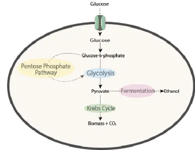
Figure 1.1 – The different metabolic pathways for glucose metabolism... 1
Figure 1.2 - Alcoholic Fermentation. ... 2
Figure 1.3 - The oxidative phase of the Pentose Phosphate Pathway. ... 3
Figure 1.4 – Cellular alterations in oleaginous fungi in nitrogen depletion conditions.. ... 6
Figure 1.5 - Proposed Sophorolipid biosynthetic Pathway. ... 7
Figure 1.6 - Mannitol cycle. ... 9
Figure 3.1 – Growth curves of wild-type and MtDH deletion mutants at 25 ºC, 30 ºC and 32.5 ºC and Mannitol, Ethanol and Glycerol yields at 30ºC, 72h.. ... 18
Figure 3.2 - Growth curves of wild-type, mtdh1Δ and mtdh1Δmtdh2Δ deletion mutants at 25ºC, 30ºC and 32.5ºC in fructose or glucose. ... 19
Figure 3.3 - Growth assays in YP supplemented with 10 g/L glucose and 10 g/L of fructose at 30 ºC and 32.5 ºC for 150 h and 100 h respectively. ... 19
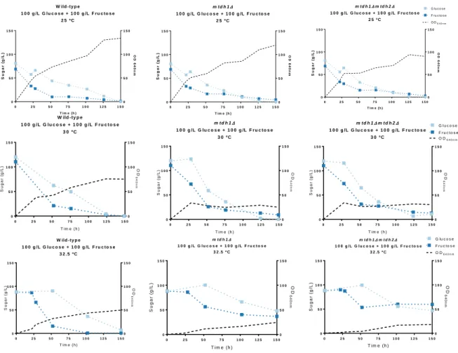
Figure 3.4 - Sugar consumption of wild-type and MtDH deletion mutants in YP supplemented with 100g/L fructose and 100g/L glucose at 25ºC, 30ºC and 32.5ºC. ... 21
Figure 3.5 - A) Fructose consumption in YP supplemented with 200g/L fructose at 25ºC, 30ºC and 32.5ºC. B) Glucose consumption in YP supplemented with 200g/L Glucose. ... 23
Figure 3.6 - Eryhtritol production in different conditions. ... 24
Figure 3.7 - Growth test on YNB supplemented with 18%(w/v) Sorbitol. ... 24
Figure 3.8 - Growth curve in YP supplemented with 1%(w/v) glucose, 1%(w/v) fructose) and 18%(w/v) sorbitol (2FG18S) at 30 ºC and 32.5 ºC. ... 25
Figure 3.9 - Growth curves of wild-type and MtDH mutants on YP supplemented with 1 % (w/v) glucose, 1% (w/v) fructose and 18% (w/v) sorbitol at 32.5 ºC. ... 26
Figure 3.10 - Growth curves of wild-type and MtDH deletion mutants in YP supplemented with 1 % (w/v) fructose, 1 % (w/v) glucose and 18 % (w/v) mannitol at 32.5ºC. ... 27
Figure 3.11 - Correlation between mannitol production in the wild-type and temperature.. ... 27
xv
Table Index
xvii
List of Abbreviations
Acl ATP-citrate lyase
AMP Adenosine monophosphate
ATP Adenosine triphosphate
Bp Base pair
CDS Coding sequence
DTT Dithiothreitol
HPLC High-performance liquid chromatography
kb Kilobase pair
Mtdh Mannitol dehydrogenase
NAD(H) Nicotinamide adenine dinucleotide (hydride)
NADP(H) Nicotinamide adenine dinucleotide phosphate (hydride)
PCR Polymerase chain reaction
PMSF Phenylmethylsulfonyl fluoride
PPP Pentose phosphate pathway
OD640nm Cell density at 640nm
W/S clade Wickerhamiella/Starmerella clade
w/v Weight per volume
Frequently used species
G. oxydans Gluconobacter oxydans
K. lactis Kluyveromyces lactis
S. cerevisiae Saccharomyces cerevisiae
Y. lipolytica Yarrowia lipolytica
St. bombicola Starmerella bombicola
Media used
YP 1 % (w/v) yeast extract and 2 % (w/v) peptone
20FG YP supplemented with 100 g/L glucose and 100g/L fructose
20Glu YP supplemented with 200 g/L glucose
20Fru YP supplemented with 200 g/L fructose
2FG YP supplemented with 10 g/L glucose and 10g/L fructose
2FG18S YP supplemented with 100 g/L glucose and 100g/L fructose and
1. Introduction
1
1. Introduction
1.1. Carbon metabolism in yeasts: general aspects
Yeasts are unicellular fungi present in terrestrial, aerial and aquatic environments, which have been applied to different industrial fields. For example, Saccharomyces species are very well-known microorganisms in the wine and beer industry, while Yarrowia lipolytica is a model organism for lipid production (Dequin, 2001; Rakicka et al., 2015). The demands for yeast’s metabolic products, not only by S. cerevisiae, require the understanding about their central carbon metabolism in order to provide possibilities to increase yields of desired products and decrease unnecessary products. This knowledge can also facilitate optimization of growth media, temperature and oxygen availability (Flores et al., 2000; Mattanovich et al., 2014).
Yeasts can use different carbon sources like polyols and alcohols but, hexoses are their preferred carbon sources, namely glucose (Flores et al., 2000). As outlined in Figure 1.1, in the cytoplasm, glucose is converted into glucose-6-phosphate by a hexokinase. After this step, Glucose-6-phosphate can be further metabolized through glycolysis being converted in the final step into pyruvate which in turn can enter the Krebs cycle or go to the fermentative pathway, which usually happens in low oxygen conditions (Rodrigues et al., 2006). Alternatively, glucose-6-phosphate can be channelled to the pentose phosphate pathway.
1. Introduction
2
1.1.1. Glycolysis
In glycolysis, the glucose uptake into the cell is converted into pyruvate through several enzymatic reactions. Until this step two ATP molecules are invested and two molecules of ATP as well as one molecule of NADH are produced per glucose molecule.
Pyruvate can have three different fates: fermentation to lactate, this usually happens in erythrocytes and in lactic acid bacteria and will not be approached in this thesis. Fermentation to ethanol that usually happen in yeasts and that is known as alcoholic fermentation, and finally respiration where pyruvate can be converted into acetyl-CoA and enter the Krebs Cycle. This happens in animals, plants and microorganisms.
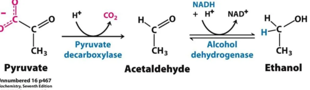
The most used yeast in fermentation is S. cerevisiae, which has an uncommon behavior denominated as Crabtree effect. Crabtree positive yeasts conduct fermentation whenever the sugar levels are high regardless of oxygen levels. Therefore, under sugar excess S. cerevisiae can convert pyruvate derived from glycolysis into ethanol and carbon dioxide, which are the final products of alcoholic fermentation (Figure 1.2) (Deken, 1966).The conversionof acetaldehyde into ethanol is conducted by the enzyme alcohol dehydrogenase and regenerates NAD+. As glycolysis needs NAD+ to continue, the regeneration
of this cofactor is very important for the normal carbon flux in this yeast and for redox homeostasis. At the niche level, the fast conversion into ethanol can represent an advantage since ethanol can discourage the growth of other microorganisms (Rozpędowska et al., 2011). However, the biomass yield is lower, so more sugar must be consumed to achieve a high cell number. Another interesting feature is the possible consumption of ethanol through gluconeogenesis when no sugar is available, however in this case oxygen is necessary.
In respiration the energy yield is higher than in fermentation. In S. cerevisiae, 16 ATP are produced in respiration while in fermentation 2 ATP are produced per glucose molecule. However, oxygen is required since it is the final electron acceptor. In this case, the final products are biomass and carbon dioxide (Verduyn et al., 1991; Barnett, 2003; Kayikci & Nielsen, 2015). Yeasts performing preferably this metabolic pathway whenever enough oxygen is present are said to be respiratory or Crabtree negative. Kluyveromyces lactis and Y. lipolytica are two examples of yeasts with this behavior (Veiga
et al., 2000).
Figure 1.2 - Alcoholic Fermentation. Conversion of pyruvate into acetaldehyde by pyruvate decarboxylase. Alcohol
1. Introduction
3
1.1.2. Pentose Phosphate Pathway
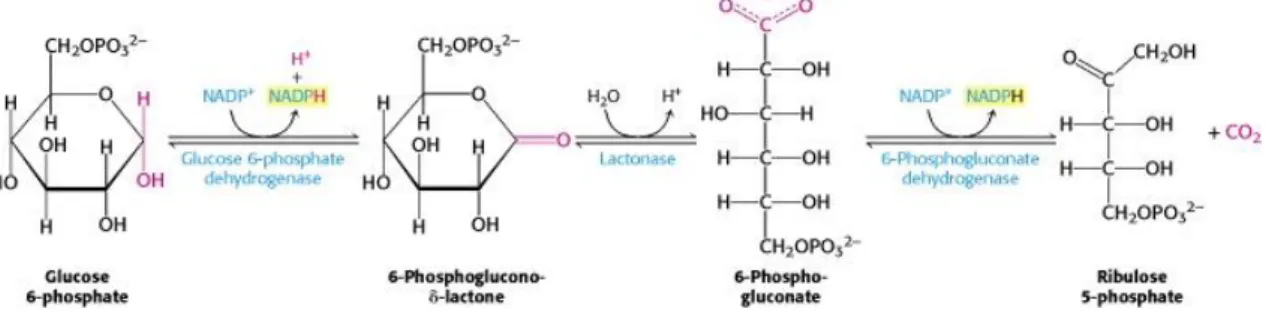
The Pentose Phosphate Pathway (PPP) is an alternative metabolic pathway branching from glycolysis -in glucose-6-phosphate - responsible for oxidation of glucose. The PPP is divided -in two phases: the oxidative phase, which is represented in Figure 1.3, that comprises oxidative reactions, where glucose-6-phosphate is converted into ribose-5-phosphate with generation of two molecules of NADPH per one molecule of glucose. This cofactor has an important role in redox homeostasis and is essential for many intermediary metabolic reactions, namely in biosynthetic pathways like synthesis of fatty acids and sterols. Also, it can be used in the reduction of oxidized nitrogen sources to ammonia. In the non-oxidative phase, a series of non-non-oxidative reversible reactions produce five-carbon sugars for nucleotide biosynthesis and convert excess five-carbon sugar into intermediates of the glycolytic pathway (Mattanovich et al., 2014; Stincone et al., 2016)
As referred in section 1.1, yeasts show differences in their central carbon metabolism. For example, while S. cerevisiae has been described as an organism with a glycolytic flux higher than the PPP flux,
K. lactis, a Crabtree negative yeast, has, on the contrary, a higher flux through the PPP than through the
glycolytic pathway (Jacoby et al., 1993; Saliola et al., 2007). In Y. lipolytica, an oleaginous yeast, it has been reported that PPP is the principal provider of NADPH in the cell, namely for fatty acid synthesis (Wasylenko et al., 2015). While in Y. lipolytica, the PPP is the main source of NADPH for lipid biosynthesis, in K. lactis that is not the main function in the cell. Instead, the PPP seems to be the main metabolic pathway used by K. lactis to oxidize glucose and the NADPH produced will be coupled to mitochondrial respiratory chain (Overkamp et al., 2002). This does not happen in Y. lipolytica or in S.
cerevisiae since their mitochondrial enzymes are not able to oxidize NADPH because they are only
specific for NADH (Kerscher et al., 1999; Overkamp et al., 2000).
In spite all the different roles that PPP or even the roles of NADPH in the cell, its main function is in the oxidative stress response and the maintenance of NADP+/NADPH ratio. Also, we must keep in mind,
that a higher flux in PPP does not mean an inexistent flux through the glycolysis or other metabolic pathway and vice-versa. Instead, both the PPP and the glycolytic pathway are dynamic and regulated processes that are interconnected, in the metabolic network that supplies energy and biomolecules needed for the biochemical processes. As environmental characteristics change, namely the availability of substrates, the metabolic adaptation must be fast. For that, metabolic reactions are regulated
Figure 1.3 - The oxidative phase of the Pentose Phosphate Pathway. Glucose-phosphate is converted into
6-Phosphogluconate-δ-lactone by a glucose-6-phosphate dehydrogenase, followed by conversion into 6-Phospho-gluconate by a lactonase. Finally, ribulose-5-phosphate will be produced by a 6-phosphogluconate dehydrogenase. The first and last reaction in the oxidative phase of PPP can produce 2 NADPH molecules per 1 glucose molecule. Image from (Berg et al., 2012)
1. Introduction
4 increasing the production of metabolites needed as well as decreasing the synthesis of metabolites that are not needed, allowing the surviving and proliferation of the species (Stincone et al., 2016).
1.2. Fructophily: a peculiar characteristic of some microorganisms
Yeast and other organisms use sugar transporters for supplying cells with energy and carbon source. In
S. cerevisiae, there are some membrane transporters, like hexose transporters, that operate through a
facilitated diffusion mechanism that supports the use of glucose as preferred carbon source, which is called glucophilic behavior (Reifenberger et al., 1995). The glucophilic behavior is related with a problem that occurs in wine fermentations. In grape juice, the amounts of glucose and fructose are approximately equal (Berthels, Otero, Bauer, Thevelein, & Pretorius, 2004). When a glucophilic yeast is used it will consume glucose preferentially over fructose resulting in big differences between fructose and glucose levels can result in stuck fermentations with a high residual concentration of fructose (Berthels et al., 2004). Since fructose is sweeter than glucose, the high amount of this sugar at the end of the fermentation can modify the palate of wine (Lee, 1987).
The preference for glucose is possibly related with the existence of the transporters encoded by the HXT (HeXose Transporter) and GAL2 (GALactose metabolism) genes that besides operating through a facilitated diffusion mechanism accept glucose as main substrate but can accept fructose and mannose too. Also, glucose sensors encoded by the SNF3 (Sucrose NonFermenting) and RGT2 (Restores Glucose Transport) genes are responsible for sensing and signalling the availability of glucose, controlling the uptake of glucose that could be a rate limiting step of glycolysis (Diderich et al., 1999; Kayikci & Nielsen, 2015). So far, there is no knowledge about the existence of an HXT gene essential for growth on glucose, instead as some of them have a high Km (low affinity) and others a low Km (high affinity)
their expression is regulated according to the amount of glucose available, but also by osmotic pressure (Özcan & Johnston, 1999), starvation (Diderich et al., 1999) and physiological state of the cells (Özcan and Johnston, 1999; Luyten et al., 2002).
All these transporters were characterized individually showing a higher affinity for glucose than for fructose, forming a likely basis for glucophily. However, a small group of yeasts has a completely different behavior, consuming fructose faster than glucose what is defined as fructophilic behavior (Sousa-Dias et al., 1996).
Fructophily was first associated to Zygosaccharomyces genus (Emmerich & Radler, 1983) but, recently was described the Wickerhamiella/Starmerella (W/S) clade within the Saccharomycotina sub phylum where are a majority of fructophilic yeasts. W/S clade encompasses species from the Wickerhamiella and Starmerella genera and closely related Candida species (Gonçalves et al., 2016). Interestingly, all the species in this clade are found in the floral niche – fructose rich environments - like fructophilic bacteria (Filannino et al., 2016). As fructophilic yeasts are able to quickly transport fructose, this could had represented an advantage to floral niche since probably it could confer a positive impact on fitness (Gonçalves et al., 2016).
Until now, fructophilic behavior was related with the presence of a membrane protein first described in the yeast Zygosaccharomyces bailii, Ffz1 (Fructose Facilitator of Zygossacharomyces). Ffz1 is a high capacity and low affinity uniporter specific for fructose (Pina et al., 2004). Interestingly, a second fructose transporter, Ffz2, was characterized in Z. rouxii, but with a difference at the substrate specificity level, since this transporter can accept both glucose and fructose as substrates (Leandro et al., 2011). However, contrary to Ffz1, Ffz2 is not essential for fructophilic behaviour (Leandro et al., 2014). So
1. Introduction
5 far, Ffz1 seems to be the only requirement for the preference of these organisms for fructose over glucose, since the deletion of the FFZ1 gene abolished the fructophilic behavior in Zygossacharomyces
rouxii (Leandro et al., 2014) and in Starmerella bombicola (Gonçalves et al., 2018).
The preference for fructose over glucose can be explained by the presence of FFZ1, but the role of fructose in the metabolism of these yeasts is still unknow. However, the knowledge of this theme could be of major importance for the wine industry as a possible solution for stuck fermentations.
1.3. St. bombicola: a frutophilic yeast and a sophorolipid producer
St. bombicola was first described in 1998 as a teleomorph of the yeast Candida bombicola (Rosa &
Lachance, 1998). This yeast has an association with bees, flowers visited by bees and can be associated with some other floriculous insects (Kurtzman et al., 2011). Phylogenetically, St. bombicola belongs to the Wickerhamiella/Starmerella clade previously referred in section 1.2. St. bombicola is a fructophilic yeast which has gained much attention due to its biotechnological potential as a producer of sophorolipids, being capable of producing up to 400g/L of this type of lipids (St. bombicola ATCC 22214) (Elshafie et al., 2015) and is amenable to genetic manipulation (Saerens et al., 2011).
Sophorolipids are biosurfactants with diverse applications such as household cleaning, personal care and cosmetics and oil industry (Bogaert et al., 2011). Also, the applications of sophorolipids in nanotechnology is starting to be explored as well as their antimicrobial activity (Kapjung et al., 2002; Singh et al., 2009) So far, sophorolipids showed antimicrobial activity against gram-positive bacteria but not against gram-negative bacteria (Kapjung et al., 2002). However, they were not used with this purpose yet (Kapjung et al., 2002). Besides being produced by non-pathogenic species, which is an important characteristic, they have several advantages compared with regular surfactants, namely their low ecotoxicity, higher biodegradability, better environmental compatibility and higher selectively and specific activity at extreme pH, temperature and salinity. In contrast, the regular surfactants have a low biodegradability, high ecotoxicity and bio-accumulation (Elshafie et al., 2015; Kapjung et al., 2002; Van Bogaert et al., 2007, 2013). For these reasons, they have been intensively studied and a pathway for sophorolipids production has been established as well as several growth medias to optimize its synthesis.
Sophorolipids are considered secondary metabolites since they are produced in the stationary phase and the absence of their production does not influence cell viability. Also, it has been suggested that sophorolipids could work as a carbon storage since they are produced upon high carbon to nitrogen ratio which is linked with their niche. Also as they have antimicrobial activity this could represent advantages against other microorganisms (Van Bogaert et al., 2013). The pathway to produce sophorolipids can be longer or shorter depending on the availability of fatty acids in the medium. When no hydrophobic substrate is present, fatty acids are synthesized from acetyl-CoA derived from glycolysis, through de
novo fatty acid synthesis. In this case, as outlined and explained in more detail in Figure 1.4, when a
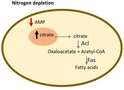
nitrogen depletion exists there are several metabolite alterations that result in an increasing of citrate in the cytoplasm. ATP-citrate lyase (Acl) will convert it into acetyl-CoA. The fatty acid synthetase (Fas) generates fatty acids and triacylglycerols (Papanikolaou, 2012; Ratledge, 2002). However, a hydrophobic substrate can be added into the medium to optimize sophorolipid synthesis like a fatty acid, a triglyceride, or a fatty acid methyl- or ethyl ester.
When this type of molecule is supplied in the medium a lipase will hydrolyze it and the fatty acid will be taken up by the cell. Also, alkanes are widely used as hydrophobic carbon source to produce
1. Introduction
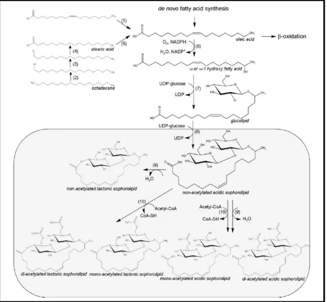
6 sophorolipids, which will be oxidized into a fatty acid too. After this, several enzymatic steps occur as represented and explained in Figure 1.5, obtaining a sophorolipid in acidic form. The majority of sophorolipids are modified by lactonization and then acetylated through an acetyl transferase acetyl-Coenzyme A dependent. In this case, an acetylated lactonized sophorolipid is obtained. A minority of the sophorolipids in acid form are acetylated through the same enzyme originating an acetylated acidic sophorolipids (Bogaert & Saerens, 2007; Bogaert et al., 2011).
St. bombicola seems to excrete the sophorolipids produced to the culture media through a multidrug
transporter (MDR) (Van Bogaert et al., 2013). However, this transporter does not seem to be essential for sophorolipid synthesis (Van Bogaert et al., 2013). So far, only two enzymes were experimentally tested and proven to be essential for sophorolipids synthesis. The deletion of CYP52M1 encoding cytochrome P450 monooxygenase totally abolished sophorolipid production (Van Bogaert et al., 2013) and deletion of the gene encoding UDP-glucosyltransferase (UGTB1) also abolished production of sophorolipids but showed accumulation of glucolipids (Saerens et al., 2011). These results suggest that both these genes encode essential enzymes for sophorolipid production (Saerens et al., 2011; Van Bogaert et al., 2013; Huang et al., 2014; Saerens et al., 2015).
Nitrogen depletion
AMP
citrate
citrate
Acl
Oxaloacetate + Acetyl-CoA
Fatty acids
Fas
Figure 1.4 – Cellular alterations in oleaginous fungi in nitrogen depletion conditions. When a nitrogen depletion exists in
the medium, the cellular AMP decreases. The lower levels of AMP lead to an alteration of Krebs cycle function. In this case, the enzymes activated by AMP decrease their activity altering the carbon flow towards accumulation of intra-mitochondrial citric acid. When citric acid levels are too high the cell secretes it to the cytoplasm. It is in the cytoplasm that ATP-citrate lyase (Acl) will convert citrate into acetyl-CoA. The fatty acid synthetase (Fas) generates fatty acids and triacylglycerols. Based on (Papanikolaou, 2012).
1. Introduction
7 As previously referred, sophorolipids are a particular type of lipid that only a few yeasts can produce. However, there are oleaginous yeasts (lipid content higher than 20 % (Boulton & Ratledge, 1981)) that produce other type of lipids biotechnologically important too due to the possibility of using them for biodiesel production. That is the case of Rhodosporidium toruloides and Rhodotorula graminis, that can produce lipids from sugars and from glycerol, however, glucose still is the preferential carbon source (Galafassi et al., 2012; Wiebe et al., 2012).
Fatty acid synthesis need large quantities of NADPH since two NADPH molecules are oxidized to NADP+ for each elongation step and one NADPH is consumed for each fatty acid desaturation reaction
(Dulermo et al., 2015). Y. lipolytica, one of the most studied oleaginous yeasts so far, is a model organism for biofuel production. Because of this, fatty acids synthesis has been intensively studied in this organism. As oleaginous organisms need large quantities of NADPH, one of the most studied aspects in lipid production in Y. lipolytica is the source of NADPH. However, the origin of NADPH for
Figure 1.5 - Proposed sophorolipid biosynthetic pathway. The first step of sophorolipid production is the conversion of
fatty acid into hydroxy fatty acid by a NADPH dependent cytochrome P450 monooxygenase. Then, through a glycosyltransferase I, glucose is glycosidically coupled to the hydroxyl group of the fatty acid. Then, a second glucose molecule is added by a glycosyltransferase II. Enzymes: 1) lipase, 2) cytochrome P450monooxygenase, 3) alcohol dehydrogenase, 4) aldehyde dehydrogenase, 5) desaturase, 6) cytochrome 0450 monooxygenase, 7) glucosyltransferase I, 8) glucosyltransferase II 9) lactonesterase, 10) acetyltransferase. Image from Van Bogaert et al., 2011
1. Introduction
8 this metabolic pathway is not completely known. Malic enzyme, which is a cytosolic enzyme that converts malate into pyruvate with production of NADPH was considered for several years the main producer of cytosolic NADPH. However, it was proven that the malic enzyme does not have a significant impact on lipid synthesis since when the gene encoding for the malic enzyme was disrupted no significant decrease in fatty acid synthesis was observed (Dulermo et al., 2015). Instead, it has been suggested that the PPP could be the main producer of cytosolic NADPH that will be used in fatty acid synthesis (Wasylenko et al., 2015). On the other hand, in K. lactis mitochondrial respiratory chain seems to have a role in NADPH regeneration what does not happen in Y. lipoytica since the external mitochondrial enzymes use NADH instead of NADPH (Harder et al., 2013; Kavscek et al., 2015; Wasylenko et al., 2015). In addition, Dulermo et al. suggest a correlation between lipid synthesis and the synthesis of mannitol. In this study, mutants unable to produce mannitol have a higher yield in lipids. Also, they observe the opposite, since when lipid synthesis decreases, through the deletion of ACL1, mannitol yield increases instead. Due to the ability of Y. lipolytica to grow on mannitol and the typical role of lipids in the cell, the authors suggested a possible role of mannitol as carbohydrate reserve, since when one of these pathways were abolished a readjustment on carbon flow occurs (Dulermo et al., 2015).
1.4 Polyols as a smart strategy to deal with high osmotic environments
In nature, the osmolarity usually changes with consequences to the integrity and hydration of the cell. When the external osmotic pressure is lower, it causes a water influx which can lead to swelling or lysis of the cell. But, when the external osmotic pressure is high, water efflux is the result leading to dehydration (Bubnová et al., 2014; Wood, 2015).
There are no active transport mechanisms for water, so microorganisms developed strategies to maintain the turgor and volume of the cell within boundaries acceptable for its normal physiology. Aspergillus
niger and Penicillium chrysogenum are capable of tolerating high external osmotic pressures using a
common strategy: synthesis or uptake of polyols, namely glycerol, erythritol and mannitol (Hult et al., 1980).
Polyols or sugar alcohols are compatible solutes widely distributed in fungi, that can have a dual function: stress response and protection against dehydration. Structurally, they are polyhydric alcohols formed by the reduction of the carbonyl group of an aldose or a ketose monosaccharide to a hydroxyl group (Solomon et al., 2007). Polyols are synthesized or taken up by yeasts, especially the ones capable of growing in high sugar or salt environments – osmotolerant yeasts (Harder et al., 2013; Bubnová et
al., 2014). Glycerol is the most common and the most studied polyol. However, there are more examples
as mannitol, sorbitol and ribitol. Different organisms can use the same sugar alcohol for different purposes. Mannitol is a good example of that, since it has been assigned different functions in fungi like carbohydrate reserve, osmoregulation, coenzyme regulation and storage of reducing power (Jennings, 1984).
1. Introduction
9
1.4.1. The importance of mannitol in fungi
1.4.1.1. Mannitol synthesis
Mannitol is one of the most abundant polyols in nature. It can be found in bacteria, plants, algae, lichens but specially in filamentous fungi. However, the pathway to synthesize mannitol is quite different between organisms (Figure 1.6). In the fungus Alternaria alternata a cycle with four enzymes was described: NADH-mannitol 1-phosphate dehydrogenase (M1pdh), NADP+-mannitol 2-dehydrogenase
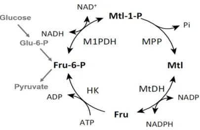
(Mtdh), mannitol 1-phophate phosphatase (Mpp) and a hexokinase (HK). The mannitol cycle is a branching from glycolysis in fructose-6-phosphate, which is converted in mannitol 1-phosphate by M1pdh and Mpp will dephosphorylated it into mannitol. Then, mannitol is converted into fructose via a reversible NADPH-mannitol dehydrogenase (Mtdh). The hexokinase is responsible for the conversion of fructose into fructose-6-phosphate. The proposed role for mannitol cycle was regeneration or production of NADPH due to the balance of the cycle: NADH + NADP+ + ATP in NAD+ + NADPH +
ADP + Pi (Hult & Gatenbeck, 1978). However, in some organisms the purpose of the production of mannitol does not seem to be the regeneration of NADP+. Instead, in the last years several functions
have been attributed to this polyol suggesting that mannitol can have different functions among organisms (Jennings, 1984; Patel & Williamson, 2016; Solomon et al., 2007).
However, in some fungi like Aspergillus niger Mtdh is expressed only in spores and M1pdh, which seems to be the major enzyme in mannitol metabolism, is expressed only in mycelia (Ruijter et al., 2003). Also, in Basidiomycetes the absence of mannitol phosphate dehydrogenase or mannitol 1-phosphatase activities is described, suggesting that mannitol metabolism is not always a cycle (Hult et
al., 1980).
In Stagonospora nodorum the absence of M1pdh causes alterations on central metabolism, since when grown on glucose, the levels of arabitol and mannitol were reduced but more trehalose was detected. When grown on fructose, mannitol production is not affected suggesting that fructose can be reduced to mannitol via Mtdh. The fact that mannitol metabolism is not always part of a cycle as well as the fact
Figure 1.6 - Mannitol cycle. Fructose-6-phosphate is converted to mannitol-1-phosphate by mannitol-1-phosphate
dehydrogenase (M1pdh). Mannitol-1-phospate is converted by mannitol-1-phosphate phosphatase into mannitol, which will be converted in fructose by mannitol dehydrogenase (Mtdh). Fructose will be phosphorylated by a hexokinase (Hk).Abbreviations: Fru: fructose; Fru-6-P_ fructose-6-phosphate; Mtl: mannitol; Mtl-1-P: mannitol-1-phosphate. Adapted from Patel and Williamson, 2016.
1. Introduction
10 that in S. nodurum the absence of M1pdh or Mtdh had effects on central metabolism and in asexual sporulation suggests a versatile role of this polyol in the cell. So, the function of mannitol is not necessarily storage compound or redox balance, instead mannitol can have different functions and its absence can have strong effects in cell physiology (Solomon et al., 2006).
1.4.1.2. The different functions of mannitol in microorganisms
One of the reported functions of mannitol is that of compatible solute (Zahid et al., 2015). To deal with low water activity – high osmotic pressure – many bacteria synthesize and accumulate polyols.
Gluconobacter oxydans lives in sugar rich environments like fruits, nectars, wine and honey syrups
being subjected to a high osmotic pressure. The osmotolerance of these bacteria is improved by production and accumulation of intracellular mannitol. Also, in different species of bacteria like
Pseudomonas fluorescence, Acinetobacter baylyi and in hetero-fermentative lactic acid bacteria it has
been reported that mannitol works as osmoprotector as in G. oxydans (Kavanagh et al., 2002; Sand et
al., 2013; Wisselink et al., 2002; Zahid & Deppenmeier, 2016).
Also, the conversion of mannitol into fructose is a unique enzymatic step that can provide a faster start of glycolysis. In Alternaria alternata, mannitol seems to have a role as storage compound too. However, it does not seem essential since spores of mannitol deletion mutants (mtdhΔ, mpdhΔ and mtdhΔmpdhΔ) could germinate. Otherwise, when mannitol levels decrease the disaccharide content increases, suggesting that mannitol is not essential as storage compound since it can be substituted by other carbohydrates, but when it is present it performs that role in the cells (Vélëz et al., 2007).
Mannitol is also used as carbohydrate reserve in pathogenic fungi (Solomon et al., 2007). Many plants are unable to metabolize mannitol, so fungal pathogens or mutualists can convert hexoses to mannitol. This limits the use of the carbon source by the plant, which is an advantage for the fungus (Solomon et
al., 2007). But, mannitol can also have another function in plant-pathogen interaction. When invaded,
plants produce Reactive Oxygen Species (ROS) in the extracellular space or apoplast which are signals to start the defense responses. As mannitol can quench the ROS produced by the plant it will block the defense signal itself. In Cladosporium fulvum, a tomato fungal pathogen, the mutants unable to produce mannitol were non-pathogenic showing that, in this case, mannitol is necessary for pathogenicity (Patel & Williamson, 2016). This can be due to the role of mannitol as quencher of ROS, but it can also be related with an intrinsic mechanism to defend the pathogen against the stresses associated with plant defense responses.
In 2006, mannitol was described for the first time as fundamental for asexual sporulation in S. nodorum. In this study, mutants unable to synthesize mannitol did not sporulate and when mannitol was added in the medium the ability to sporulate was restored. Although it has not been proven so far, mannitol could work as a signalling molecule for the initiation of asexual conidiation in S. nodorum (Solomon et al., 2006).
However, mannitol can have yet another role in sexual structures in fungi. In Aspergillus fischeri, the deletion mutant unable to convert fructose-6-phosphate into mannitol-1-phosphate exhibit delayed formation of ascospores and some of ascospores were incompletely formed. Also, conidia germination after exposure to stress conditions was lower than wild-type showing that conidia were more sensitive to heat and oxidative stress. However, there were no differences in vegetative growth indicating that mannitol seems to be fundamental for conidia germination (Wyatt et al., 2014).
1. Introduction
11 Some authors described a dual function for mannitol in the rust fungus Uromyces fabae. In this case, mannitol was found in spores but disappeared rapidly upon infection structure formation leading the authors to suppose that mannitol could have a role as carbohydrate storage. But mannitol also seems to work as a scavenger of Reactive Oxygen Species (ROS) in this species. This shows that mannitol has different functions not only depending on the organism, but it can also have different roles in the same organism emphasizing its versatility, which by itself is very interesting (Voegele et al., 2005).
In summary, so far mannitol has been described to have roles in stress tolerance in high osmotic environments, can work as a quencher of ROS, as carbohydrate reserve or it can have a role in sexual and asexual development in fungi. Several microorganisms also produce high amounts of mannitol including yeasts belonging to the W/S clade (Gonçalves, 2018). But, so far, the role of mannitol has not been established. Candida magnoliae, a yeast belonging to the W/S clade, has been described as a producer of high amounts of mannitol. Still its function was not elucidated (Lee et al., 2003; Song et al., 2002). Probably due to the presence of this species in nectar where the osmotic pressure is high, mannitol could be involved in osmoprotection. As well as other yeasts belonging to W/S clade (Gonçalves, 2018),
St. bombicola produces high amounts of mannitol and lipids. In turn, Y. lipolytica produces high amounts
of lipids, a pathway that has already be showed (see section 1.3) to be interconnected with mannitol synthesis in this yeast. In this way, the mannitol and lipid synthesis could also be interconnected in St.
bombicola.
1.5. Objectives
Ffz1 is essential for fructophilic behavior, however it does not seem to be the only requirement for the preference for fructose over glucose, so it is possible that fructose dependent metabolic pathways may play a role in fructophilic yeasts. W/S clade yeasts, namely St. bombicola, produces high amounts of mannitol from fructose through Mtdh. Since this is a fructose dependent pathway, the first objective of this study was to understand if the absence of this pathway could affect the fructophilic behavior of St.
1. Introduction
2. Materials and Methods
13
2. Materials and Methods
2.1. Yeast strains
St. bombicola PYCC 5882 (will be referred in this work as wild-type strain) was isolated from a flower
of Ornithogalum narbonense in Cascais and is deposited in the Portuguese Yeast Culture Collection (PYCC) in Caparica, Portugal. All the deletion mutants used in this work were constructed in this strain background. The culture was maintained in YPD medium (1% (w/v) yeast extract (Difco), 2% (w/v) peptone (Difco), 2% (w/v) glucose and 2% (w/v) agar (Labchem).
2.2. Growth experiments with St. bombicola and deletion mutants
For growth assays, the strains (wild-type, mtdh1Δ, mtdh1Δ mtdh2Δ and for some assays mtdh2Δ and
mtdh1Δ sor1Δ) were pre-grown overnight (o/n) in 100 mL flasks containing 20 mL of culture medium
at the temperature used for the growth assay. The inoculum was set at OD640nm=0.2 in 30 mL of medium
YP (1% (w/v) yeast extract and 2% (w/v) peptone) supplemented with a carbon source in 250 mL flasks. The cell density was measured at 640nm and was monitored for 100-150h and supernatant samples were collected for further metabolites analyses.
2.2.1 Assimilation tests
To test if sorbitol could be assimilated as a carbon source, wild-type and the deletion mutant available at the lab sor1Δ (sorbitol dehydrogenase) were grown on 100 mL flasks containing 20 mL of Yeast Nitrogen Base medium (YNB, Difco) supplemented with 2% (w/v) or 18% (w/v) sorbitol. Growth was monitored for 7 days.
To test if mannitol could be assimilated as a carbon source wild-type and MtDH deletion mutants were grown in the YP supplemented with 18% (w/v) mannitol. Growth was monitored for 7days.
2.3. Metabolite Analyses
2.3.1. Analysis of extracellular metabolites: quantification by HPLC
Extracellular metabolites were analysed by HPLC. 1 mL of culture was centrifugated for 1min at 16000g and the supernatant was collected. If necessary, the sample was diluted in a sodium azide solution in distilled water (0.025% w/v) prior to filtration through Q-Max RR Nylon Filters with 0.22µm pores (Frisenette). 10µL was injected into a Dionex P680 instrument equipped with an ASI-100 Automatic
2. Materials and Methods
14 Sample Injector and a 2142 Differential Refractometer. For these analyses, the eluent was a sodium azide solution in bi-distilled water (0.025%) at a flux of 0.6 mL.min-1. The Aminex HPX-87P (300mm
x 7.8 mm, BioRad) column was operated at 80 ºC. Solutions containing glucose, fructose, ethanol, glycerol, erythritol and mannitol with concentrations between 10 and 15 g/L were sequentially diluted for construction of a calibration curve.
To detect citric acid, the column used was Aminex HPX-87H (300 mm x 7.8 mm, Biorad) at 65 ºC with an Ultimate 3000 Diode Array Detector at 210 nm. The mobile phase a solution of 5 mM of Sulfuric Acid at 0.6 mL.min-1 in bi-distilled water. The pure standard of citric acid was 5 g/L which was
sequentially diluted for construction of a calibration curve.
The results were treated and integrated using the software Chromeleon v6.8 (Dionex). Two replicates were analysed.
2.3.2. Determination of metabolite yields
After the concentration in the growth medium was determined by HPLC, the yield of each metabolite was calculated per gram of sugar consumed. For that, the sugar consumed was considered and equation used is shown:
Equation 2.1 𝑀𝑒𝑡𝑎𝑏𝑜𝑙𝑖𝑡𝑒 𝑌𝑖𝑒𝑙𝑑 (𝑔/𝑔 𝑠𝑢𝑔𝑎𝑟 𝑐𝑜𝑛𝑠𝑢𝑚𝑒𝑑) = 𝑀𝑒𝑡𝑎𝑏𝑜𝑙𝑖𝑡𝑒 (𝑔/𝐿)
𝑆𝑢𝑔𝑎𝑟 𝑐𝑜𝑛𝑠𝑢𝑚𝑒𝑑 (𝑔/𝐿)
2.3.3. Analysis of Intracellular Metabolites
2.3.3.1. Preparation of crude extracts for intracellular metabolite analysis
Wild-type and MtDH deletion mutants were grown in duplicate for 72 h in YP supplemented with 20% (w/v) of fructose and glucose (1:1) at the temperatures 20 ºC, 25 ºC and 30 ºC at 180 r.p.m (rotations per minute). After this time, cells were harvest by centrifugation (4 ºC, 10 min, 8000g) and 1mL of supernatant was collected for HPLC analysis. Then, the cells were washed twice with 100 mM phosphate buffer pH = 7 and stored at -20 ºC. After thawing, the cells were resuspended in 400 µL of TRIS Lysis buffer (0.1 M triethanolamine hydrochloride, 2 mM MgCl2, 1 mM DTT and 1 µM PMSF)
and 200 µL glass beads (212-300µm). The cells were lysed by six alternate cycles of 1 min vortexing followed by 1 min cooling on ice. The cell debris were removed by centrifugation (4ºC, 20min, 9000 g) and the supernatant collected for intracellular analysis by HPLC. For protein quantification 10 µL of the supernatant was collected.
2.3.3.2. Protein Quantification
2. Materials and Methods
15
2.3.3.3. Analysis of Intracellular Metabolites: Quantification by HPLC
The intracellular samples were analysed at the Biological and Chemical Analysis Facility at UCIBIO. The samples (10µL) were eluted with NaOH 0.6 M at a flux 0.4 mL/min at 25 ºC in a CarboPac MA1 column and the peaks were identified with a pulsed amperometric detector. The presence of glucose, fructose, ethanol, glycerol, erythritol and mannitol was analysed. Two replicates were analysed.
2.3.4. Determination of intracellular metabolites
After quantification of intracellular metabolites, the metabolites produced were analysed, however, the amounts of intracellular metabolites could be influenced for the efficiency of the lysis. So, to control the efficiency of cell lysis, the total protein was measured as explained in section 2.3.3.2. Thus, to be able to compare the intracellular results from the three temperatures, we decided to consider the total protein of the crude extracts (described in 2.3.3.1). Therefore the follow formula was used:
2. Materials and Methods
3. Results
17
3. Results
The results obtained during this thesis are partially confidential. Therefore, the results presented in this section are only a part of the work performed which correspond to the non-confidential results.
3.1. Understanding the function of mannitol and mannitol dehydrogenase in St.
bombicola
The preference for fructose over glucose as carbon source was previously shown to be related with the presence of the Ffz1 fructose transporter (Pina et al., 2004). In St. bombicola, as well as in Z. rouxii, the deletion of the gene encoding for this transporter totally abolished the fructophilic behavior, however the relevance of fructose in their metabolism has not been elucidated yet (Gonçalves et al., 2018; Leandro et al., 2014). In addition to being a carbon source, fructose can also be a final electron acceptor to re-oxidation of NAD(P)H in fructophilic bacteria, resulting in the formation of mannitol (Zaunmüller
et al., 2006).
Candida magnoliae, a species from the W/S clade, has already been described as a producer of high
amounts of mannitol from fructose (Lee et al., 2003; Song et al., 2002). Moreover, the mannitol yield of several species from the W/S clade was determined and all of them, with the exception of St.
bacillaris, seem to produce high amounts of mannitol when grown on fructose as carbon source
(Gonçalves, 2018).
In St. bombicola, mannitol seems to be produced directly from fructose through the enzyme mannitol dehydrogenase which uses NADPH but not NADH as a cofactor (Gonçalves, 2018). Also, in the genome of St. bombicola two genes encoding for two mannitol dehydrogenase (Mtdh) were found. Single and double deletion mutants were previously constructed (mtdh1Δ, mtdh2Δ, mtdh1Δmtdh2Δ). In order to understand if mannitol dehydrogenase could have an impact in fructophily, the different mutants were grown in YP medium supplemented with 100 g/L glucose and 100 g/L of fructose (henceforth this medium will be named 20FG) since it is a medium where fructophily is apparent. In figure 3.1 where the growth curves are represented it is possible to see that the deletion mutants mtdh1Δ and
mtdh1Δmtdh2Δ are not able to reach the same cell densities as the wild-type. However, the mtdh2Δ
deletion mutant grows as well as the wild-type suggesting that Mtdh1 is the major enzyme involved in mannitol synthesis. Measuring the mannitol production, we were able to observe that mtdh1Δ and
mtdh1mtdh2Δ do not synthesized mannitol, while mtdh2Δ is able to produce mannitol what corroborates
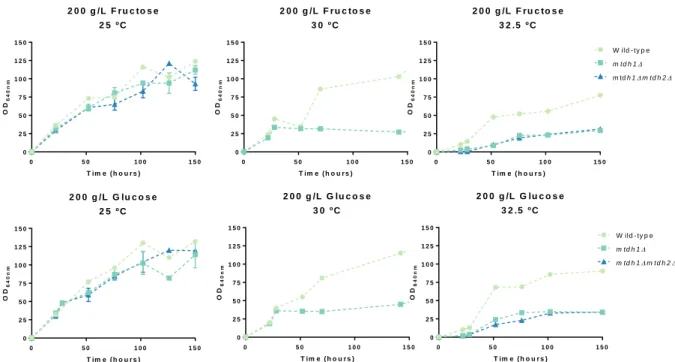
the previous results that Mtdh1 is the major enzyme involved in mannitol synthesis in St. bombicola. To try to explain why these deletion mutants reach lower cell densities, we first assessed whether this effect was also observed when a single sugar was present (either glucose or fructose). MtDH encoded an enzyme that converts fructose into mannitol, so the deletion of this gene could have more impact on the metabolism of fructose. To evaluate that, the osmotic pressure was maintained (20 % (w/v)), but we added either only glucose or only fructose to the growth medium (Figure 3.2). As it is possible to observe, in both media tested, we saw that the MtDH deletion mutants did not reach the same cell densities as the wild-type independently of the sugar, suggesting that the lower cell densities of the
3. Results
18
MtDH deletion mutants are not related with one specific sugar, since their behavior is observable in
presence of both glucose or fructose.
Among the different functions that mannitol can have in the cell, one of the most studied so far is its role as osmoprotector. Gluconobacter oxydans accumulates mannitol as a compatible solute under osmotic stress conditions. But, when a double deletion mutant lacking the two mannitol dehydrogenases is grown in a high osmotic medium, the deletion mutant presents a defective growth and has an intracellular mannitol amount lower than the wild-type. These results suggests that mannitol synthesis in G. oxydans is involved in osmoprotection. (Zahid et al., 2015; Zahid & Deppenmeier, 2016). In our experiments with St. bombicola the partial growth impairment of the mannitol dehydrogenase mutants could be related with the high osmolarity of the medium (20 % (w/v) sugar). To test this, the wild-type strain and the MtDH deletion mutants were cultivated at the temperatures where the growth impairment was more pronounced, in this case 30 ºC and 32.5 ºC and in lower sugar concentrations where osmotic pressure is reduced, namely YP medium supplemented with 10 g/L fructose and 10 g/L glucose, henceforth referred to as 2FG.
Figure 3.1 – Growth curves of wild-type and MtDH deletion mutants at 25 ºC, 30 ºC and 32.5 ºC and Mannitol, Ethanol and Glycerol yields at 30 ºC, 72 h. The strains were grown in YP supplemented with 100 g/L Glucose and 100 g/L Fructose
at 25 ºC, 30 ºC and 32.5 ºC for 150 h. The yields represented were determined at 72 h at 30 ºC.The experiments were performed with two biologicals replicates in two independent experiments and the error bars represent the standard deviation n=4.
3 0 º C Y ie ld ( g /g o f s u g a r c o n s u m e d ) Ma nn ito l Eth an ol Gly ce rol 0 .0 0 .2 0 .4 0 .6 W ild - ty p e m td h 1 m td h 2 m td h 1m td h 2 2 5 º C 0 5 0 1 0 0 1 5 0 0 2 5 5 0 7 5 1 0 0 1 2 5 1 5 0 T im e ( h o u r s ) O D6 4 0 n m 3 0 º C 0 5 0 1 0 0 1 5 0 0 2 5 5 0 7 5 1 0 0 1 2 5 1 5 0 T im e ( h o u r s ) O D6 4 0 n m 3 2 .5 º C 0 5 0 1 0 0 1 5 0 0 2 5 5 0 7 5 1 0 0 1 2 5 1 5 0 T im e ( h o u r s ) O D6 4 0 n m W ild - ty p e m td h 1 m td h 1m td h 2 m td h 2
3. Results
19 In figure 3.3 the growth curves of wild-type and MtDH deletion mutants in 2FG are represented. It is observable that at 30 ºC there are no differences in cell densities comparing with the wild-type. However, when grown at 32.5 ºC in the same medium, MtDH deletion mutants are not even able to grow, suggesting that the absence of mannitol/mannitol dehydrogenase in 2FG medium at 32.5 ºC is indispensable for growth. On the other hand, these results suggest that mannitol itself or mannitol synthesis is not involved in protection against osmotic stress in St. bombicola.
So far, we know that MtDH deletion mutants do not reach the same cell densities as the wild-type, but the reason for a lower cell density is not understood yet. Also, it is not known whether only the biomass yield is affected or the growth rate is also affected by the absence of mannitol/mannitol dehydrogenase. To test this we determined the growth rates during the exponential phase. Table 3.1 lists the specific growth rate of MtDH deletion mutants in different media and temperatures.
Figure 3.2 - Growth curves of wild-type, mtdh1Δ and mtdh1Δmtdh2Δ deletion mutants at 25 ºC, 30 ºC and 32.5 ºC in fructose or glucose. The strains were grown on YP supplemented with 200 g/L Fructose (first row) and 200 g/L glucose
(second row) at 25 ºC, 30 ºC and 32.5 ºC for 150 h. The error bars represent the standard deviation of two biological replicates
Figure 3.3 - Growth assays in YP supplemented with 10 g/L glucose and 10 g/L of fructose at 30 ºC and 32.5 ºC for 150 h and 100 h respectively. The error bars represent the standard deviation of 4 assays which were performed twice with
two biological replicates (n=4).
3 2 .5 º C 0 5 0 1 0 0 0 1 0 2 0 3 0 4 0 5 0 T im e ( h o u r s ) O D 6 4 0 n m W ild - ty p e m td h 1 m td h 2 m td h 1m td h 2 3 0 º C 0 5 0 1 0 0 1 5 0 0 1 0 2 0 3 0 4 0 5 0 T im e ( h o u r s ) O D 6 4 0 n m 2 0 0 g /L F r u c to s e 2 5 º C 0 5 0 1 0 0 1 5 0 0 2 5 5 0 7 5 1 0 0 1 2 5 1 5 0 T im e ( h o u r s ) O D6 4 0 n m 2 0 0 g /L F r u c to s e 3 0 º C 0 5 0 1 0 0 1 5 0 0 2 5 5 0 7 5 1 0 0 1 2 5 1 5 0 T im e ( h o u r s ) O D6 4 0 n m 2 0 0 g /L F r u c to s e 3 2 .5 º C 0 5 0 1 0 0 1 5 0 0 2 5 5 0 7 5 1 0 0 1 2 5 1 5 0 T im e ( h o u r s ) O D6 4 0 n m W ild - ty p e m td h 1 m td h 1m td h 2 2 0 0 g /L G lu c o s e 2 5 º C 0 5 0 1 0 0 1 5 0 0 2 5 5 0 7 5 1 0 0 1 2 5 1 5 0 T im e ( h o u r s ) O D6 4 0 n m 2 0 0 g /L G lu c o s e 3 0 º C 0 5 0 1 0 0 1 5 0 0 2 5 5 0 7 5 1 0 0 1 2 5 1 5 0 T im e ( h o u r s ) O D 6 4 0 n m 2 0 0 g /L G lu c o s e 3 2 .5 º C 0 5 0 1 0 0 1 5 0 0 2 5 5 0 7 5 1 0 0 1 2 5 1 5 0 T im e ( h o u r s ) O D6 4 0 n m W ild - ty p e m td h 1 m td h 1m td h 2
3. Results
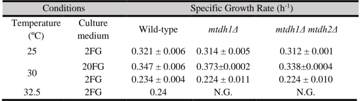
20 In Table 3.1 are represented the specific growth rates in different conditions, 20FG at 30 ºC and 2FG at 25 ºC, 30 ºC and 32.5 ºC. Interestingly, looking into the specific growth rates of MtDH deletion mutants, they grow at the same rate as the wild-type in all conditions tested. This could indicate that mannitol or mannitol dehydrogenase could influence the biomass yield but not the growth rate. Also, comparing the specific growth rates at 30 ºC between 20FG and 2FG it is possible to observe that the specific growth rate is higher in wild-type and in MtDH deletion mutants in a high sugar concentration medium.
Table 3.1 – Specific growth rates of wild-type and MtDH deletion mutants. Standard deviation results from two different
experiments performed with biological replicates. NG means that the strains did not grown in the condition tested.
Conditions Specific Growth Rate (h-1)
Temperature (ºC) Culture medium Wild-type mtdh1Δ mtdh1Δ mtdh2Δ 25 2FG 0.321 ± 0.006 0.314 ± 0.005 0.312 ± 0.001 30 20FG 0.347 ± 0.006 0.373±0.0002 0.338±0.0004 2FG 0.234 ± 0.004 0.224 ± 0.011 0.224 ± 0.010 32.5 2FG 0.24 N.G. N.G.
As mannitol is synthesized directly from fructose, the sugar consumption in 20FG at 25 ºC, 30 ºC and 32.5 ºC was analysed (Figure 3.4) in order to understand if the absence of this metabolic pathway could alter the consumption of fructose. The preference for fructose over glucose is observable at all temperatures and all strains tested. However, the MtDH deletion mutants does not consume fructose as the wild-type. For example, at 30 ºC at 124 h the wild-type consumed all the sugar in the medium, while the MtDH deletion mutants still have about 10 g/L of residual fructose available. This is more noticeable at 32.5 ºC where the MtDH deletion mutants could not consume all the fructose in the medium in 150 h when the wild-type consumes all fructose in 102 h. The consumption of glucose seems to be different too but only at 32.5 ºC. From 102 h MtDH deletion mutants barely consume the sugars, namely fructose. Besides that, at 150 h in the MtDH deletion mutants there is approximately 50 g/L of glucose in the medium, while the wild-type consumed all the glucose present in the medium. The absence of Mtdh seem to attenuate the fructophilic behavior. So, to understand if this only happen when both sugars are present or if it happens even when only one sugar is present, the sugar consumption profile in medium containing 200 g/L fructose or 200 g/L glucose was analysed (Figure 3.5).