Vol.49, n. 6 : pp. 881-888, November 2006
ISSN 1516-8913 Printed in Brazil BRAZILIAN ARCHIVES OF BIOLOGY AND TECHNOLOGY A N I N T E R N A T I O N A L J O U R N A L
Purification and Characterization of
ββββ
-1, 3-Glucanase from
the Secretion of
Simira glaziovii
Colleters (Rubiaceae)
Felipe Almeida Vieira1, Maura da Cunha2, Denise Espellet Klein2, André de Oliveira Carvalho1and Valdirene Moreira Gomes1*
1Laboratório de fisiologia e Bioquímica de Microrganismos; 2Laboratório de Biologia Celular e Tecidual; Centro
de Biologia e Biotecnologia; Universidade Estadual Norte Fluminense Darcy Ribeiro; Rua Alberto Lamego, 2000; Horto; 28015-620; [email protected]; Campos de Goytacazes - RJ - Brasil
ABSTRACT
In this study, β-1,3-glucanase was isolated from Simira glaziovii secretion. The purification process was achieved by a combination of chromatographic methods and was analyzed by SDS-PAGE. The purified enzyme presented an estimated molecular mass of 35 kDa. The optimum pH of enzyme was 5.2
Key words: β-1,3-glucanase, PR protein, chitinases, secretory structure
INTRODUCTION
Although plants are naturally exposed to several phytopathogenic microorganisms, they present tolerance to these pathogens, due to their different structures (cuticles, trichomes, stomata and tyloses) and biochemical mechanisms (such as phenols, phytoalexins, cyanogenic glycosides, protease inhibitors and hydrolases) (Pascholati and Leite, 1994; Oliveira et al., 2003; Caramori et al., 2004). Previous studies have reported that PR proteins are implied in the defense of the plant against viral, bacterial and fungal phytopathogens (Pegg and Young, 1981; Young and Pegg, 1982; Boller, 1987; Boller and Mêtraux 1988; Yun et al., 1987); the PR proteins from families 2 and 3 are
β-1,3-glucanases and chitinases, respectively. Regard hydrolytic enzymes, it has also been reported that these increased in concentration in some plants infected by fungi, such as tomato (Pegg, 1977), tobacco (Kauffmam et al., 1987;
Legrand et al., 1987), potato (Kombrick et al., 1988), cucumber (Boller and Metraux, 1988) and beans (Vôgeli et al., 1988; Mauch and Staechelin, 1989). These proteins have been implicated in the resistance mechanisms of plants against pathogens and insects (Gomes et al., 1994).
secretion of the Simira glaziovii colleter (Fig. 1), was isolated and characterized a β-1,3-glucanase, in order to understand the mechanisms of defense of the plants present in secretion structures.
MATERIALS AND METHODS
Plant materials
Shoot apex from Simira glaziovii (K. Schum.) Steyerm, were collected in the Atlantic rain forest of Barrage of Saracuruna, Duque de Caxias, RJ.
Scanning electron microscopy
Stipules were fixed for two hours in 2.5 % Glutaraldehyde, 4.0 % paraformaldehyde buffered with 50 mmol L-1 cacodylate buffer, pH 7.2. Subsequently the material was post-fixed for two hours at room temperature with 1.0 % osmium tetroxide in 50 mmol L-1 cacodylate buffer, pH 7.2. The post-fixed material was dehydrated in a graded series of acetone solutions, critical point dried in CO2, covered with 20 nm gold and
observed with a ZEISS 962 digital scanning electron microscope.
Protein determination
Total protein was quantified using protein micro assays based on the Bradford assay (1976).
ββββ-1,3-glucanase enzyme activity
The determination of the β-1,3-glucanase activity in secretion samples was made according to the methodology described by Fink et al., (1988). The reaction mixture was composed of 50 µL of the secretion sample, 125 µL laminarin (2 mg mL-1 in Buffer sodium acetate 50 mmol L-1, pH 5.0) and mixed to a final volume of 500 µL in a buffer of sodium acetate 50 mmol L-1, pH 5.0. The reaction was incubated at 37 ° C for 12 h. After the incubation period, 500 µL of the copper reagent and this mixture was warmed to 100° C for 10 min. The mixture was left to return to room temperature and 500 µL of arsen-molybdate
Detection of peroxidase activity
Peroxidase activity was determined by spectrophotometry at 470 nm (Leon et al., 2002). The reaction mixture consisted of 0.53 % (v/v) guaiacol and 0.042 % (v/v) H2O2 diluted in 100
mmol L-1 citrate-phosphate buffer, pH 5.5. The reaction mixture was added to a final volume of 3 mL. The activity unit (a.u.) was defined as 0.01 absorbance increase in reaction mixture per 3 min /µg protein.
Detection of chitinase activity
Chitinase activity was determined by the fluorescence released by the cleavage of the fluorogenic substrate, 4-methylumbelliferyl-β-D -N, N', N''-triacetylchitotrioside (Sigma Chemical Co.) (O’Brien and Colwell, 1987). The standard reaction was performed by incubating 0.25 mmol L-1 substrate with 50 µL of sample (0.1 mg mL-1) in 2 mL of 50 mmol L-1 acetate buffer (pH 4.0) and the liberated methylumbelliferyl (MU) was measured fluorimetrically employing 320 nm primary and 460 nm secondary filters in a Hitachi F4500 fluorescence spectrophotometer. In order to relate fluorescence output to the concentration of released product a calibration curve was constructed using 4MU-sodium salts. One unit of enzyme activity corresponded to one nmol methylumbeliferone liberated per min.
Enzyme extraction
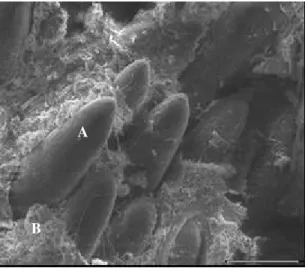
Figure 1- Simira glaziovii colleters, showing numerous colleters and secretion (100 X). A - colleter; B- secretion.
Enzyme purification
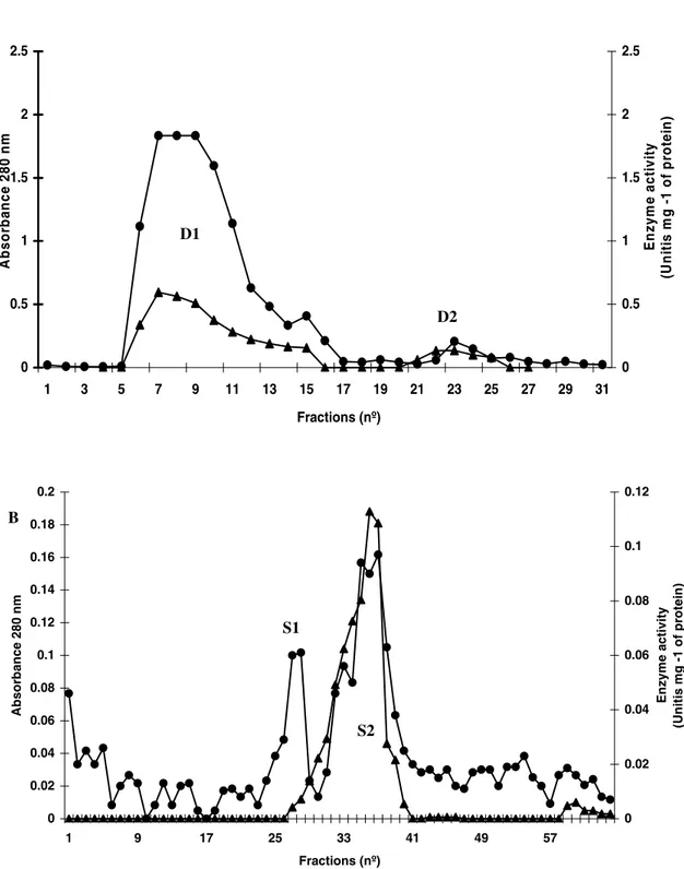
For β-1,3-glucanase purification, a DEAE-Sepharose column (2.5 × 25 cm) equilibrated with 100 mmol L-1 Tris-HCl (pH 8.0) was employed for separation of proteins from the F/0-90 fraction. Elution was initially achieved by the equilibrium buffer, when we obtained fraction D1. Bound proteins (D2) were eluted with 1 mol L-1 NaCl in the same buffer. D1 fraction (which shows high enzyme activity) was pooled and submitted to one cycle of gel filtration chromatography in Sephcryl S-100 (column of 2.3 x 60.5 cm) equilibrated and developed in 50 mmol L-1 acetate buffer (pH 8.0). Fractions enriched in β-1,3-glucanase activity were pooled and recovered after dialysis in distilled water and freeze-drying.
SDS-PAGE
A discontinuous SDS gel electrophoresis of 15% acrylamide was performed using a vertical mini-gel system (Bio-Rad Inc., USA) with a 0.75mm thickness. The gel was prepared basically according to Laemmli (1970).
Detection of ββββ-1,3-glucanase extract from SDS-PAGE
Detection of the β-1,3-glucanase activity in SDS-PAGE was made after gel electrophoresis, according to the methodology described by Trudel and Asselin (1989). At the end of the run, the gel was carefully taken from the glass plates and incubated for 24 h at 37 °C with 0.1 mol L-1
incubation, the gel was washed in 0.1 mol L-1 sodium acetate, pH 5.0, and the β-1,3-glucanase activity was immediately determined, as described in item, β-1,3-glucanase enzyme activity
RESULTS
Fig. 1 shows the scaning electron microscopy of the colleters from S. glaziovii found on the adaxial surface of stipules. This revealed the presence of numerous colleters with a large quantity of secretion may be seen.
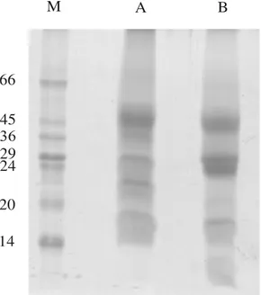
F/0-70 fraction analyzed by SDS-PAGE (Fig. 2) showed that the secretion from S. glaziovii was a mixture of proteins with molecular masses covering a range approximately from 45 to 14 kDa (Fig. 2 lane A). When treated with reducing agents, inter or intra-chain disulphide linkages for some of these proteins could be seen (Fig. 2 lane B).
To identify the presence of proteins in the secretion that were related to plant defense, different enzymatic assays were performed. Peroxidase and chitinase activity was not detected under the conditions assessed. Since the presence of the β-1,3-glucanase activity was observed (Table 1). The purification of a β-1,3-glucanase from S. glaziovii secretion was achieved through the utilization of ion exchange and size exclusion chromatographies. The 0/90 ammonium sulphate fraction was initially fractionated by DEAE-Sepharose, which demonstrated the presence of
A
exclusion chromatography in Sephacryl S–100 and separated into two new fractions denominated S1 and P2 (Fig. 3B). The analyses of β-1,3-glucanase activity demonstrated its activity mainly in the S2 fraction, although a small proportion was found
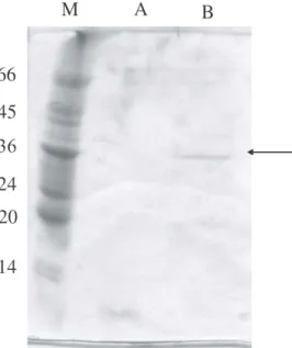
The S2 fraction presented a single band of the approximately 35 kDa. This band, when submitted to enzymatic assay, showed a high specific activity for β-1,3-glucanase.
M A B
66
45
29 24
20
14
Figure 2 - Denaturing SDS-PAGE of fraction F/0-90 obtained from secretion of S. glaziovii
colleters. A - Sample without treatment with β-mercaptanoethanol; B - sample treated with β-mercaptanoethanol; M - markers (kDa).
Table 1 - Activity of β-1,3-glucanase, chitinase and peroxidase enzymes found in the F/0-90 fraction from S. glaziovii
secretion. Enzyme activities were measured in the freeze-dried secretion recovered. Results are the means of three determinations.
Activity analysis in the crude secretion
Enzymes Activity
(Units mg-1 protein)
Standard deviation
β-1,3-glucanase 3.160 0.00972
Chitinase 0 0
Figure 3 - A- DEAE-Sepharose chromatography of the (F/0-90). The column was equilibrated with 0.1 molL-1 Tris-HCl buffer (pH 8.0) and eluted with 1 mol L-1 NaCl. Protein absorbance at 280 nm (•); β-1,3-glucanase activity (). B – Chromatography of the D1 fraction in the Sephacryl S-100 column. The column was equilibrated and eluted with 0.05 mol L-1 sodium acetate buffer, pH 5.0, at a constant flow rate of 30 mLh-1. Protein absorbance at
A
D1
D2
0 0.5 1 1.5 2 2.5
1 3 5 7 9 11 13 15 17 19 21 23 25 27 29 31
Fractions (nº)
E
n
z
y
m
e
a
c
tiv
it
y
(U
n
iti
s
m
g
-1
o
f p
ro
te
in
)
0 0.5 1 1.5 2 2.5
A
b
so
rb
an
ce 280 n
m
B
0 0.02 0.04 0.06 0.08 0.1 0.12 0.14 0.16 0.18 0.2
1 9 17 25 33 41 49 57
Fractions (nº)
Ab
so
rb
an
ce 280 nm
0 0.02 0.04 0.06 0.08 0.1 0.12
En
z
y
m
e
activity
(Uni
ti
s
mg -1
of pr
ote
in)
S1
24
20
14
Figure 4 - SDS-PAGE, under denaturing conditions, of the fractions obtained after Sephacryl S-100
chromatography. A - S1; B - S2; M - markers (kDa).
DISCUSSION
β-1,3-glucanase has been purified from many plant species, usually by multi-step procedures employing ion exchange columns followed by gel filtration and hydrophobic interaction (Young and Pegg, 1982; Kauffmann et al., 1987; Mauch et al., 1988; Balance and Sevendsen, 1988; Vogeli et al., 1988; Kurosaki et al., 1991). In this study, the presence of a β-1,3-glucanase in the secretion of the Simira glaziovii colleter was investigated. Similar techniques as used in this work have been used for the purification of β-1,3-glucanase in celery (Stephen and Rebecca, 1993), tomato seeds (Yukio and Hisashi, 2000), pepper stems (Young and Byung, 1997) and young barley leaves (Maria and Geoffrey, 1993).
The physical-chemical characteristics, found in S. glaziovii β-1,3-glucanase, were very similar to the properties of celery β-1,3-glucanase and of those of other species of plants, with a molecular mass of 35 kDa, an acidic isoelectric point (4.1) and optimum pH of 5.2-5.5 (Stephen and Rebecca, 1993; Young and Pegg, 1982; Kauffmann et al., 1987; Mauch et al., 1988).
Secretory structures, such as trichomes (Williams et al., 1982), nectaries (Arumugasamy et al., 1990) and salt and resin glands (Subramarian et al., 1989), in addition to being involved in pollination processes, osmorregulation and reduction of the perspiration in several organs, are also involved in
the defense of the plant against herbivore and pathogen attacks (Fanh, 1987; Thomas, 1991). Detection of β-1,3-glucanase exuded in the secretion of the S. glaziovii colleter suggested that this structure could be related to the defense of the plant against pathogen attacks. Plants exude a variety of substances through their aerial parts, roots and germinating seeds. Many of these substances are used by the plants to create adverse conditions or may attack the pathogen directly (Beart et al., 1985; Friend, 1991).
Similarly to the secretion of S. glaziovii, only one soluble β-1,3-glucanase exists in celery (Stephen and Rebecca, 1993), unlike in other species of plants, where different isoforms of Chitinase and
RESUMO
Uma β-1,3-glucanase foi purificada a partir da secreção de Simira glaziovii, através de vários processos cromatográficos, tendo a análise do perfil protéico acompanhado de SDS-PAGE. A enzima purificada apresentou uma massa molecular estimada de 35 kDa. O pH ótimo obtido para a enzima foi de 5,2.
ACKNOWLEDGEMENT
We acknowledge the financial support from the Brazilian agencies, CNPq and FAPERJ, and from the Universidade Estadual do Norte Fluminense-Darcy Ribeiro.
REFERENCES
Alexopoulos, C. J.; Mims, C. W. and Blackwell, W. (1996), In Introductory mycology: characteristics of fungi. 4th ed. New York: John Wiley & Sons. v. 2. pp. 26-60.
Arumugasamy, K.; Subramanian, R. B. and Inamdar, J. A. (1990), Struture, ontogeny and histochemistry of cyathial nectaries in euphorbia-heterophylla L. (euphorbiaceae). Acta Societatis Botanicorum Poloniae, 59, 3-8.
Ballance, G. M. and Svendsen, I. B. (1988), Purification and amino acid sequence determination of an endo-β -1,3-glucanase from barley. Carlsberg Research Communication, 53, 411-419.
Beart, J. E.; Lilley, T. H. and Haslam, E. (1985), Plant polyphenols secondary metabolism and chemical defense: some observations. Phytochemistry,24, 8-33. Boller, T. (1987), In plant-microbe interactions:
molecular and genetic perspectives: hydrolytic enzymes in plats disease resistance. New York: MacMillan. v. 2. pp. 385-413.
Boller, T. and Mêtraux, J. P. (1988), Extracellular localization of chitinase in cucumber. Physiology and Molecular Plant Pathology,33, 11-16.
Bradford, M. M. (1976), A rapid sensitive method for the quantification of microgram quantities of protein utilising the principle of dye binding. Biochemistry,
72, 248-254.
Caramori, S. S.; Lima, C. S. and Fernandes, K. F. (2004), Biochemical characterization of selected plant species from Brasilian savanas. Brazilian Archives of Biology and Technology, 47, 253-259.
Chye, M. L. and Cheung, K. Y. (1995), β-1,3-glucanase is highly-expressed in laticifers of Hevea brasiliensis.
Darvill, A. G. and Albersheim, P. (1984), Phytoalexins an their elicitors - a defense against microbial infection in plants. Annual Review of Plant Physiology, 35, 243-275.
Fahn, A. (1987), Extrafloral nectaries of Sambucus niger L. Annals of Botany, 60, 299-308.
Fink, W.; Liefland, M. and Mendgen, K. (1988), Chitinases and β-1,3-glucanases in the apoplastic compartment of oat leaves (Avena sativa L.). Plant Physiology, 88, 270 - 275.
Friend, J. (1991), The Biochemistry and Cell Biology of Interaction. Advances in Plant Pathology, 7, 85-129. Gomes, V. M. and Xavier-Filho, J. (1994), Biochemical
defenses of plants. Brazilian Archives of Biology and Technology, 37, 371-383.
Kauffmann, S.; Legrand, M.; Geoffroy. P. and Fritig, B. (1987), Biological function of pathogenesis-related protein: Four PR proteins of tobacco-leaves have β -1,3-glucanase activity. EMBO Journal, 6, 3209-3212. Klein, D. E.; Gomes, V. M.; Silva-Neto, S. J. and
Cunha, M. (2004), The structure of colleters in several species of simira (Rubiaceae). Annals of Botany, 94, 733-740.
Kombrik, E.; Schröder, M. and Hahlbrock, K. (1988). Several pathogenesis-related proteins in potato are β -1,3-glucanases and chitinases. Proceedings of the National Academy of Science USA., 85, 782-786. Kurosaki, U.; Tokitoh, Y. and Nishi, A. (1991),
Purification and characterization of wall-bound β -1,3-glucanase in cultured carrot cells. Plant Science, 77, 21-28.
Laemmli, U. K. (1970), Cleavage of structural proteins during the assembly of the head of bacteriophague T4. Nature, 227, 680-685.
Legrand, M.; Kauffmann, S.; Geoffroy. P. and Fritig, B. (1987), Biological function of pathogenesis-related protein is chitinases. Proceedings of the National Academy of Science USA, 64, 6750-6754.
Leon, I. C.; Alpeeva, I. S.; Shubar, T. A.; Galaev, I. Y.; Csoregi, E. and Sakhar, O. V. I. Y. (2002), Purification and substrate specificity of peroxidase from sweet potato tuber. Plant Science, 663, 1111-1119.
Hrmova, M. and Fincher, G. B. (1993), Purification and properties of three (1-3)-β-D-glucanase isoenzymes from young leaves of barley (Hordeum vulgare).
Biochemical Journal, 289, 453-461
Mauch, F.; Hadwiger, L. A. and Boller, T. (1988), Antifungal hydrolases in pea tissue. 1. Purification and characterization of two chitinases and β -1,3-glucanase differentially regulated during development and in response to fungal infection. Plant Physiology,
87, 325-333.
Oliveira, A. S.; Xavier-Filho, J. and Sales, M. P. (2003), Cysteine proteinases and cystatins. Brazilian Archives of Biology and Technology, 46, 91-104. Pascholati, S. F. and Leite, B. (1994), Mecanismos
bioquímicos de resistência às doenças. Revisão Anual de Patologia de Plantas, 2, 1-51.
Pegg, G. F. (1977), Glucanohydrolases of higher plants: A possible defense mechanism against parasitic fungi, in Cell wall Biochemistry Related to Specificity in Host Plant-Pathogen interaction. (Osio: Universitetsforiage). pp. 371-382.
Pegg, G. F. and Young, D. H. (1981), Changes in glycosidase activity and their relationship to fungal colonization during infection of tomato by
Verticillium albo-atrum. Physiology and Plant Pathology, 19, 371-382.
Robbrecht, E. (1988), Tropical woody Rubiaceae. Characteristic features and progressions. Contributions to a new subfamilial classification.
Opera Botanica Belgica, 1, 1-271.
Subramanian, R. B.; Murugan, V.; Mohan, J. S. S. and Inamdar, J. A. (1989), Optical microscopic studies on the structure and secretion of resin glands in some Apocynacea. Proceedings of the Indian Academy of Sciences-Plant Science, 99, 423-429.
Stephen, L. K. and Rebecca, G. (1993), Affinity purification and characterization of a β-1,3-glucanase from celery. Plant Science, 93, 31-39.
Thomas, V. (1991), Review, Structural, functional and phylogenetic aspects of the colleter. Journal of Botany, 68, 278-305.
Trudel, J. and Asselin, A. (1989), Detection of chitinase activity after polyacrylamide gel electrophoresis.
Analytical Biochemistry., 178, 362-366.
Vögeli, V.; Meins, P. and Boller, T. (1988), Co-ordinated regulation of chitinase and β-1,3-glucanase in been leaves. Planta, 174, 364-372.
Vögeli-Lange, R.; Fründt, C.; Hart, C. M.; Beffa, R.; Nagy, F. and Meins Jr., F. (1994), Evidence for a role of β-1,3-glucanase in dicot seed germination. Plant Journal, 5, 273-278.
The genesis of form in Oleander (Nerium oleande L.).
Australian Journal of Botany, 30, 677-687.
Young, D. H. and Pegg, G. F. (1982), The action of tomato and Verticillium albo-atrum glycosidases on the hyphal wall of V. albo-atrum. Physiology and Plant Pathology, 21, 411-423.
Young, J. K. and Byung, K. H. (1997), Isolation of a basic 34-kiloDaltons β-1,3-glucanase with inhibitory activity against Phytophthora capsici from pepper stems. Physiology and Molecular Plant Pathology,
50, 103-115.
Yun, D. J.; Bressan, R. A. and Hasegawa, P. M. (1987), Plant antifungal proteins, in: J. Janick (Ed),
Horticultural Reviews, Willey, New York, 14, 39-88. Morohashi, Y. and Matsushima, H. (2000),
Development of ß-1,3-glucanase activity in germinated tomato seeds. Journal of Experimental Botany, 51, 1381-1387.