Periodontics
366 Braz Oral Res., (São Paulo) 2012 Jul-Aug;26(4):366-72
Hilana Paula Carillo Artese(a) Celso Oliveira de Sousa(a) Maria Cynésia Medeiros de Barros Torres(a)
Carina Maciel Silva-Boghossian(b) Ana Paula Vieira Colombo(b)
(a) Department of Dental Clinic, Division of Graduate Periodontics, School of Dentistry, Federal University of Rio de Janeiro, Rio de Janeiro, RJ, Brazil.
(b) Department of Medical Microbiology, Institute of Microbiology, Federal University of Rio de Janeiro, Rio de Janeiro, RJ, Brazil.
Corresponding author:
Hilana Paula Carillo Artese E-mail: [email protected]
Received for publication on Jan 31, 2012 Accepted for publication on May 08, 2012
Effect of non-surgical periodontal
treatment on the subgingival microbiota
of patients with chronic kidney disease
Abstract: This study investigated the effect of non-surgical periodontal therapy on the composition of subgingival microbiota of patients with chronic kidney disease (CKD). Sixteen CKD pre-dialysis individuals (CKD) and 14 individuals without clinical evidence of kidney disease (C) presenting chronic periodontitis were treated by scaling and root plan-ing. Subgingival samples were collected from each patient and analyzed for their composition by checkerboard at baseline and 3 months post-therapy. Signiicant differences between groups at baseline were sought by the Mann-Whitney and χ2 tests. Changes over time were examined by
the Wilcoxon test. At baseline, the CKD group had signiicantly lower counts of E. faecalis compared to the C group (p < 0.05). After treat-ment, the levels of a greater number of species were reduced in the C group. Higher levels of A. israelii, C. rectus, F. periodonticum, P. micra, P. nigrescens, T. forsythia, N. mucosa, and S. anginosus (p < 0.05) were found in the CKD group compared to the C group. Also, non-responsive sites in CKD individuals harbored signiicantly higher levels of patho-genic species (T. forsythia, P. gingivalis, T. denticola, Fusobacterium
spp., D. pneumosintes, E. faecalis and S. aureus; p < 0.05) than sites that responded to therapy, as well as non-responsive sites in the C group. The periodontitis-associated subgingival microbiota of CKD and systemical-ly healthy individuals was similar in composition. However, high levels of pathogenic species persisted in the subgingival microbiota of patients with CKD after treatment.
Descriptors: Periodontitis; Dental Scaling; Root Planing; Microbiology; Kidney Failure, Chronic.
Introduction
Chronic kidney disease (CKD) is a public health problem worldwide,1
and its prevalence has been increasing.2 CKD is deined as glomerular
iltration rate (GFR) reduction or kidney damage, relected as abnor-mal urine sediment or abnorabnor-malities in the renal anatomy.1 Periodontitis
has emerged as a non-traditional risk factor and a prediction model for CKD.3 The link between periodontal disease and CKD may be due to
concomitant infection and inlammation.4 The periodontal inlammatory
state may add to the chronic inlammation present in CKD,5 decreasing
renal function.6 Periodontal therapy reduces inlammation and improves
endothelial function,7 leading to more effective kidney microcirculation
and iltration. CKD patients with periodontitis can also be effectively treated by periodontal mechanical therapy.8,9 In a previous study, we demonstrated that
non-surgical mechanical treatment led to periodon-tal clinical improvement, as well as an improvement in GFR levels.8 However, limited data are available
regarding the subgingival microbial proile of pre-dialysis CKD patients and the effect of therapy on the oral microbiota. Therefore, the aim of this study was to investigate the effect of mechanical non-sur-gical periodontal therapy on the subgingival micro-biota of CKD patients with chronic periodontitis.
Methodology
The present study was a two-arm monocentric clinical trial with a 3-month follow-up period. The study was conducted according to the principles outlined in the Declaration of Helsinki on experi-mentation involving human individuals, and was approved by the Human Research Committee/Insti-tute for Community Health Studies at the Federal University of Rio de Janeiro, protocol 113/2006. To be enrolled, patients were informed about the na-ture of the proposed treatment, its risks and bene-its, and signed informed consent forms. The sample population has been described in a previous paper.8
From January to November 2006, 16 pre-dialysis (CKD) patients were recruited from the Nephrol-ogy Division of Clementino Fraga Filho University Hospital, and 14 systemically healthy individuals (C) were recruited from the General Medicine de-partment of the same hospital. This study used the GFR to deine the groups, according to the renal function stages proposed by the National Kidney Foundation.1 The CKD group consisted of patients
with clinical diagnosis of renal failure, having GFR between 89 and 15 mL/min, and receiving conserva-tive treatment (pre-dialysis). The C group consisted of patients seeking general medical care without signs and symptoms of renal disease and having GFR > 90 mL/min. All study participants were be-tween 35 and 76 years of age, presented at least 15 teeth, and were diagnosed as having chronic peri-odontitis, i.e., the presence of ≥ 4 sites in 3 different teeth with clinical attachment level (CAL) ≥ 4 mm and bleeding on probing (BOP). A detailed medical
history was obtained from all participants at base-line, as well as information about age, gender, eth-nicity, and smoking habit. Exclusion criteria includ-ed HIV infection, pregnancy, lupus erythematosus, rheumatoid arthritis, need for antibiotic prophylaxis for periodontal procedures, periodontal treatment, and/or use of antibiotics in the preceding 6 months. A full-mouth periodontal clinical examination was performed at 6 sites per tooth (excluding third mo-lars) by one calibrated examiner (C.O.S.) at both vis-its. Periodontal assessment included probing depth (PD) and CAL, measured to the nearest millimeter with a periodontal probe (UNC-15, Hu-Friedy, Chi-cago, USA), the presence or absence of BOP, supra-gingival bioilm (VP), supra-gingival marginal bleeding (GB), and suppuration (SUP). Intra-class correla-tion coeficients > 0.90 were obtained for PD and CAL. The periodontal treatment was performed by a single experienced periodontist (H.P.C.A.). Both groups received non-surgical periodontal therapy consisting of oral hygiene instructions, and supra- and subgingival scaling and root planing with hand instruments (Gracey curettes; Hu-Friedy) under local anaesthesia. One sextant was instrumented at each dental visit (1- to 2-hour session), and the therapy was completed within 6-8 weeks.
Microbiological assessment
in-368 Braz Oral Res., (São Paulo) 2012 Jul-Aug;26(4):366-72
dividual sterile Gracey curettes (Hu-Friedy), and were placed in individual tubes. Bacterial cells un-derwent lysis, and denatured DNA was ixed on a nylon membrane (GE Healthcare Life Science, São Paulo, Brazil) using the checkerboard slot blot de-vice (Minislot 30, Immunetics, Cambridge, USA). Digoxigenin-labeled (Roche Applied Science, São Paulo, Brazil) whole-genomic DNA probes were hy-bridized at 90° to the lanes of the plaque samples in the slot blot device (Miniblotter 45, Immunetics). After hybridization, the membranes were washed at high stringency, bound probes were detected us-ing phosphatase-conjugated antibody to digoxigen-in (Roche Applied Science), and luorescence was captured by an imaging system (Storm 860, GE Healthcare Life Science). Signals were evaluated vi-sually by comparison with the standards at 105 and
106 bacterial cells for the test species on the same
membrane. They were recorded as:
• 0, not detected;
• 1, < 105 cells;
• 2, approximately 105 cells;
• 3, 105 to 106 cells;
• 4, approximately 106 cells; and
• 5, > 106 cells.
The sensitivity of the assay was adjusted to per-mit the detection of 104 cells of a given species by
adjustment of the concentration of each DNA probe. The microbiological test was read by a single cali-brated examiner (C.M.S.B.).
Statistical analysis
Statistical analyses were performed with SPSS software (SPSS, release 17.0, Chicago, USA). Micro-bial data were presented as mean levels (× 105
bacte-rial cells). The levels (scores 0 to 5) of each species in a sample were converted to absolute numbers, and the mean counts were computed for each pa-tient and averaged within the groups. Moreover, the mean counts of bacteria in sites that did not improve or presented disease progression, i.e., PD increase and/or attachment loss at 3 months post-therapy, were compared to sites that improved (PD reduction and CAL gain) after treatment. Signiicant differ-ences in demographic, clinical, and microbiological parameters between groups were determined by the Mann-Whitney and χ2 tests. Differences in clinical
and microbiological changes between groups over time were evaluated by the Wilcoxon Signed-Rank test. The level of signiicance for all analysis was 5%.
Results
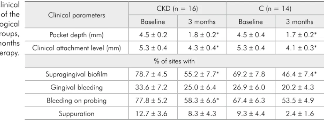
The majority of individuals in both groups were females (9 in the CKD and 10 in the C group), white, and non-smokers; however, no signiicant differences for these parameters were observed be-tween groups. In contrast, those in the CKD group were signiicantly older (58.8 ± 10.8 years) than those in the C group (52.0 ± 3.3 years; p = 0.014, Mann-Whitney test). Regarding the clinical features of the sampled sites (Table 1), there were no signii-cant differences between groups for all periodontal
Clinical parameters CKD (n = 16) C (n = 14)
Baseline 3 months Baseline 3 months Pocket depth (mm) 4.5 ± 0.2 1.8 ± 0.2* 4.5 ± 0.4 1.7 ± 0.2* Clinical attachment level (mm) 5.3 ± 0.4 4.3 ± 0.4* 5.3 ± 0.4 4.1 ± 0.3*
% of sites with
Supragingival biofilm 78.7 ± 4.5 55.2 ± 7.7* 69.2 ± 7.8 46.4 ± 7.4* Gingival bleeding 33.6 ± 7.2 25.0 ± 6.4 26.9 ± 6.0 20.2 ± 4.3 Bleeding on probing 77.8 ± 5.2 58.3 ± 6.6* 67.4 ± 6.3 53.5 ± 4.9 Suppuration 12.7 ± 3.6 8.3 ± 4.3 9.3 ± 4.4 2.4 ± 1.6 CKD: chronic kidney disease pre-dialysis individuals; C: systemically healthy individuals; *Refers to significant differences between baseline and 3 months post-therapy within the groups (p < 0.05, Wilcoxon test). Table 1 - Periodontal clinical
parameters at baseline and at 3 months post-therapy (p > 0.05, Mann-Whitney test). Both groups showed signiicant clinical improvement in those sites for PD, CAL, and VP after treatment (p < 0.05, Wil-coxon test). The CKD group also showed signiicant improvement in BOP. The subgingival microbial proiles of both groups at baseline and 3 months af-ter therapy are depicted in Figure 1. In general, the C group showed absolute higher levels of many test-ed species, especially members of the green and or-ange complexes. However, only the species Entero-coccus faecalis was detected in signiicantly higher mean counts in C compared to CKD individuals at baseline (p = 0.025; Mann-Whitney test). Levels of most species decreased signiicantly after treatment in the C group, whereas, in the CKD group, signii-cant reductions were observed only for the species
Actinomyces gerencseriae, Actinomyces oris, A.
ac-tinomycetemcomitans, Fusobacterium nucleatum polymorphum, Streptococcus constellatus, Lepto-trichia buccalis, Dialister pneumosintes, Enterics, and Staphylococcus aureus. Moreover, a signiicant increase in mean counts was observed for Prevotella nigrescens in the CKD group (p < 0.05, Wilcoxon test). At 3 months post-therapy, signiicantly higher levels of Actinomyces israelii, Campylobacter rec-tus, Fusobacterium periodonticum, Parvimonas micra, Prevotella nigrescens, Tannerella forsythia, Neisseria mucosa, and Streptococcus anginosus
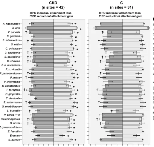
were found in the CKD group compared with the C group (p < 0.05, Mann-Whitney test). The sub-gingival microbiota of sites that did or did not show clinical improvement after treatment were also ana-lyzed. A total of 42 sites (35.6%) from 13 members of the CKD group and 31 sites (28.2%) from 12 members of the C group presented increases in PD
Figure 1 - Mean levels (x 105 cells) of bacterial species in the two clinical groups (CKD: chronic kidney disease pre-dialysis individuals; C: systemically healthy controls) at baseline and 3 months post-therapy.
A.a.: Aggregatibacter actinomycetemcomitans; *refers to significant difference between groups at baseline (p = 0.025, Mann-Whitney test); †refers to significant differences within groups over time (p < 0.05, Wilcoxon test); ‡refers to significant differences between groups at 3 months post-therapy (p < 0.05, Mann-Whitney test).
Figure 1
0 2 4 6 8 10 12
A. gerencseriae A. israelii A. naeslundii I A. oris A. odontolyticus V. parvula S. gordonii S. intermedius S. mitis S. oralis S. sanguinis A.a. C. gingivalis C. ochracea C. sputigena E. corrodens C. rectus C. showae E. nodatum F. n. nucleatum F. n. polymorphum F. n. vicentii F. periodonticum P. micra P. intermedia P. nigrescens S. constellatus T. forsythia P. gingivalis T. denticola E. saburreum G. morbilorum L. buccalis N. mucosa P. acnes I + II P. melaninogenica S. anginosus S. noxia A. baumannii D. pneumosintes E. faecalis Enterics P. aeruginosa S. aureus
0 2 4 6 8 10 12
A. gerencseriae A. israelii A. naeslundii I A. oris A. odontolyticus V. parvula S. gordonii S. intermedius S. mitis S. oralis S. sanguinis A. actinomycetemcomitans C. gingivalis C. ochracea C. sputigena E. corrodens C. rectus C. showae E. nodatum F. n. nucleatum F. n. polymorphum F. n. vicentii F. periodonticum P. micra P. intermedia P. nigrescens S. constellatus T. forsythia P. gingivalis T. denticola E. saburreum G. morbilorum L. buccalis N. mucosa P. acnes I + II P. melaninogenica S. anginosus S. noxia A. baumannii D. pneumosintes E. faecalis Enterics P. aeruginosa S. aureus Ba se line 3 m on ths CKD (n = 16)
Mean counts x 105 cells
C (n = 14)
Mean counts x 105 cells
*
‡
‡
Baseline 3 months
370 Braz Oral Res., (São Paulo) 2012 Jul-Aug;26(4):366-72
and/or loss of clinical attachment in spite of treat-ment. The number of non-respondent sites did not differ between groups (p > 0.05, χ2 test; data not
shown). Figure 2 shows the microbial composition of successfully treated and non-responsive sites in both groups at 3 months post-therapy. Those in the CKD group had signiicantly higher levels of sev-eral species in sites that did not respond to therapy compared with sites that did, particularly the patho-genic species T. forsythia, Porphyromonas gingi-valis, Treponema denticola, Fusobacterium spp., and non-oral species including D. pneumosintes,
E. faecalis, and S. aureus (p < 0.05, Wilcoxon test). Conversely, no signiicant differences regarding the levels of all tested species between sites that did or did not respond to therapy were observed in the C group (Figure 2). Comparisons of non-responsive sites between groups demonstrated signiicantly lower levels of Veillonella parvula, Streptococcus intermedius, Capnocytophaga sputigena, Eikenella corrodens, Campylobacter showae, F.
periodon-ticum, P. micra, T. forsythia, Eubacterium sabur-reum, L. buccalis, Prevotella melaninogenica, and
Selenomonas noxia in individuals in the C group than in those in the CKD group (p < 0.05, Mann-Whitney test).
Discussion
Limited data are available regarding the compo-sition of the periodontal microbiota of individuals with CKD, as well as the impact of mechanical peri-odontal therapy on their microbiota. In this investi-gation, we showed that the periodontal microbiota of persons with chronic periodontitis and CKD and that of systemically healthy individuals was similar in composition, except that E. faecalis was found in higher counts in the C group. Although not con-sidered a periodontal pathogen, E. faecalis produc-es various virulence factors that may be related to periodontal inlammation, tissue destruction, and neutrophil impairment.11 Moreover, this species is a
bioilm-forming bacterium, capable of adhering and
Figure 2 - Bar chart of bacterial species that differed significantly in mean levels (in log 10 ± SEM) at 3 months post-therapy between sites that responded (pocket depth reduction/attachment gain) or not (pocket depth increase/attachment loss) to periodontal therapy in the two clinical groups (CKD: chronic kidney disease pre-dialysis individuals; C: systemically healthy controls).
*Refers to significant differences between sites that responded or not to therapy within the CKD group (p < 0.05, Wilcoxon test); †refers to significant differences between groups regarding sites that did not respond to therapy (p < 0.05, Mann-Whitney test). Figure 2
CKD
(n sites = 42) (n sites = 31)C
Mean counts in log 10 Mean counts in log 10
* * * * * * * **
* *
* * * *
* *
**
*
* *
* * **
* *
* †
† †
†
†
†
†
† †
† †
† †
†
† † †
† †
†
†
†
† †
PD increase/ attachment loss
invading soft tissues, which enables this organism to co-aggregate with many oral species.12,13E. faecalis
may also enhance pathogenicity in mixed infections with anaerobic bacteria.14 Previous studies by our
group have shown an association between E. faeca-lis and chronic periodontitis.15,16 However, it is
difi-cult to explain why this species was detected in high-er levels in the phigh-eriodontitis bioilm of systemically healthy individuals compared with those with CKD. Due to the immunosuppression usually present in CKD patients,17 as well as the common association
of E. faecalis and kidney infections, one would ex-pect to ind this species in higher levels in the CKD group. Changes in the bacterial levels from baseline to 3 months after therapy showed that a larger num-ber of species diminished signiicantly in the C com-pared with the CKD group. In addition, one species of the orange complex, P. nigrescens, showed a sig-niicant increase in the CKD group after treatment. However, few species differed between groups at 3 months, including species of the red and orange complexes, which were found in higher levels in the CKD group. Further analyses comparing responsive and non-responsive sites showed that individuals with CKD presented higher levels of many species, such as T. forsythia, P. gingivalis, T. denticola, Fu-sobacterium sp., D. pneumosintes, E. faecalis, and
S. aureus, in sites with PD increase and attachment loss after therapy, whereas no differences between those sites were observed for those in the C group. Moreover, non-responsive sites in the CKD group presented higher counts of V. parvula, S. intermedi-us, C. sputigena, E. corrodens, C. showae, F. peri-odonticum, P. micra, T. forsythia, E. saburreum, L. buccalis, P. melaninogenica, and S. noxia than non-responsive sites in the C group. Species of the red complex and D. pneumosintes have been recently associated with treatment failure or periodontal at-tachment loss, as well as non-responsive sites in
gen-eralized aggressive periodontitis.18,19 The persistence
of high levels of many pathogenic species in CKD patients compared with systemically healthy indi-viduals after treatment could be related to the im-munocompromised state associated with uremia in CKD patients.20 Conceivably, the uremia can cause
an indirect effect on the microbiota by modifying the host inlammatory or immune response, and by changing the host-parasite balance, favoring a rapid re-colonization by pathogenic species after mechan-ical therapy. One should consider, however, that this was a short-term post-therapy study consisting of a small sample population. Further longitudinal investigations are needed to evaluate how the per-sistence of high levels of periodontal pathogens will affect the eficacy of mechanical periodontal treat-ment of individuals with both CKD and chronic periodontitis.
Conclusions
The microbial composition of the periodonti-tis-associated subgingival bioilm of individuals with CKD was very similar to that of systemically healthy individuals. Nevertheless, fewer bacterial species were affected by mechanical periodontal therapy in the CKD than in the C group. In addi-tion, pathogenic species persisted in high levels in non-responsive sites of CKD individuals compared with C patients.
Acknowledgments
This study was supported in part by the Nation-al Council for Scientiic and TechnologicNation-al Develop-ment (CNPq), the Coordination of ImproveDevelop-ment of Higher Education Personnel (CAPES), Brasilia, Bra-zil; and by the Foundation for Research Financial Support in the State of Rio de Janeiro (FAPERJ), Rio de Janeiro, Brazil.
References
1. National Kidney Foundation. K/DOQI Clinical Practice Guidelines for Chronic Kidney Disease: Evaluation, classi-fication, and stratification. Am J Kidney Dis. 2002 Feb;39(2 Suppl 1):S1-S266.
372 Braz Oral Res., (São Paulo) 2012 Jul-Aug;26(4):366-72 3. Fisher MA, Taylor GW. A prediction model for chronic
kidney disease includes periodontal disease. J Periodontol. 2009 Jan;80(1):16-23.
4. Kshirsagar AV, Craig RG, Beck JD, Moss K, Offenbacher S, Kotanko P, et al. Severe periodontitis is associated with low serum albumin among patients on maintenance hemodialysis therapy. Clin J Am Soc Nephrol. 2007 Mar;2(2):239-44. 5. D’Aiuto F, Nibali L, Parkar M, Patel K, Suvan J, Donos N.
Oxidative stress, systemic inflammation, and severe periodon-titis. J Dent Res. 2010 Nov;89(11):1241-6.
6. Fried L, Solomon C, Shlipak M, Seliger S, Stehman-Breen C, Bleyer AJ, et al. Inflammatory and prothrombotic markers and the progression of renal disease in elderly individuals. J Am Soc Nephrol. 2004 Dec;15(12):3184-91.
7. Seinost G, Wimmer G, Skerget M, Thaller E, Brodmann M, Gasser R, et al. Periodontal treatment improves endothelial dysfunction in patients with severe periodontitis. Am Heart J. 2005 Jun;149(6):1050-4.
8. Artese HP, Sousa CO, Luiz RR, Sansone C, Torres MC. Ef-fect of non-surgical periodontal treatment on chronic kidney disease patients. Braz Oral Res. 2010 Oct-Dec;24(4):449-54. 9. Graziani F, Cei S, La Ferla F, Vano M, Gabriele M, Tonetti M.
Effects of non-surgical periodontal therapy on the glomerular filtration rate of the kidney: an exploratory trial. J Clin Peri-odontol. 2010 Jul;37(7):638-43.
10. Socransky SS, Smith C, Martin L, Paster BJ, Dewhirst FE, Levin AE. “Checkerboard” DNA-DNA hybridization. Bio-techniques. 1994 Oct;17(4):788-92.
11. Jett BD, Huycke MM, Gilmore MS. Virulence of enterococci. Clin Microbiol Rev. 1994 Oct;7(4):462-78.
12. Al-Ahmad A, Müller N, Wiedmann-Al-Ahmad M, Sava I, Hübner J, Follo M, et al. Endodontic and salivary isolates of Enterococcus faecalis integrate into biofilm from human
sali-vary bacteria cultivated in vitro. J Endod. 2009 Jul;35(7):986-91.
13. Johnson EM, Flannagan SE, Sedgley CM. Coaggregation in-teractions between oral and endodontic Enterococcus faecalis and bacterial species isolated from persistent apical periodon-titis. J Endod. 2006 Oct;32(10):946-50.
14. Brook I. Effect of Streptococcus faecalis on the growth of Bacteroides species and anaerobic cocci in mixed infections. Surgery. 1988 Jan;103(1):107-10.
15. Souto R, Colombo AP. Prevalence of Enterococcus faecalis in subgingival biofilm and saliva of subjects with chronic periodontal infection. Arch Oral Biol. 2008 Feb;53(2):155-60. 16. Da Silva-Boghossian CM, Souto RM, Luiz RR, Colombo AP.
Association of red complex, A. actinomycetemcomitans and non-oral bacteria with periodontal diseases. Arch Oral Biol. 2011 Sep;56(9):899-906.
17. Kaysen GA. The microinflammatory state in uremia: causes and potential consequences. J Am Soc Nephro. 2001 Jul;12(7):1549-57.
18. Colombo AP, Boches SK, Cotton SL, Goodson JM, Kent R, Haffajee AD, et al. Comparisons of subgingival microbial profiles of refractory periodontitis, severe periodontitis, and periodontal health using the human oral microbe identifica-tion microarray. J Periodontol. 2009 Sep;80(9):1421-32. 19. Heller D, Varela VM, Silva-Senem MX, Torres MC,
Feres-Filho EJ, Colombo AP. Impact of systemic antimicrobials combined with anti-infective mechanical debridement on the microbiota of generalized aggressive periodontitis: a 6-month RCT. J Clin Periodontol. 2011 Apr;38(4):355-64.