EVALUATION OF THE RESPIRATORY
FUNCTION IN MYASTHENIA GRAVIS
A N IMPORTANT TOOL FOR CLINICAL FEATURE AND DIAGNOSIS OF THE DISEASE
PAULO A. P. SARAIVA*, JOSÉ LAMARTINE DE ASSIS**, PAULO E. MARCHIORI**
ABSTRACT - Myasthenic gravis may affect both inspiratory and expiratory muscles. Respiratory involvement occurred in almost all patients with myasthenia gravis in all clinical forms of the disease: 332 lung function tests done in 324 myasthenic patients without respiratory symptoms (age 34.6 ± 18.3 years) were examined. Lung volumes analysis showed that all the patients of both sexes with generalized or ocular myasthenia gravis showed "myasthenic pattern". Male patients with "ocular" form only presented the "myasthenic pattern" with lung impairment and had, from the lung function point of view, a more benign behaviour. Female patients with the "ocular" form exhibited a behaviour of respiratory variables similar to that of the generalized form. It was not observed modification of the variables that suggested obstruction of the higher airways. The "myasthenic pattern" was rarely observed in other neuromuscular diseases, except in patients with laryngeal stenosis.
KEY WORDS: myasthenia gravis, lung function tests, myasthenic pattern.
Avaliação da função respiratória na miastenia gravis: importância na caracterização clínica e no diagnóstico da doença
RESUMO - O comprometimento respiratório é fator limitante na evolução clinica da miastenia gravis (MG) e as formas clínicas mais graves apresentavam acometimento bulbar e respiratório. Para avaliar a reserva respiratória foram examinados em 324 pacientes com MG (forma ocular 62, generalizada 246 e timomatosa 16) as seguintes variáveis da prova de função pulmonar (PFP): capacidade vital forçada (FVC); volume onde o fluxo expiratório é igual a 1 litro por segundo (VF=1); volume expiratório forçado no primeiro segundo (FEV1); fluxo expiratório forçado medido entre 0,2 e 1,2 litros (FEF); fluxo médio expiratório forçado, medido entre 25 e 75% da FVC (FMF); intervalo de tempo entre 25 e 75% da FVC (FMFT); tempo médio de trânsito na expiração forçada (MTT); capacidade pulmonar total (TLC); volume residual (RV); curva fluxo-volume para pesquisa do "padrão miastênico". A análise estatística realizada foi: "t pareado" entre paciente e seu padrão e "t não pareado" entre grupos. Conclusões: Todos os pacientes apresentaram o padrão miastênico e esta alteração da curva fluxo-¬ volume sugeriu disfunção dos músculos da laringe. Nos pacientes com formas clinicamente localizadas as PFP também se mostraram alteradas revelando a generalização da sintomatologia mais frequentemente no sexo feminino. Não foi observada modificação das variáveis que indicam obstrução das vias aéreas decorrente do uso de anticolinesterásicos no tratamento da MG nem aumento de incidência de asma brônquica com o uso de drogas anticolinesterásicas.
PALAVRAS-CHAVE: miastenia grave, provas funcionais respiratórias, padrão miastênico.
Acquired myasthenia gravis (MG) is a disease of neuromuscular transmission in which antibodies to the nicotinic acetylcholine receptor (AAChR) play an important role in the
Laboratory of Respiratory Function of the Orthopedic Department and Department of Neurology, Hospital das Clínicas, São Paulo University Medical Scool (FMUSP): *Chief of Pulmonary Test of the Orthopedic Department; **Associated Professor, Department of Neurology. Aceite: 19-juIho-1996.
pathogenesis'-4,8
. The clinical forms are: severe, which patients have respiratory involvement; accentuated, patients with bulbar symptoms without respiratory involvement; moderate, with only a motor dysfunction. Further, there are systemic or localized (ocular) forms4 1 1 1 5
. Diagnosis is based upon the clinical findings, the electromyography (EMG) and on the serum levels of A A C h R9 1 0 1 4 1 9
. The seric levels of AAChR detectable in 68% of our myasthenic patients are important for diagnosis and responsible for the loss of the endplate acetylcholine receptor6 1 2
. Unusually low levels of false positive serum concentration of AAChR were also found in other diseases. Furthermore, there is no specific , definitive and consensual diagnostic test for MG. Myasthenia gravis may affect both inspiratory and expiratory muscles20
.
Aiming to study the impairement of other muscles in the localized or systemic forms of myasthenic patients lacking respiratory symptoms, an evaluation of lung functions was undertaken. Lung function tests (LFT) assure the concrete determination of the respiratory functional reserve through the analysis of different respiratory variables2
-3 -7
.
PATIENTS AND METHODS
Three-hundred-thirty-two LFT of MG patients, confirmed by EMG and serum concentration of AAChR measuring the flow-volume curve were analysed and 324 of these patients were chosen as considered clinically stable, from a ventilatory point of view. Of those, 62 patients had a predominantly ocular form (POF), 27 were male (age 39±19 years) and 35 female (age 33±18 years); 246 had the generalized form, 68 were male (age 38±17 years) and 178 female (age 32±14 years); 16 had the thymomatous form, 8 male (age 38±26 years) and 8 female (age 44±21 years) (Table 1). Three patients with oculopharyngeal dystrophy, 2 with inflammatory myositis, 2 with chronic demyelinating polyneuropathy, one with pseudomyasthenic syndrome, and one with upper stenosis of trachea were also evaluated.
To plot the curves a vitalograph spyrometer was used and to analyse the shape of the curves the Hewlett Packard electronic Vertek spyrograph was used.
75% of FVC (FMF); 6. Time lapse between 25 and 75% of FVC (FMFT); 7. Average transit time of expiration (MTT); 8. Total lung capacity (TLC); 9. Residual volume (RV); 10. "Myasthenic pattern".
The airways closure volume (CIV) has been used for many years - demanding expensive and specialized equipment and also a major participation of the patients, not guaranteed in many instances. It was observed that airway closure capacity was more reliable, although patient and equipment requirements were the same. We noted that they might undergo a different reference: instead of the end of FVC, not always stable, the beginning of the forced expiratory curve, and thus define a point called 'T'-where the expiratory flow is equal to one which presents a close correlation to function of total lung capacity (TLC) and airways closure capacity (CIC). The volume corresponding to point "J" was designated VF=1 with the same defining strenght of CIC, however much easier to measure and not requiring sophisticated and expensive equipment.
Statistical analysis used was: a) descriptive statistics; b) paired Student t test between the values of each different variable and its standard; c) unpaired Student t test between the different clinical forms. The number Cruncher Statistical System was used.
RESULTS
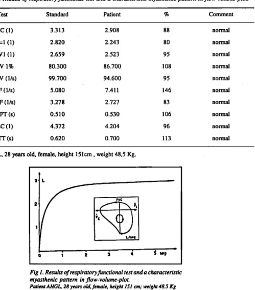
Table 1 shows the distribution of the patients. Table 2 shows the distribution of the diverse clinical forms according to sex, number of studied cases and presence or absence of really significant differences (RSD). Table 3 shows the results of the studied variables. Table 4 shows the results of the lung test of MG patients. Table 5 shows a respiratory functional proof in a chronic demyelinating polyneuropathy (CIDP). Figure 1 shows the characteristic flow-volume loop in this patient with MG. Figure 2 shows the flow-volume in a patient with CIDP.
DISCUSSION
From the respiratory involvement depend on severity of MG5
Myasthenic Pattern
The typical spirometric pattern in patients with respiratory muscles weakness is a restrictive ventilatory defect with fairly well-preserved forced expiratory flow-rates16
. Among all variables of respiratory function evaluated we observed that the myasthenic pattern is characterized by the modification of the flow-volume curve which appears flattened, both at inspiration, resembling, at calm breathing, more to a rectangle than to a lozenge, and even more evident at forced movements. These modifications were also observed in some cases of neurologic disease where the motricity of the pharynx and of the larynx is impaired. This pattern is similar to the changes found in patients with larynx, trachea and/or large bronchi stenosis. Its finding in MG suggested an eventual fatigue of the laryngeal muscles, with reduction of its useful gauge at the vocal chords level.
Differences between clinical forms and sex
The ocular form of MG has peculiar clinical, antigenic and therapeutic characteristics different from those of the systemic f o r m1 3
"1 8
. Although frequently reported the authors of this paper question this characterization, as RSD were found: in myasthenic pattern and, in females, also of the FVC, VF=1 and RV, thus suggesting that in the ocular form other muscle groups also undergo changes.
Male patients with "ocular" form only presented the "myasthenic pattern" with lung impairment and had, from functional respiratory test (FRT) point view, a more benign clinical finding. Female patients with the "ocular" form exhibit a behaviour of the respiratory variables similar to that of the systemic form. Male patients with "ocular" exhibit a different behaviour pattern form that of female patients.
The bronchoconstriction effect of anticholinesterasic agent
The anticholinesterasic agent was associated with an increase in airway resistance by muscarinic stimulation of bronchial smooth muscle by acetylcholine.
The acute changes after anticholinesterasic therapy are: improved global respiratory muscle function (increased PImax and PEmax), increased FRC because of greater respiratory muscle tone, increased static compliance, increased maximal transdiaphragmatic pressure, minimal changes in lung volume, and improved ventilatory response to hypercapnia
It is known that anticholinesterase agents may trigger by their vagal action a greater resistance of the airways to the gas flow, particularly in asthmatic subjects. It should be expected that in MG this would be more frequent with a more evident modification of the VF= 1, FEV1, FEF, FMF, FMFT, MTT variables. However, there is no such evidence. The incidence of asthma was not higher than that found in the overall population cared for at the LFL-IOT.
CONCLUSION
All clinically stable myasthenic patients, without clear respiratory symptoms presented the so called "myasthenic pattern". A change in the flow-volume curve which characterizes "myasthenic pattern", observed in the generalized forms as well as in the localized (ocular) form, suggests malfunction of other muscle groups. The observed modifications reveal persistent and early impairment of the laryngeal muscles.
A flow-volume curve of myasthenic patients disclosed a pattern resembling that of upper airways obstruction, thus suggesting that there is at least some impairment of laryngeal and tracheal muscles.
Male patients with the "ocular" form only presented the "myasthenic pattern" with lung impairment.
Female patients with the "ocular" form exhibit a behaviour of the respiratory variables similar to that of the systemic form.
Male patients with the ocular form exhibited a different behaviour pattern from that of female patients.
No change of the variables that indicate obstruction of the airways due to use of acetylcholinesterase agents employed in the treatment of MG was found.
The involvement of the muscle of vocal chord evaluated by FRT, the dysfunction of the stapedius muscle and dysfunction of some type of the muscle fiber of the ocular motor system can reveal a wide dysfunction of the polysynaptic neuromuscular junction of MG5 1 3
.
REFERENCES
1. Assis JL. Miastenia grave. São Paulo: Sarvier, 1990.
2 Bever CT Jr., Aquino AV, Penn AS, Lovelace RE, Rowland LLP. Prognosis of ocular myasthenia. Ann Neurol 1983; 14:516-519. 3. Contamin F, Doubrere JF, Thorel S. Les formes oculo-motrices de 1a myasthenic J Fr Ophthalmol 1984;7:57-62. 4. Drachman DB, Silva S, Ramsay D.Pestronk A. Humoral pathogenesis of myasthenia gravis. Ann NY Acad Sci 1987;505:90-105. 5. Ferguson I, Murphy RP, Lascellles RG. Ventilatory failure in myasthenia gravis. J Neurol Neurosurg Psychiatry 1982;45:
217-222.
6. Garlepp MJ, Kay PH, Dawkins RL. The diagnostic significance of autoantibodies to the acetylcholine receptor. J Neuroimmunol 1982;337-350.
7. Grassino A, Fracchia C, Rampulla C, Zocchi L. Respiratory muscles in chronic obstructive pulmonary disease. London: Springer-Verlag, 1988.
8. Lennon VA. Immunology of the acetylcholine receptor. Immunol Commun 1976;5:323-544. 9. Lindstrom J, Selton D, Fujii Y. Myasthenia gravis. Adv Immunol 1988;42:233-283.
10. Lindstrom JM, Seybold ME, Lennon VA, Whittinghan S, Duane DD. Antibody to acetylcholine receptor in myasthenia gravis. Neurology 1976;26:1054-1059.
11. Lisak RP, Barchi RL. Myasthenia gravis. Philadelphia: Saunders, 1982.
12. Marchiori PE. Anticorpo anti-receptor de acetilcolina em miastenia grave. Tese, Faculdade de Medicina - Universidade de São Paulo; São Paulo, 1985.
13. Mier-Jedrzejowicz AK, Bophy C, Green M. Respiratory muscle function in myasthenia gravis. Am Rev Respir Dis 1988; 138:867-873.
14. Negrin P, Fardin P. La prova delia stimolazione ripetitiva nella diagnosi delle sindromi di fatica neuro-muscolare: revisione critica dei resultati a proposito de 62 casi. Riv Neurol 1979;4:297-309.
15. Oosterhuis HIGH. Myasthenia gravis. Edinburgh: Churchill Livingstone, 1984.
16. Rochester DF, Arora NS. Respiratory muscle failure. Med Clin North Am 1983 ;67:573-597.
17. Ruff R, Kaminski H, Maas E, Spiegel P. Ocular muscles: physiology and structure-function correlations. Bull Soc Beige Ophthalmol 1989;237:321-352.
18. Schmidt D. Ocular myasthenia: differences between the ocular and the generalized form of myasthenia. Bull Soc Beige Ophthalmol 1989;237:353-380.
19. Stalberg E. Clinical electrophysiology in myasthenia gravis. J Neurol Neurosurg Psychiatry 1980;43:622-633.