ISSN 0104-6632 Printed in Brazil
www.abeq.org.br/bjche
Vol. 28, No. 01, pp. 37 - 44, January - March, 2011
Brazilian Journal
of Chemical
Engineering
EVALUATION OF GROWTH, CARBAZOLE
BIODEGRADATION AND ANTHRANILIC ACID
PRODUCTION BY
Pseudomonas stutzeri
A. L. Larentis
1*, H. C. C. Sampaio
1, C. C. Carneiro
1, O. B. Martins
2and T. L. M. Alves
11Universidade Federal do Rio de Janeiro, Programa de Engenharia Química / COPPE, Laboratório de Bioprocessos,
Phone: + (55) (21) 2562-8343, Fax: + (55) (21) 2562-8300, C.P. 68502,
Centro de Tecnologia, (CT), Bloco G, Ilha do Fundão, CEP: 21945-970 - Rio de Janeiro - RJ, Brasil. E-mail: *[email protected]; [email protected]
2
Universidade Federal do Rio de Janeiro, Instituto de Bioquímica Médica, Laboratório de Biologia Molecular, C.P. 68041, Centro de Ciências da Saúde, (CCS), Bloco D, Ilha do Fundão, CEP: 21941-590, Rio de Janeiro - RJ, Brasil.
(Submitted: April 10, 2010 ; Revised: August 8, 2010 ; Accepted: September 27, 2010)
Abstract - The proportion of nitrogenated compounds such as carbazole in heavy fractions of crude oil is higher in Brazil than in other parts of the world. The degradation of this compound by microorganisms has already been described for bacteria such as Pseudomonas stutzeri ATCC 31258. Assays were undertaken to assess the influence of different carbazole concentrations on cell growth, carbazole degradation and the formation of anthranilic acid (an intermediate in the carbazole degradation pathway). The results indicated that there was an accumulation of anthranilic acid in the medium with the higher concentration of substrate (10 g/L), which could be related to the inhibition of Pseudomonas stutzeri growth in an excess of carbazole. With 1 g/L of carbazole, growth was found to be ten times greater (0.37 g dry cell weight/L) and there was no accumulation of anthranilic acid (formation of around 7 mg/L), with complete carbazole degradation after three days.
Keywords: Carbazole; Anthranilic acid; Biodegradation; Biodenitrogenation; BDN.
INTRODUCTION
Carbazole and dibenzopyrroles are nitrogenated aromatic heterocyclic compounds that are commonly found in crude oil, as is the case of Brazilian crude (Leite et al., 2005), which are recalcitrant to removal. The environmental problems associated with the presence of these compounds in oil and other fuels include the generation and emission of oxides of nitrogen (NOx), which are active in the formation of
acid rain and the destruction of the ozone layer. Research into their degradation has been intensified in the last decade as the increasingly strict environmental regulations have forced countries to reduce their emission levels. Also, nitrogen compounds have an economic impact on oil refining processes, because they poison the catalysts used for
cracking, inhibit hydrodesulfurization (HDS), and alter the quality of the products derived from them (Benedik et al., 1998; Kilbane II, 2006). Currently, hydroprocessing is used to remove nitrogen and sulfur heteroatoms (HDN and HDS, respectively). These processes require high temperatures and pressures and affect the other constituent parts of oil, which could be overcome by coupling this with biodegradation pathways, due to the selectivity and mild conditions required for biorefining (Bressler et al., 2003; Larentis, 2005; Kilbane II, 2006).
described as Gram-negative rod bacteria, such as those presented in Table 1, and there are still others that are being isolated and studied. The literature also contains descriptions of Gram-positive strains capable of degrading carbazole (Table 1).
In the study by Hisatsuka and Sato (1994), a Gram-negative strain was isolated and identified as
Pseudomonas stutzeri (deposited under code ATCC
31258), which grows well aerobically with carbazole as a sole source of carbon and nitrogen. Anthranilic acid, an intermediate in the biosynthesis of L-tryptophan, was identified as a metabolite in the degradation of carbazole by the bacteria and its accumulation in large quantities was observed in the culture medium. After four days’ growth at 30°C
with 10 g/L carbazole in a culture medium containing non-ionic surfactants, around 4 g/L of anthranilic acid was produced.
With a view to undertaking biodegradation assays using Pseudomonas stutzeri ATCC 31258, growth curves were obtained for two initial carbazole concentrations: 10 g/L (10.000 ppm) and 1 g/L (1000 ppm). The assays were undertaken in order to measure carbazole degradation and anthranilic acid formation over time. The purpose of the higher concentration was to compare with the biodegradation test described by Hisatsuka and Sato (1994), while the second concentration was chosen to see how well the strain would grow in a culture medium with a ten times lower concentration of carbazole.
Table 1: Carbazole-degrading bacteria.
Strain Gram References
Pseudomonas resinovorans CA06 and CA10 negative Ouchiyama et al., 1993; Habe et al., 2001
Pseudomonas stutzeri ATCC 31258 / INCQS 00520 negative Hisatsuka and Sato, 1994; Larentis, 2005
Pseudomonas sp. KUKK-1,2,3,8; Escherichia coli KUKK-6; Serratia sp.
KUKK-7 negative Kobayashi et al., 1995
Pseudomonas cepacia F297 negative Grifoll et al., 1995
Pseudomonas sp. LD2 negative Gieg et al., 1996
Burkholderia cepacia CB1; Xanthamonas sp. CB2 negative Shotbolt-Brown et al., 1996
Sphingomonas CB3, formerly Pseudomonas negative Shotbolt-Brown et al., 1996; Shepherd and Lloyd-Jones, 1998
Pseudomonas stutzeri OM1 negative Ouchiyama et al., 1998
Sphingomonas sp. CDH-7 negative Kirimura et al., 1999
Ralstonia sp. RJGII.123, formerly Xanthomonas ampelina negative Grosser et al., 1991; Schneider et al., 2000
Pseudomonas putida ATCC 17484 negative Loh and Yu, 2000
Novosphingobium sp. KA1, formerly Sphingomonas sp. KA1 negative Habe et al., 2002; Inoue et al., 2004; Gai et al, 2010
Pseudomonas rhodesiae KK1 negative Yoon et al., 2002
Sphingomonas sp. GTIN11 negative Kilbane II et al., 2002
Pseudomonas sp. C3211 negative Jensen et al., 2003
Neptuniibacter sp. CAR-SF negative Fuse et al., 2003; Nagashima et al., 2010
Sphingomonas sp. CP19 negative Bressler et al., 2003
Pseudomonas sp. XLDN4-9 negative Li et al., 2004; Li et al., 2006
Pseudomonas sp. K23, K22, K15 and J11; Janthinobacterium sp. J3 and J4;
Pantoea sp. J14; Novosphingobium sp. J30; Sphingomonas sp. J40 and M2 negative Inoue et al., 2004
Acinetobacter sp. IC001; Pseudomonas sp. IC017; Sphingomonas sp. IC033, IC075, IC081, IC097 and IC145; Burkholderia sp. IC049, IC129 and IC138;
Achromobacter sp. IC074; Erythrobacter sp. IC114; Janthinobacterium sp. IC161; Stenotrophomonas sp. IC193; Marinobacterium sp. IC961 and IC977
negative Inoue et al., 2005
Burkholderia sp. IMP5G negative Castorena et al., 2006
Sphingomonas sp. XLDN2-5 negative Gai et al., 2007; Gai et al., 2010
Novosphingobium sp. NIY3 negative Ishihara et al., 2008
Sphingomonas sp. VKM B-2434 negative Baboshin et al., 2008
Klebsiella sp. LSSE-H2 negative Li et al., 2008
Kordiimonas sp. OC3, OC6S, OC9 and OC11S; Erythrobacter sp. OC4 and OC8S; Hyphomonas sp. OC5; Sphingosinicella sp. OC5S; Caulobacter sp. OC6 and OC10; Lysobacter sp. OC7
negative Maeda et al., 2009a; Maeda et al., 2009b; Maeda et al., 2010
Sphingomonas sp. JS1 negative Yang et al., 2009
CBZ-21 unidentified Baboshin and Golovleva, 2010
Bacillus sp. KUKK-4,5 positive Kobayashi et al., 1995
Janibacter sp. YY-1 positive Yamazoe et al., 2004a; Yamazoe et al., 2004b
Nocardioides aromaticivorans IC177 positive Inoue et al., 2005; Inoue et al., 2006
Gordonia sp. F.5.25.8 positive Santos et al., 2006
Arthrobacter sp. P1-1 positive Seo et al., 2006
Bacillus sp. T2.3 to T2.6, T3.1 and T3.3, T4.1 to T4.3, T6.1 to T6.6 and T7.0 positive Cunha et al., 2006
Dietzia cinnamea P4 positive Von der Weid et al., 2007
MATERIALS AND METHODS
The strain Pseudomonas stutzeri ATCC 31258 was deposited at the Laboratório de Materiais de Referência/Departamento de Microbiologia/ INCQS/ Fiocruz under code INCQS 00520.
Growth Conditions and Culture Medium Composition
Pseudomonas stutzeri ATCC 31258 / INCQS
00520 was grown in 100 mL at 30ºC and with rotation of 200 rpm for three days. A minimal growth medium was used, which comprised: 10 g carbazole, 10 g Na2HPO4.12H2O, 5.5 g KH2PO4,
0.25 g MgSO4.7H2O and 0.01 g FeSO4.7H2O in 1 L
distilled water, plus 200 μL Tween 20, according to the description in Hisatsuka and Sato (1994). The surfactant was added to increase the dispersion of carbazole in water, to improve accessibility to this compound by the strain. Another substrate concentration was tested, using 1 g carbazole with the same composition for 1 L of culture medium.
Cell Growth Measurements
Cell growth was measured every 12 hours in the experiments with 10 g/L and 1 g/L of carbazole by absorbance at 600nm (Abs600nm) and by counting
colony-forming units (CFU) on LB agar plates [1% (m/v) NaCl, 1% (m/v) bacto-tryptone and 0.5% (m/v) yeast extract, pH 7.5, adjusted with NaOH, and 1.5% (m/v) agar]. After plating 10 μL of the culture medium diluted 1010-fold, the plates were incubated for around 18h at 37°C to obtain isolated colonies.
The conversion from absorbance measured at 600 nm (Abs600nm) to the dry cell weight of
Pseudomonas stutzeri ATCC 31258 was obtained for
the points after three days of cell growth and samples were taken in duplicate. For each 30 mL of 3-day culture medium, 0.0112 g was obtained, giving a concentration of 0.37 g dry cell weight /L.
Carbazole Determination by Gas Chromatography After Extraction with Ethyl Acetate
Carbazole was extracted from the culture medium in two stages, using 4 mL ethyl acetate in an acidic medium for each 2 mL of culture medium at each stage. It was detected by gas chromatography (Varian 3380 with an FID detector and CP-SIL5CB capillary column measuring 15 m in length, 0.25 mm external diameter and 0.25 μm internal diameter),
using the following temperatures: 250°C at the injector, 300°C at the detector, column heated to 150-250°C / 8 min and a 1:8 split (volume in the column:volume discharged), with nitrogen as the carrier gas at 60 kPa. Areas detected in FID-GC from known carbazole concentrations were used as standard for substrate determination. The surfactant addition minimizes sampling errors inherent to irregular dispersion of the insoluble substrate in the medium.
Determination of Anthranilic Acid
Anthranilic acid was determined using Ehrlich’s reagent, which consists of a solution of 1 g p -dimethyl-aminobenzaldehyde, 50 mL of 25% HCl and 5 mL ethanol, and analyzed by absorbance at 450 nm, as described in Hisatsuka and Sato (1994); 100 μL of Ehrlich’s reagent was used in 1 mL. A molar absorption coefficient was obtained for determining anthranilic acid in an aqueous medium (minimal growth medium
for Pseudomonas stutzeri) by the linear correlation
(R2=0.99): Abs450nm = 0.0011 AA (μM).
RESULTS AND DISCUSSION
The results for cell growth, carbazole degradation and anthranilic acid formation over three days’ growth of Pseudomonas stutzeri ATCC 31258 at two different carbazole concentrations (10 g/L and 1 g/L) are discussed below. Results for 10 g/L carbazole are presented in Table 2 and Figure 1.
The results obtained for 10 g/L carbazole were similar to those obtained by Hisatsuka and Sato (1994), with an accumulation of anthranilic acid as the strain grew, although at a lower concentration than identified by these authors (after three days, around 1 mM or 140 mg/L anthranilic acid (MWAA = 136 g/gmol) was
measured). The carbazole data under these assay conditions were not deemed satisfactory because of the inefficient extraction using ethyl acetate caused by the excess substrate. The growth curve and product formation are shown in Figure 1 (a).
It was found that, under these conditions, the absorbance measurements at 600 nm (Abs600nm)
suffered interference from the excess carbazole in the culture medium, and it correlated poorly with the count of CFUs, as can be see in Figure 1 (b).
Table 2: Growth of Pseudomonas stutzeri ATCC 31258 in 10g/L carbazole in a minimal medium.
Points Time (h) Abs600nm CFU (1010) AA (μM)
0 0 0.093 13 54.5
1 14 0.285 11 120.5
2 24 0.342 46 212.7
3 38 0.635 70 561.4
4 48 0.505 70 738.6
5 63 0.520 60 1056.8
6 75 0.350 60 1109.1
0 10 20 30 40 50 60 70 80
0 20 40 60 80
Time (h) CF U ( 1 0 10 ) 0 200 400 600 800 1000 1200 A n th ra nilic a cid ( μ M) CFU AA
CFU = 121 Abs600nm
R2 = 0.73
0 10 20 30 40 50 60 70 80 90
0 0.2 0.4 0.6 0.8
Abs600nm CF U ( 1 0 10) (a) (b)
Figure 1: (a) Growth (CFU) and anthranilic acid formation for Pseudomonas stutzeri ATCC 31258 in 10 g/L
carbazole in a minimal growth medium. (b) Correlation between Abs600nmand count of CFUs on LB agar plates
for Pseudomonas stutzeri ATCC 31258 growth in 10 g/L carbazole.
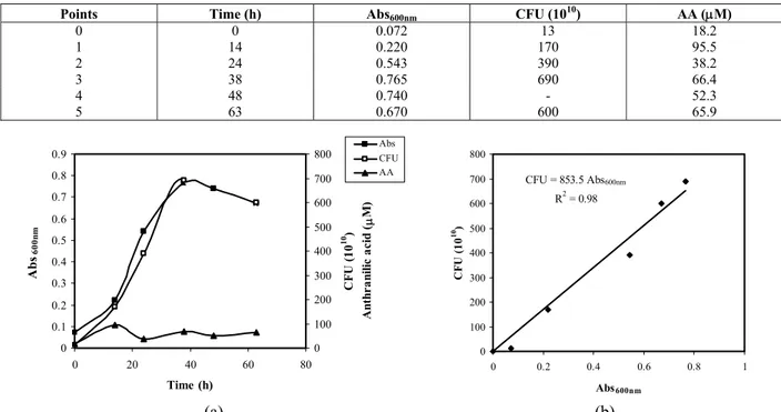
Table 3: Growth of Pseudomonas stutzeri ATCC 31258 in 1g/L carbazole in minimal medium.
Points Time (h) Abs600nm CFU (1010) AA (μM)
0 0 0.072 13 18.2
1 14 0.220 170 95.5
2 24 0.543 390 38.2
3 38 0.765 690 66.4
4 48 0.740 - 52.3
5 63 0.670 600 65.9
0 0.1 0.2 0.3 0.4 0.5 0.6 0.7 0.8 0.9
0 20 40 60 80
Time (h)
Ab
s 60
0n m 0 100 200 300 400 500 600 700 800 CF U ( 1 0 10 ) A n th r a n ili c a c id (μ M) Abs CFU AA
CFU = 853.5 Abs600nm
R2 = 0.98
0 100 200 300 400 500 600 700 800
0 0.2 0.4 0.6 0.8 1
Abs600nm CF U ( 1 0 10) (a) (b)
Figure 2: (a) Growth (Abs600nm and CFU) and anthranilic acid formation for Pseudomonas stutzeri ATCC 31258
in 1 g/L carbazole in minimal growth medium. (b) Correlation between Abs600nmand count of colony formation
The carbazole analysis by gas chromatography indicated that, for the lower initial carbazole concentration (1 g/L), there was significant substrate uptake, and around 60% of the carbazole was degraded in 48h, 75% at 63h and complete degradation was observed after three days. This is a very promising biodegradation assay and is comparable with the best results for other carbazole-degrading strains described in the Introduction section. Under these assay conditions, no accumulation of anthranilic acid was found in the culture medium, as can be seen in Figure 2 (a); its levels remained very low (around 50 μM or 7 mg/L) throughout the entire growth period.
According to the data presented in Table 3, in the culture medium with the lower carbazole concentration, growth of around 600 CFUs (Abs600nm ~ 0.7) was observed after three days of
cell growth, corresponding to 0.37 g/L dry cell weight. At this concentration, a high correlation was identified between the colony count (CFU) and the absorbance at 600 nm (Abs600nm), indicating
that the latter measurement can be reliably used (Figure 2b).
A comparison of the results in Figures 1 and 2 shows that growth in the culture medium with the lower carbazole concentration (1 g/L) was around ten times greater than in the medium with the higher concentration (10 g/L). The production of anthranilic acid was assessed for both initial carbazole concentrations and it was found that, in the lower concentration, around 7 mg/L was obtained, while in the culture medium with a high carbazole concentration there was an accumulation of anthranilic acid (nearly 140 mg/L after three days). These results indicate that the accumulation of anthranilic acid in the culture medium may be related to the inhibition of the growth of Pseudomonas
stutzeri in a medium with excess amounts of
carbazole.
CONCLUSIONS
With a view to undertaking biodegradation assays, curves for Pseudomonas stutzeri ATCC 31258 growth, carbazole degradation and anthranilic acid formation were assessed for two different carbazole concentrations (10 g/L and 1 g/L). After three days, 0.37 g/L cells (dry weight) were grown in the 1 g/L culture medium, complete degradation of the initial carbazole was observed, and 7 mg/L of anthranilic acid were formed, confirming carbazole as a sole source of carbon and energy for the bacteria. When the
carbazole concentration was higher, the growth was ten times lower and the excess carbazole led to an accumulation of 140 mg/L anthranilic acid, which inhibited the growth of the bacteria.
The 1 g/L (1000 ppm) assay results for
Pseudomonas stutzeri ATCC 31258 carbazole
biodegradation are very promising for the application of this strain in biorefining of Brazilian crudes, which contain more nitrogenated compounds than in other parts of the world (Leite et al., 2005).
NOMENCLATURE
AA anthranilic acid concentration Abs450nm absorbance measured at 450 nm
Abs600nm absorbance measured at 600 nm
ATCC American Type Culture Collection BDN biodenitrogenation
CFU colony-forming unit
HDN hydrodenitrogenation HDS hydrodesulfurization
INCQS Instituto Nacional de Controle de Qualidade em Saúde / Fiocruz
LB Luria Bertani
MW molecular weight
ppm part per million
ACKNOWLEDGMENTS
This study was supported by the Conselho Nacional de Desenvolvimento Científico e Tecnológico (CNPq) and PETROBRAS.
REFERENCES
Baboshin, M. A., Akimov, V.N., Baskunov, B. P., Born, T. L., Khan, S. U. and Golovleva, L. A., Conversion of Polycyclic Aromatic Hydrocarbons by Sphingomonas sp. VKM B-2434. Biodegradation, 19, 567 (2008).
Baboshin, M. A. and Golovleva, L. A., The Strategy of Strain Selection for a Mixed Culture Performing Rapid Conversion of a Mixture of Polyaromatic Compounds. Microbiology, 79, No. 1, 73 (2010).
Benedik, M .J., Gibbs, P. R., Riddle, R. R. and Willson, R. C., Microbial denitrogenation of fossil fuels. Trends in Biotechnology, 16, 390 (1998).
of carbazole by combined biological and catalytic treatment. American Chemical Society, Petroleum Chemistry Division Preprints, 48, No. 1, 44 (2003).
Castorena, G., Mugica, V., Le Borgne, S., Acuña, M. E., Bustos-Jaimes, I. and Aburto, J., Carbazole biodegradation in gas oil/water biphasic media by a new isolated bacterium Burkholderia sp. strain IMP5GC. Journal of Applied Microbiology, 100, No. 4, 739 (2006).
Cunha, C. D., Rosado, A. S., Sebastián, G. V., Seldin, L. and Von der Weid, I., Oil biodegradation by Bacillus strains isolated from the rock of an oil reservoir located in a deep-water production basin in Brazil. Applied Microbiology and Biotechnology, 73, 949 (2006). Fuse, H., Takimura, O., Murakami, K., Inoue, H. and
Yamaoka, Y., Degradation of chlorinated biphenyl, dibenzofuran, and dibenzo-p-dioxin by marine bacteria that degrade biphenyl, carbazole, or dibenzofuran. Bioscience, Biotechnology and Biochemistry, 67, No. 5, 1121 (2003).
Gai, Z., Yu, B., Li, L., Wang, Y., Ma, C., Feng, J., Deng, Z. and Xu, P., Cometabolic degradation of dibenzofuran and dibenzothiophene by a newly isolated carbazole-degrading Sphingomonas sp. strain. Applied and Environmental Microbiology, 73, No. 9, 2832 (2007).
Gai, Z., Wang, X., Liu, X., Tai, C., Tang, H., He, X., Wu, G., Deng, Z. and Xu, P., The Genes Coding for the Conversion of Carbazole to Catechol Are Flanked by IS6100 Elements in Sphingomonas sp. Strain XLDN2-5. PLoS ONE, 5, No. 4, e10018 (2010).
Gieg, L.M., Otter, A. and Fedorak, P. M., Carbazole Degradation by Pseudomonas sp. LD2: Metabolic Characteristics and the Identification of Some Metabolites. Environmental Science & Technology, 30, No. 2, 575 (1996).
Grifoll, M., Selifonov, S. A., Gatlin, C. V. and Chapman, P. J., Actions of a versatile fluorene-degrading bacterial isolate on polycyclic aromatic compounds. Applied and Environmental Microbiology, 61, No. 10, 3711 (1995).
Grosser, R. J., Warshawsky, D. and Vestal, J. R., Indigenous and Enhanced Mineralization of Pyrene, Benzo[a]pyrene, and Carbazole in Soils. Applied and Environmental Microbiology, 57, No. 12, 3462 (1991).
Guo, W., Li, D., Tao, Y., Gao, P. and Hu, J., Isolation and description of a stable carbazole degrading microbial consortium consisting of
Chryseobacterium sp. NCY and Achromobacter
sp. NCW. Current Microbiology, 57, 251 (2008).
Habe, H., Ide, K., Yotsumoto, M., Tsuji, H., Hirano, H., Widada, J., Yoshida, T., Nojiri, H. and Omori, T., Preliminary examinations for applying a carbazole-degrader, Pseudomonas sp. strain CA10, to dioxin-contaminated soil remediation. Applied Microbiology and Biotechnology, 56, No. 5-6, 788 (2001).
Habe, H., Ashikawa, Y., Saiki, Y., Yoshida, T., Nojiri, H. and Omori, T., Sphingomonas sp. strain KA1, carrying a carbazole dioxygenase gene homologue, degrades chlorinated dibenzo-p -dioxins in soil. FEMS Microbiology Letters, 211, 43 (2002).
Hisatsuka, K. and Sato, M., Microbial Transformation of Carbazole to Anthranilic Acid by Pseudomonas
stutzeri. Bioscience, Biotechnology and
Biochemistry, 58, 213 (1994).
Inoue, K., Widada, J., Nakai, S., Endoh, T., Urata, M., Ashikawa, Y., Shintani, M., Saiki, Y., Yoshida, T., Habe, H., Omori, T. and Nojiri, H., Divergent structures of carbazole degradative car operons isolated from Gram-negative bacteria. Bioscience, Biotechnology and Biochemistry, 68, No. 7, 1467 (2004).
Inoue, K., Habe, H., Yamane, H., Omori, T. and Nojiri, H., Diversity of carbazole-degrading bacteria having the car gene cluster: isolation of a novel Gram-positive carbazole-degrading bacterium. FEMS Microbiology Letters, 245, No. 1, 145 (2005).
Inoue, K., Habe, H., Yamane, H. and Nojiri, H., Characterization of novel carbazole catabolism genes from Gram-positive carbazole degrader
Nocardioides aromaticivorans IC177. Applied
and Environmental Microbiology, 72, No. 5, 3321 (2006).
Ishihara, A., Dumeignil, F., Aoyagi, T., Nishikawa, M., Hosomi, M., Qian, E. W. and Kabe, Y., Degradation of carbazole by Novosphingobium sp. strain NIY3. Journal of the Japan Petroleum Institute, 51, No. 3, 174 (2008).
Jensen, A.-M., Finster, K. W. and Karlson, U., Degradation of carbazole, dibenzothiophene, and dibenzofuran at low temperature by Pseudomonas sp. strain C3211. Environmental Toxicology and Chemistry, 22, No. 4, 730 (2003).
Kilbane II, J. J., Daram, A., Abbasian, J. and Kayser, K. J., Isolation and characterization of Sphingomonas sp. GTIN11 capable of carbazole metabolism in petroleum. Biochemical and Biophysical Research Communications, 297, 242 (2002).
Kirimura, K., Nakagawa, H., Tsuji, K., Matsuda, K., Kurane, R. and Usami, S., Selective and Continuous Degradation of Carbazole Contained in Petroleum Oil by Resting Cells of Sphingomonas sp. CDH-7. Bioscience, Biotechnology and Biochemistry, 63, No. 9, 1563 (1999).
Kobayashi, T., Kurane, R., Nakajima, K., Nakamura, Y., Kirimura, K. and Usami, S., Isolation of Bacteria Degrading Carbazole under Microaerobic Conditions, i.e. Nitrogen Gas Substituted Conditions. Bioscience, Biotechnology and Biochemistry, 59, No. 5, 932 (1995).
Larentis, A. L., Clonagem e Expressão em
Escherichia coli dos Genes da Rota de
Degradação de Carbazol de Pseudomonas
stutzeri. Tese de Doutorado, Universidade
Federal do Rio de Janeiro, COPPE, Programa de Engenharia Química (2005).
Leite, L. F., Neto, J. N. N. and Bevilaqua, J. V., Biorefineries and Biofuels: Current Activities and Future Vision of Petrobras. ACS Division of Fuel Chemistry, 50, 726 (2005).
Li, L., Xu, P. and Blankespoor, H.D., Degradation of carbazole in the presence of non-aqueous phase liquids by Pseudomonas sp. Biotechnology Letters, 26, 581 (2004).
Li, L., Li, Q., Li, F., Shi, Q., Yu, B., Liu, F. and Xu, P., Degradation of carbazole and its derivatives by a Pseudomonas sp. Applied Microbiology and Biotechnology, 73, 941 (2006).
Li, Y. G., Li, W. L., Huang, J. X., Xiong, X. C., Gao, H. S., Xing, J. M. and Liu, H. Z., Biodegradation of carbazole in oil/water biphasic system by a newly isolated bacterium Klebsiella sp. LSSE-H2. Biochemical Engineering Journal, 41, 166 (2008).
Loh, K.-C. and Yu, Y.-G., Kinetics of carbazole degradation by Pseudomonas putida in presence of sodium salicylate. Water Research, 34, No. 17, 4131 (2000).
Maeda, R., Nagashima, H., Widada, J., Iwata, K. and Omori, T., Novel marine carbazole-degrading bacteria. FEMS Microbiology Letters, 292, 203 (2009a).
Maeda, R., Nagashima, H., Zulkharnain, A. B., Iwata, K. and Omori, T., Isolation and characterization of a car gene cluster from the naphthalene, phenanthrene, and carbazole-degrading marine isolate Lysobacter sp. strain OC7. Current Microbiology, 59, 154 (2009b). Maeda, R., Ishii, T., Ito, Y., Zulkharnain, A. B., Iwata,
K. and Omori, T., Isolation and characterization of the gene encoding the chloroplast-type ferredoxin component of carbazole 1,9a-dioxygenase from a
putative Kordiimonas sp. Biotechnology Letters, 32, 1725 (2010).
Nagashima, H., Zulkharnain, A. B., Maeda, R., Fuse, H., Iwata, K. and Omori, T., Cloning and Nucleotide Sequences of Carbazole Degradation Genes from Marine Bacterium Neptuniibacter sp. Strain CAR-SF. Current Microbiology, 61, 50 (2010).
Nojiri, H. and Omori, T., Carbazole Metabolism by Pseudomonads. In: Ramos, J.-L. and Filloux, A. (Eds), Pseudomonas, vol. 5, p. 107-145. Springer, New York (2007).
Ouchiyama, N., Zhang, Y., Omori, T. and Kodama, T., Biodegradation of Carbazole by Pseudomonas spp. CA06 and CA10. Bioscience, Biotechnology and Biochemistry, 57, No. 3, 455 (1993).
Ouchiyama, N., Miyachi, S. and Omori, T., Cloning and nucleotide sequence of carbazole catabolic genes from Pseudomonas stutzeri strain OM1, isolated from activated sludge. Journal of General and Applied Microbiology, 44, 57 (1998).
Santos, S. C. C., Alviano, D. S., Alviano, C. S., Pádula, M., Leitão, A. C., Martins, O. B., Ribeiro, C. M. S., Sassaki, M. Y. M., Matta, C. P. S., Bevilaqua, J. V., Sebastián, G. V. and Seldin, L., Characterization of Gordonia sp. strain F.5.25.8 capable of dibenzothiophene desulfurization and carbazole utilization. Applied Microbiology and Biotechnology, 71, 355 (2006).
Schneider, J., Grosser, R. J., Jayasimhulu, K., Xue, W., Kinkle, B. and Warshawsky, D., Biodegradation of carbazole by Ralstonia sp. RJGII.123 isolated from a hydrocarbon contaminated soil. Canadian Journal of Microbiology, 46, No. 3, 269 (2000).
Seo, J., Keum, Y., Cho, I. K. and Li, Q. X., Degradation of dibenzothiophene and carbazole by
Anthrobacter sp. P1-1. International Biodeterioration
& Biodegradation, 58, 36 (2006).
Shepherd, J. M. and Lloyd-Jones, G., Novel Carbazole Degradation Genes of Sphingomonas CB3: Sequence Analysis, Transcription, and Molecular Ecology. Biochemical and Biophysical Research Communications, 247, No. 1, 129 (1998).
Shotbolt-Brown, J., Hunter, D. W. F. and Aislabie, J., Isolation and description of carbazole-degrading bacteria. Canadian Journal of Microbiology, 42, No. 1, 79 (1996).
Von der Weid, I., Marques, J. M., Cunha, C. D., Lippi, R. K., Santos, S. C. C., Rosado, A. S., Lins, U. and Seldin, L., Identification and biodegradation potential of a novel strain of
Dietzia cinnamea isolated from a
Yamazoe, A., Yagi, O. and Oyaizu, H., Degradation of polycyclic aromatic hydrocarbons by a newly isolated dibenzofuran-utilizing Janibacter sp. strain YY-1. Applied Microbiology and Biotechnology, 65, 211 (2004a).
Yamazoe, A., Yagi, O. and Oyaizu, H., Biotransformation of fluorene, diphenyl ether, dibenzo-p-dioxin and carbazole by Janibacter sp. Biotechnology Letters, 26, 479 (2004b).
Yang, M., Li, W., Guo, X., Qu, Z., Zhu, X. and Wang,
X., Isolation and identification of a carbazole degradation gene cluster from Sphingomonas sp. JS1. World Journal of Microbiology and Biotechnology, 25, 1625 (2009).