The defensive behavior of animals is regarded as an evo-lutionary adaptation, which is defined as an evolutive pro-cess that encompasses genetic changes, new phenotypic traits and enhancement of individuals’ fitness (Barrows 2000). In particular, Lepidoptera larvae have developed a series of stra-tegic defenses against predators like ants, that often co-oc-cur in its feeding sites (Kaminski et al. 2009). These defensive strategies are divided into two classes: primary defenses that prevent the encounter between predator and larvae and sec-ondary defenses that prevent the attack after detection of a potential predator by the larva (Gross 1993). Examples of primary defenses include the construction of shelters and bridges which serves as a mechanical barrier that decreases the likelihood of being located by ant predators, and the chemical camouflage of the larvae that allows it to become undetectable by the ants due to their resemblance to the sub-strate (Portugal & Trigo 2005). On the other hand, second-ary defenses comprise strategies like: biting, struggling, spitting, leaping from the leaf to remain hanged by a thread of silk, and the epidermal specializations. The latter adapta-tions range from simple bristles to more complex structures in the form of thorn, or even specialized glands that secrete
harmful substances to potential predators and parasitoids (Stehr 1987; Salazar & Whitman 2001).
In this study we address a defensive adaptation in Lepi-doptera of the genus Heliconius Kluk, 1780. The genus Heliconius forms one of the most successful groups of neotropical butterflies. They feature several evolutionary ad-aptations such as Müllerian mimicry, nocturnal aggregation in dormitories (Turner 1981; Salcedo 2011), unpalatability, advantageous mating events (Turner 1981), host plant speci-ficity, wide visual spectrum and large brain (Swihart & Swihart 1970); refined behavior, learning ability and sense of spatial location (Gilbert 1975), nectar and pollen feeding habits which maximizes the longevity, fecundity and fertility of eggs (Gil-bert 1972; Dunlap-Pianka et al. 1977), non-predation of eggs by larvae relatives (De Nardin & Araújo 2011), among others. Heliconius erato phyllis Fabricius, 1775 (Lepidoptera, Nymphalidae, Heliconiinae) is one of the 29 subspecies of Heliconius erato Linnaeus, 1758, the most variable species of the genus. It presents a wide distribution in South America, where it occurs from the northeast to the south of Brazil, Bo-livia, northeastern Argentina, Uruguay (Kaminski et al. 2002), and Paraguay (Holzinger & Holzinger 1994).
Defensive behavior associated with secretions from the prosternal paired
glands of the larvae of
Heliconius erato phyllis
Fabricius
(Lepidoptera, Nymphalidae)
1Eliane de Oliveira Borges2, Marcelo Eduardo Borges3 & Paulo Henrique Gorgatti Zarbin4
1 Contribution of the Department of Zoology, Universidade Federal do Paraná, Brazil.
2 Departamento de Fisiologia, Universidade Federal do Rio Grande do Sul, Campus Centro, 90050–170, Porto Alegre-RS, Brasil.
3 Departamento de Zoologia, Universidade Federal do Paraná, Centro Politécnico, Jardim das Américas, 81531–990, Curitiba-PR, Brasil.
4 Departamento de Química, Universidade Federal do Paraná, Centro Politécnico, Jardim das Américas, 81531–990, Curitiba-PR, Brasil.
ABSTRACT. Defensive behavior associated with secretions from the prosternal paired glands of the larvae of Heliconius erato phyllis Fabricius (Lepidoptera, Nymphalidae). Our work presents for the first time, the defensive behavior associated with the release of the product of the prosternal paired glands of the larva of Heliconius erato phyllis Fabricius, 1775 (Lepidoptera, Nymphalidae, Heliconiinae). The prosternal glands were first described for larvae of H. erato phyllis. They are formed by two types of glandular structures: the impair gland and the paired glands. The prosternal glands are located within the conical integumentary sac, which in turn is situated on the individual’s prosternum. The main goal of this study is to analyze the exist-ence of any secretion from the prosternal paired glands, and check the action mode of this secretion. The methodology used for chemical analysis of the glands included the aeration and, analysis in gas chromatography and gas chromatography-mass spec-trometry. The results show that the prosternal glands do not produce volatiles. Bioassays were conducted with simulated and natural attacks and revealed that the prosternal paired glands produce secretions of defense together with silk produced by labials glands as a defense strategy, described for the first time, against ants. The strategy consists in wrapping the ant with silk threads, the entire wrapped object moved to the end of the body, with the aid of the legs and prolegs, and possibly fixed in a nearby place. Evidence for the existence of a conical integumentary sac in larvae of other species and families of Lepidoptera allows us to propose the possibility of occurrence of prosternal paired glands with defensive function in these other groups as well.
Females of H. erato phyllis oviposit only on plants of the family Passifloraceae, which suggests a coevolution with their host plants (Benson et al. 1976). The passion fruit plants are well known to have extrafloral nectaries on the edge of the leaves and bracts, and they are associated with several ant species (Gilbert 1975; Benson 1976; Smiley 1985, 1986). Despite that the resources provided by plants constitute the main food item of several arboreal ants, many species also behaving as opportunistic predators (Floren et al. 2002; Davidson et al. 2003) or even having a high degree of spe-cialization (Morais 1994). Since larvae of H. erato phyllis feed on the same plant species, they are most likely to be encountered and predated by ants, as well as the eggs (Gil-bert 1975; Smiley 1985, 1986). Given this dominance over the foliage it is reasonable to think that ants must have a strong impact on the biology of herbivorous insects.
Defensive strategies in adults and larvae of H. erato phyllis include the presence of cyanogenic glycosides in their tis-sues, which determines the degree of toxicity in adults and makes them unpalatable to vertebrate predators. It is known that the adult produces these substances from aminoacids precursors (Nahrstedt & Davis 1983; Spencer 1988), and these aminoacids are derived from the pollen ingested by them (Gilbert 1991). These compounds could be hijacked or modified from the host plants, and alternatively, synthesized by the larvae (Borges et al. 2010). In larvae of H. erato phyllis, it is suggested that the presence of thorns along the body prevents the oviposition by parasitoids. The prosternal glands, a recently described trait, may be related to defensive mecha-nisms as well as odoriferous abdominal glands of adults (Borges et al. 2010).
The prosternal exocrine glands of the larva of H. erato phyllis are present in the five larval instars and occur in two different types. The first type consists of a pair of ellipsoidal units called ‘paired glands’. They are associated with a coni-cal integumentary sac that is projected in the prosternum. It expels a secretion through ducts that open into fine pores on the lateral surfaces of the sac, which is everted by internal hydrostatic pressure. The other type of gland is formed by a single secretion pouch in the anterior portion of prothorax, called ‘impair gland’. It opens on the body surface through a transverse slit at the anterior portion of the prosternum. The edges of the slit have several sensilla, which may probably function as proprioceptor. It is also supposed that the prosternal glands may exist in the larva of other species and families due to previous investigations that demonstrate the existence of the conical integumentary sac on the prosternum in these other studied species (Peterson 1962; Osborn & Jaffé 1998; Paluch et al. 2001; Furtado & Campos-Neto 2004; Souza et al. 2006). A histological analysis of glands and sur-face area (light microscopy and transmission electron mi-croscopy) demonstrates that the glandular units of the paired glands are composed of simple tall columnar epithelium, and the unit of the impair gland consists of simple low columnar epithelium. Paired glands secretion is amorphous, acidophilic, stored in the secretory units.
The function of these glands may involve a defense mechanism for the larvae, due to observations of changes in its posture when the anterior portion of their body is physi-cally stimulated. The observation of this behavior suggested the hypothesis of involvement in defense against attacks from potential predators (Borges et al. 2010). From this scenario, we established the goal to describe and elucidate the func-tion of the paired prosternal glands in larvae of H. erato phyllis. In this article we present the results of chemical analy-sis of aerations, and a description of the behavior related to the use of the prosternal paired glands when the larvae face different threats.
MATERIAL AND METHODS
In order to verify if the prosternal paired glands have a defensive function in the five larval instars, specimens from all instars of H. erato phyllis were obtained for analytical experiments, chemical analyzes and behavioral bioassays. We cultivated its host plant Passiflora edulis Sims, and their seed-lings were transplanted to pots and kept under natural condi-tions in the open in a reserved area of the Universidade Federal do Paraná (UFPR), Curitiba, Paraná, Brazil. Leaves and branches were used as larval food source in the rearing room. Potted plants of P. edulis and Passiflora actinia Hooker were kept in semi-natural conditions in the insectary built in the same area, whose leaves and branches served as oviposi-tion site for adults. Adults of H. erato phyllis used in this study were collected in the cities of Curitiba and Quatro Barras, Paraná, Brazil, and maintained in the insectary that was built in UFPR. Both the renewal of the artificial diet and the collection of the eggs were made daily. The collected eggs were kept in seminatural conditions in a rearing room set-ting, and the hatched larvae on leaves of P. edulis were kept in plastic bottles containing water. The whole setup was wrapped in porous cloth sacs.
We then verified if the prosternal paired glands of the larva released volatiles, and if these volatiles were associ-ated with defensive responses to the threat of potential preda-tors. We assessed the existence of possible volatile compounds extracted in different scenarios, as well as its concentration and chemical composition. The methodology used for chemi-cal analysis of possible volatiles of the prosternal glands in-cluded the aeration and analysis in gas chromatography (GC) and gas chromatography-mass spectrometry (GC MS).
used for aeration, with adsorbent filter at one end and a con-stant source of filtered air at the other. Volatiles were col-lected in time intervals of 24, 48 and 72 hours. In the aeration system A, four and fifth instar larvae received provocative artificial stimuli from bristles of a brush in the dorsal region of the larva on alternate times for 1 hour. The artificial stimuli were maintained until the larva displayed an aggressive be-havior. In aeration system B, four larvae individualized by porous cloth were kept in a glass tube during aeration. Fourth and fifth instar larvae were grouped separately. Eighteen samples of volatiles were collected from the first aeration system, and eight samples of volatiles were obtained from the second aeration system. The extracts were then obtained by washing the filter adsorbent with hexane and subsequent concentration (50mL or 50%) for chemical analyzes in gas chromatography.
The chemicals were analyzed in a gas chromatography/ mass spectrometry (GC/MS), model Shimadzu QP-2010 Plus equipped with a capillary column RTX-5 (30 mm × 0.25 mm × 0.25 mm) (Restek, Bellefonte, Pennsylvania, USA), with detector flame ionization (FID). The initial column tempera-ture was 50°C, held for three minutes. After this time, the temperature was increased by 7 °C/min until it reached 270°C. This max temperature was then kept constant through a pe-riod of 5 minutes.
The use of prosternal paired glands was also analyzed along with behavioral responses to threat scenarios. In the bioassays, we recorded video footage of the behavior of nine larvae in stress situations with artificial provocation stimuli for posterior analysis. We built a setup where a beam of light was projected on the larva of the fifth instar, and filmed it during 30s with camcorder model GZ-HM320 SUB Silver Full HD. A black background was used for contrast. We first used an artificial provocation stimuli that consisted of touch movements on the head of the larvae with a fine brush, as the individual remained in a horizontal position on a leaf of P. edulis. In a second scenario, we used specimens of the ant Tapinoma melanocephalum Fabricius, 1793 to conduct the behavioral bioassays. The observations of natural provoca-tive stimuli were made by putting all instars in direct contact with the living specimens of T. melanocephalum. Workers of T. melanocephalum were collected with bait fish in edible oil in the rearing room. This ant species is very generalist, with a diet ranging from sugary food sources to dead arthropods (Osborne et al. 1995; Scheurer & Liebig 1998). The external morphology and behaviors were studied primarily on fresh material. The drawings followed the stan-dard procedures of Borges et al. (2010) and Vegliante and Hasenfuss (2011). All illustrations were made with the aid of a stereoscopic microscope coupled to a camera lucida.
RESULTS
Airings. The analysis of the chromatograms shows that
in aeration system A it was collected only the plant’s volatiles, and in the aeration system B it was collected only volatiles
from the porous cloth. These results indicate that no defen-sive volatiles are produced by the larvae.
Bioassays – Behavioral Experiments
Direct observations and video footage revealed that the prosternal paired glands act together with the labial silk glands. The product of glandular secretion is a jet, which initially is suggested to be in a liquid state. These glands seem to promote a chemical-mechanical defense that is used as a strategy of active defense against predators, specifically ants. In the rearing room it was found that workers of the ant T. melanocephalum preyed on eggs, larvae and weakened adults with reduced flight space.
Responses to artificial stimuli. All larval instars
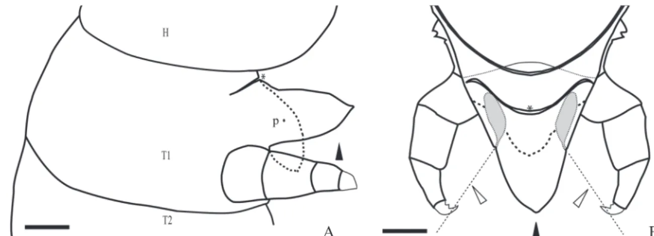
dis-played the same behavioral sequence of events when artifi-cially stimulated. The description of the response of fifth instar larva to artificial provocation happen as follows: the larva initially detects the potential predator, and then it folds its thorax several times with regular movements. The legs remain free and it turns its head towards the disturbing sub-ject. In this initial stage the larva has slow movements and also tries to move through the substrate. If the attack move-ments persist, the larva becomes increasingly active by dis-playing more incisive movements of its thorax. After a few minutes of constant attack it is possible to observe that the conical integumentary sac inflates (Fig. 1A). The sac is pre-sented as a protrusion, similar in color to the head capsule, and its length is subequal to the length of the fore legs. After that, four different possible outcomes of behavior are ob-served. In some occasions, the larvae moved through the substrate while maintaining the inflated sac. On a second possible outcome, it was observed the larvae directing jets of glandular secretion from pores of the prosternal paired glands. In the observation of the film images it was noted that the secretion of both pores are eliminated at the same direction of the brush’s bristles, which is probably interpreted as a potential predator (Fig. 1B).
In a third possible response, the larva releases silk from their labial glands toward the predator during the artificial attack (Fig. 2A). The larva positioned itself with a lifted tho-rax and loose legs, and then moves the thotho-rax in an attempt to hit the target (Fig. 2B). The jets are directed to the base of the object (Fig. 2C). After a few minutes of constant artifi-cial attacks and intense defensive behavior of the larva, it folds its thorax back to its maximum in order to gain mo-mentum. Soon after the larva delivers a sharp blow with the head toward the artificial predator (Fig. 2D) while still re-leasing secretion from labial glands and possibly also from the prosternal paired glands.
In a fourth behavior outcome, the larva resists initially to the threat. Then, it raises its thorax and directs its head to the fake predator, to regurgitate and bite the threating object.
Responses to natural stimulation
Fig. 1. Schematic representation of fifth instar prosternal glands of Heliconius erato phyllis. A. Lateral view of the prosternal sac during extrusion; fixed sac (dotted line) and sac in natural extrusion (closed arrowhead); head (H); prothorax (T1); mesothorax (T2), paired glandular opening (p), impair glandular opening (*). B. Antero-dorsal internal view of the prosternal sac during natural extrusion; fixed sac (dotted line) and sac in natural extrusion (closed arrowhead); prosternal paired glands (solid gray), impair glandular opening (*). The direction of the secretion release during artificial and natural attack is indicated by the open arrowhead. Scale bar = 50µm.
Fig. 2. Fifth instar larva of Heliconius erato phyllis when threatened by artificial stimuli. A. The larva releases silk threads from its labial glands to the base of the object. B. The larva raises its thorax and moves it around the target. C. The larva directs jets of the prosternal gland secretions and silk threads to the base of the object. D. The larva raises its thorax, bends it backwards, and then releases a strike in direction of the object. It also releases silk threads and possibly secretion of the prosternal paired glands.
A B
A
D C
stimulus. However, one larva of the third instar and one in the fourth instar also showed an additional behavior. After the display of the sequence of behavioral events described above, they approached the predator and with the aid of the legs wrapped the ant with threads of secretion forming a small sphere. Then the sphere was conducted with the legs and pro-legs towards the last larval segment (Fig. 3).
and opens on a tapered protrusion of the movable body wall (Povel & Beckers 1982) with a pair of muscles placed near the opening (Schaffer 1889). In other families it is an invagi-nation of the body wall around a hole, which forms a vesti-bule. It can be everted by an increase in the hydrostatic pressure and retracted by the action of muscles originated in the head or in the segment T1 (Detwiler 1922; Marti & Rogers 1988). In some Noctuoidea and Nymphalidae, there are ex-trinsic muscles inserted in the structure (Klemensiewicz 1883; Marti & Rogers 1988) which can be actively everted by some Nymphalidae (Fanfani & Dazzini 1989). The chemical analy-ses that resulted from the experiments shows that the prosternal glands of H. erato phyllis do not produce volatiles, even when the individual in threatened. The elimination of impair gland secretion was also not observed. In other Nymphalidae species, the conical integumentary sac diffuses volatiles repellents to ants and unpleasant to human olfac-tion (Devries & Martinez 1993; Osborn & Jaffé 1998; Osborn et al. 1999). On the other hand, some representatives of the family Rodinidae use it for communication with symbiotic ants (Devries et al. 2004; Kaminski 2008).
In Lepidoptera larvae, the chemical composition of the secretions of the conical integumentary sac is diverse. It con-sists of carboxylic acids in Nymphalidae (Osborn & Jaffé 1998) or formic acid as already described in Notodontidae and Noctuidae (Marti & Rogers 1988; Nakamura 1998). In these families they can also be composed by alcohols and esters (Hallberg & Poppy 2003) or single-chain hydrocar-bons and terpenes (Severson et al. 1991). In Heliconiinae, chemical analysis in larvae of Dione juno Cramer, 1779 and Abananote hylonome Doubleday, 1844 demonstrated the existence of defensive compounds such as acetic, linoleic and palmitic acids (Osborn & Jaffé 1998).
The observed behaviors of this study demonstrates for the first time that specifically the prosternal paired glands in H. erato phyllis have clearly a defensive function. This is evi-denced when the larva inflates its conical integumentary sac when threatened, and then secretes against the target through the pores of the glands if the threat persists. The same pattern of behavior related to the use of the conical integumentary sac has also been described in other species of Notodontidae, Nymphalidae and Noctuidae (Klemensiewicz 1883; Detwiler 1922; Percy & Macdonald 1979; Marti & Rogers 1988; Fanfani & Dazzini 1989; Osborn et al. 1999). It is also known that the secretion of the conical integumentary sac is added to the silk that form the cocoons of Cerura vinula Linnaeus, 1758 (Notodontidae) (Byers & Hinks 1976). Our field and labora-tory observations suggest that all larval instars of H. erato phyllis exhibit similar behavioral responses to both artificial and natural attacks. The glandular units of the prosternal paired glands can also operate in combination with the silk glands of the head. However, the success of the strategy depends on dif-ferent factors like the larval instar, its physiology, size, and the number of predators. It is very likely that the emission of a discharge of silk toward the base of the object that disturbs the larva suggests an initial goal of restricting movements of the
DISCUSSION
In this paper we proved the defensive behavior related to the use of prosternal paired glands in larvae of Heliconius erato phyllis. The prosternal glands are located within the conical integumentary sac, which in turn is situated on the individual’s prosternum (Borges et al. 2010). To the best of our knowledge, there are no studies in other species attesting the existence of an internal gland within this structure. There-fore, in these cases we will reference only the conical integu-mentary sac. The conical integuintegu-mentary sac is a very common structure in species of the family Nymphalidae (Peterson 1962; Stehr 1987; Miller 1991), and has a scattered system-atic distribution in other groups like Yponomeutidae, Noctuoidea, Hesperiidae and Papilionoidea (Bourgogne 1951; Peterson 1962; Miller 1991; Devries & Martinez 1993). It is important to consider that the structure displays mor-phological plasticity in different groups, which in turn can be associated with a variety of physiological and adaptive functions as well (Vegliante & Hasenfuss 2011). In Caligopsis seleucida Hewitson, 1877 (Nymphalidae, Brassolinae), the conical integumentary sac is shaped as a red projection in front of the prothoracic legs (Furtado & Campos-Neto 2004). Particularly in Yponomeutidae, the structure is not protruded
Fig. 3. Illustration of the behavior of fifth instar larva of Heliconius erato phyllis after facing a natural predator. The picture represents the larva conducting the silk sphere containing a worker ant with the help of its legs and prolegs towards the last larval segment (based in Vegliante & Hasenfuss 2011).
A F
B
C
D
E
G
H
I
predators. The strike with the head over the immobilized preda-tor can also facilitate the next step that consists in wrapping the prey with the secreted threads along with the aid of the legs. The conduction of the wrapped enemy toward the last larval segment through movements of the legs to the prolegs suggests that the sphere containing the immobilized predator is likely tied in a place nearby with additional threads pro-duced by the larva’s spinnerets.
According to the classification of Gross (1993), prosternal paired glands are secondary defense strategies that prevent the attack after the detection of a potential predator by the larvae. Evidence for the existence of a conical integumen-tary sac in larvae of other species and families of Lepidoptera allows us to propose the possibility of occurrence of prosternal paired glands with defensive function in these other groups as well. Furthermore, in these cases the possibility of the adoption of similar defensive behavior in also very plau-sible. We look forward for future studies where addressing this topic may help elucidate these new assumptions.
ACKNOWLEDGEMENTS
Thanks are due to Vitor Antonio Nardino, Cinthia Sayuri Tamura, Luis Anderson Ribeiro Leite and Dayana Bonfantti (Universidade Federal do Paraná) for helping in the video recording and in editing the figures. This study was supported by Conselho Nacional de Desenvolvimento Científico e Tecnológico (CNPq) (Grant no. 134753/2010-0 to the first author).
REFERENCES
Barrows, E.M. 2000. Animal Behavior Desk Reference. A Dictionary of Animal Behavior, Ecology, and Evolution. 2nd ed. Boca Raton, C.R.C. Press, 936 p.
Benson, W.W., Brown Jr., K.S. & Gilbert, L.E.1975. Coevolution of plants and herbivores: passion flower butterflies. Evolution29: 659–680. Borges, E. de O.; Faccione-Hauser, M.C. & Moreira, G.P.R. 2010.
Morphology of the prosternal glands of Heliconius erato (Lepidoptera: Nymphalidae). Psyche: Article ID 892960, 8 pages. Doi 10.1155/ 2010/892960
Bourgogne, J. 1951. Ordre des Lépidoptères, p. 174–448. In: Grassé, P. P. (ed.). Traité de Zoologie: Anatomie Systématique, Biologie. 10.
Paris,Masson.
Byers, J.R. & Hinks, C.F. 1976. Fine structure of the midventral abdomi-nal tracheal glands in banded wolly bear caterpillars (Arctiidae: Lepidoptera). Canadian Journal of Zoology54:1824–39. Davidson, D.W., Cook, S.C., Snelling, R.R. & Chua, T.H. 2003. Explaining
the abundance of ants in lowland tropical rainforest canopies. Science 300: 969–972.
De Nardin, J. & Araújo, A.M. 2011. Kin recognition in immatures of
Heliconius erato phyllis (Lepidoptera; Nymphalidae). Journal of Ethology 29: 499-503.
Devries, P.J. & Martinez, G.E. 1993. The morphology, natural history, and behavior of the early stages of Morpho cypris (Nymphalidae: Morphinae)—140 years after formal recognition of the butterfly.
Journal of the New York Entomological Society 101: 515–30. Devries, P. J., Cabral, B.C. & Penz, C.M. 2004. The early stages of
Apodemia paucipuncta (Riodinidae): myrmecophily, a new caterpillar ant-organ and consequences for classification. Milwaukee Public Museum Contributions in Biology and Geology102: 1–13. 7.
Detwiler, J.D. 1922. The ventral prothoracic gland of the red-humped apple caterpillar (Schizura concinna Smith and Abbot). Canadian Entomologist54:175–91.
Dunlap-Pianka, H., Boggs, C.L. & Gilbert, L.E. 1977. Ovarian dynamics in heliconiini butterflies: programmed senescence versus eternal youth. Science197: 487–490.
Fanfani, A. &, Dazzini, M.V. 1989. The jugular gland of Inachis io L. larva (Lepidoptera: Nymphalidae: Heliconiinae). Pubblicazioni dell’Istituto di Entomologia dell’Università di Pavia38:1–8. Floren, A., Biun, A. & Linsenmair, K.E. 2002. Arboreal ants as key
predators in tropical lowland rainforest trees. Oecologia131: 137– 144.
Furtado, E. & Campos-Neto, F.C. 2004. Caligopsis seleucida (Hewitson) e seus estágios imaturos. (Lepidoptera, Nymphalidae, Brassolinae).
Revista Brasileira de Zoologia21: 593–597.
Gilbert, L.E. 1972. Pollen feeding and reproductive biology in Heliconius
butterflies. Proceedings of the National Academy of Science of the United States of America 69: 1403–1407.
Gilbert, L.E. 1975. Ecological consequences of a coevolved mutualism between butterflies and plants, p. 210–240. In: Gilbert, L.E. & Raven, P.H. (eds.). Coevolution of animals and plants. Austin, University of Texas, xiii+246 p.
Gilbert, L.E. 1991. Biodiversity of a Central American Heliconius
community: pattern, process, and problems, p. 403–427. In: Price, P.W., Lewinsohn, T.M., Fernandez, G. and Benson, W.W. (eds.). Plant-Animal Interactions. Evolutionary Ecology in Tropical and Temperate Regions. New York, John Wiley and Sons.
Gross, P. 1993. Insect behavioral and morphological defenses against parasitoids. Annual Review of Entomology38: 251–273.
Hallberg, E. & Poppy, G. 2003. Exocrine glands: chemical communication and chemical defense, p. 361–75. In: Kristensen, N.P. (ed.). Handbook of Zoology/Handbuch der Zoologie, volume 4. Artropoda: Insecta.
Lepidoptera, Moths and Butterflies, Part 2: Morphology, Physiology, and Development. De Gruyter, xii+564 p.
Holzinger, H. & Holzinger, R. 1994. Heliconius and related genera.
Venette, SciencesNaturelles, 328 p.
Kaminski, L.A. 2008. Immature stages of Caria plutargus (Lepidoptera: Riodinidae), with discussion on the behavioral and morphological defensive traits in nonmyrmecophilous riodinid butterflies. Annals of the Entomological Society of America 101: 906–914.
Kaminski, L.A., Tavares, M., Ferro, V.G. & Moreira, G.R.P. 2002. Morfologia externa dos estágios imaturos de heliconíneos neotropicais. III. Heliconius erato phyllis (Fabricius) (Lepidoptera, Nymphalidae, Heliconiinae). Revista Brasileira de Zoologia19: 977–993. Kaminski, L.A., Sendoya, S.F., Freitas, A.V. & Oliveira, P.S. 2009.
Ecolo-gia comportamental na interface formiga-planta-herbívoro: interações entre formigas e lepidópteros. Oecologia Brasiliensis13: 27–44. Klemensiewicz, S. 1883. Zur näheren Kenntnis der Hautdrüsen bei den
Raupen und bei Malachius. Verhandlungen der Zoologisch-Botanischen Gesellschaft in Wien 32: 459–474.
Marti, O.G. & Rogers, C.E. 1988. Anatomy of the ventral eversible gland of fall armyworm, Spodoptera frugiperda (Lepidoptera: Noctuidae), larvae. Annals of the Entomological Society of America 81: 308– 17.
Miller, J. S. 1991. Cladistics and classif ication of the Notodontidae (Lepidoptera: Noctuoidea) based on larval and adult morphology.
Bulletin of the American Museum of Natural History no. 204, p. 1–230.
Morais, H.C. 1994. Coordinated group ambush: a new predatory behavior in Azteca ants (Dolichoderinae). Insectes Sociaux41: 339–342. Nahrstedt, A. & Davis, R.H. 1983. Occurrence, variation and biosynthesis
of the cyanogenic glucosides linamarin and lotaustralin in species of the Heliconiini (Insecta: Lepidoptera). Comparative Biochemistry and Physiology Part B75: 65–73.
Osborn, F. & Jaffé, K. 1998. Chemical ecology of the defense of two nymphalid butterfly larvae against ants. Journal of Chemical Ecology 24: 1173–1186.
Osborn, F., Sánchez, F. & Jaffé, K. 1999. Ultrastructure of the spines and neck gland of Abananote hylonome Doubleday, 1844 (Lepidoptera: Nymphalidae). International Journal of Insect Morphology and Embryology 28: 321–330.
Osborne, L.S., Peña, J.E. & Oi, D.H. 1995. Predation by Tapinoma melanocephalum (Hymenoptera: Formicidae) on two-spotted spider mites (Acari: Tetranychidae) in Florida greenhouses. Florida Entomologist78: 565–570.
Paluch, M., Casagrande, M.M. & Mielke, O.H.H. 2001. Estágios imatu-ros de Actinote carycina Jordan (Lepidoptera, Nymphalidae, acraeinae). Revista Brasileira de Zoologia18: 883–896.
Percy, J. & MacDonald, J.A. 1979. Cells of the thoracic defensive gland of the red-humped caterpillar, Schizura concinna (J. E. Smith) (Lepidoptera: Notodontidae): ultrastructural observations. Canadian Journal of Zoology 57: 80–94.
Peterson, A. 1962. Larvae of Insects, An Introduction to Nearctic Species. Part 1. Lepidoptera and Plant Infesting Hymenoptera.
4th ed. Columbus, Ohio State University.
Portugal, A.H.A. & Trigo, J.R. 2005. Similarity of cuticular lipids between a caterpillar and its host plant: a way to make prey undetectable for predatory ants? Journal of Chemical Ecology31: 2551–2561. Povel, G.D.E. & Beckers, M.M.L. 1982. The prothoracic “defensive” gland
of Yponomeuta-larvae (Lepidoptera, Yponomeutidae). Proceedings of the Koninklijke Nederlandse Akademie van Wetenschappen Series C 85: 393–98.
Salazar, B.A. & Whitman, D.W. 2001. Defensive tactics of caterpillars against predators and parasitoids, p. 161–207. In: Ananthakrishnan, T.N. (ed.). Insects and Plant Defence Dynamics. Plymouth, Science Publishers, Inc., 253 p.
Salcedo, C. 2011. Evidence of predation and disturbance events at
Heliconius (Insecta: Lepidoptera: Nymphalidae) nocturnal
aggregations in Panama and Costa Rica. Journal of Natural History 45: 1715–1721.
Schäffer, C. 1889. Beiträge zur Histologie der Insekten. Zoologische Jahrbücher Abteilung für Anatomie und Ontogenie der Thiere 3:611–652.
Scheurer, S. & Liebig, G. 1998. Tapinoma melanocephalum Fabr. (Formicidae,Dolichoderinae) in buildings – Observations on its biology and control. Anzeiger für Schädlingskunde Pflanzenschutz Umweltschutz71: 145–148.
Severson, R.F., Rogers, C.E., Marti, O.G., Gueldner, R.C. & Arrendale, R.F. 1991. Ventral eversible gland volatiles from larvae of the fall armyworm, Spodoptera frugiperda (J. E. Smith) (Lepidoptera: Noctuidae). Agricultural and Biological Chemistry 55: 2527–2530. Smiley, J.T. 1985. Heliconius caterpillar mortality during establishment
on plants with and without attending ants. Ecology66: 845–849. Smiley, J.T. 1986. Ant constancy at Passiflora extrafloral nectaries: effects
on caterpillar survival. Ecology67: 516–524.
Souza, N.A., Veiga, A.F.S.L., Casagrande, M.M. & Gondim Jr., M.G.C. 2006. Morfologia externa de imaturos de Caligo teucer (Linnaeus) (Lepidoptera, Nymphalidae). Revista Brasileira de Zoologia 23: 1243–1250.
Spencer, K.C. 1988. Chemical mediation of coevolution in the Passiflora
-Heliconius interaction, p. 167–240. In: Spencer, K.C. (ed.). Chemical Mediation of Coevolution. San Diego, Academic Press.
Stehr, F.W. 1987. Order Lepidoptera, p. 288–305. In: Stehr, F.W. (ed).
Immature Insects. Vol. 1, Dubuque, Kendall-Hunt, xiv+754 p. Swihart, C.A. & Swihart, S.L. 1970. Colour selection and learned feeding
preferences in the butterfly Heliconius charitonius Linn. Animal Behaviour18: 60–64.
Turner, J.R.G. 1981. Adaptation and evolution in Heliconius: a defense of NeoDarwinism. Annual Review of Ecology and Systematics 12: 99–121. Vegliante, F. & Hasenfuss, I. 2011. Morphology and diversity of exocrine glands in lepidopteran larvae. Annual Review of Entomology 57:187– 204.