Vânia Sofia Fidalgo Pobre
Dissertation presented to obtain the Ph.D degree in Biology
Instituto de Tecnologia Química e Biológica | Universidade Nova de Lisboa
Oeiras,
Vânia Sofia Fidalgo Pobre
Dissertation presented to obtain the Ph.D degree
in Biology
Instituto de Tecnologia Química e Biológica |
Universidade Nova de Lisboa
RNAs Structural Determinants in the
ii
Work financed by
:
Fundação para a Ciência e Tecnologia (FCT) PhD Fellowship - SFRH/BD/43206/2008.
Work performed at:
Control of Gene Expression Laboratory Instituto de Tecnologia Química e Biológica Av. da República
Estação Agronómica Nacional 2780-157 Oeiras
Portugal
Tel.: +351 21 446 95 48 Fax.: +351 21 441 12 77
Professora Doutora Cecília Maria Pais de Faria de Andrade Arraiano
Investigadora Coordenadora do Instituto de Tecnologia Química e Biológica, Universidade Nova de Lisboa.Co-Supervisor:
Doutor José Eduardo Marques Andrade
Investigador do Instituto de Tecnologia Química e Biológica, Universidade Nova de Lisboa.
President of the Jury:
Doutora Maria Helena Dias dos Santos
Professora Catedrática do Instituto de Tecnologia Química e Biológica da Universidade Nova de Lisboa, por delegação.
Examiners:
Professor Doutor Kenneth John McDowall
Astbury Center for Structural Molecular Biology / Faculty of Biological Sciences, Universty of Leeds, United Kingdom.
Professor Doutor Arsénio do Carmo Sales Mendes Fialho
Professor Associado com Agregação do Instituto Superior Técnico, Universidade Técnica de Lisboa.
Professora Doutora Maria da Glória Calado Inglês Esquível
Professora Auxiliar do Instituto Superior de Agronomia, Universidade Técnica de Lisboa.
Doutor Pedro Menano Lobo Fernandes
Investigador do Instituto Gulbenkian de Ciência, Fundação para a Ciência e Tecnologia.
Professora Doutora Claudina Amélia Marques Rodrigues
To my mother,
my father and
“If we knew what it was we were doing, it would not be called research, would it?”
xi I am really thankful to all the people that have directly and indirectly contributed
to this work. I would not finish this Thesis without you all. This work would not
have been possible without the support of:
Instituto de Tecnologia Química e Biológica, Universidade Nova de Lisboa (ITQB/UNL) for providing the work conditions and scientific environment for the execution of my Doctoral work.
Fundação para a Ciência e a Tecnologia (FCT) for the financial support of my Ph.D. fellowship (SFRH/BD/43206/2008).
My supervisor, Professor Cecília Arraiano, for all the support given during all these years. I am very grateful for her trust in me and cheering me up when things
were not working. Most of all I am thankful for the constant encouragement to
learn new things and for all the opportunities she gave me to pursue with my
work.
My co-supervisor, Doctor José Andrade, for teaching and guiding me since I enter the lab. I am extremely grateful for all the patience he always showed. Most
importantly I am thankful for his friendship. The long hours spent in the lab are
xii
Inês Silva and Filipa Reis, for all the friendship, the good moments shared and the support. We started together and are finishing together!
To all my friends in other labs in the institute, for sharing your scientific knowledge, for the coffee breaks and mostly for turning the institute into a
pleasant place, a second home!
To all my other friends outside the ITQB, for showing me a life outside of science. For the small talks, the good laughs and the nights spend out that help me relax!
To my house colleagues Marta and Meire, for all the support, especially these last few months. For the shared meals, the long talks and all the friends introduced
that allowed me to know people from so many different countries and cultures.
To my family, in special to my parents and my brother, for always be there for me. For all the support, for never letting me give up of anything in my life. For
listening to me about my work even knowing you don’t understand it. What I have
today I owed to you!
Abstract xv
Resumo xix
List of Publications xxiii
Thesis Outline xxvii
Chapter 1– Introduction 1
Chapter 2– The crucial role of PNPase in the degradation 63 of small RNAs that are not associated with Hfq
Chapter 3– Small RNA modules confer different 109 stabilities and selectively choose among different targets
Chapter 4– Analysis of the global role of Escherichia coli 167
exoribonucleases by RNA-Seq
Chapter 5– Discussion 219
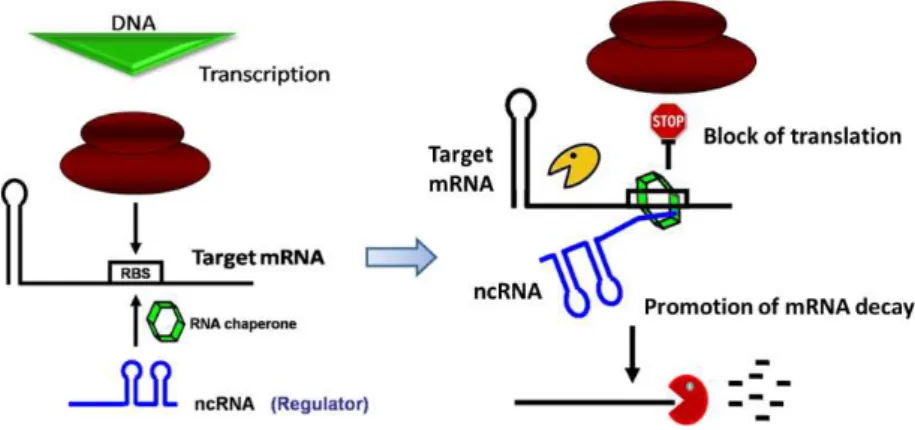
Small non-coding RNAs (sRNAs) are regulatory molecules that typically are
not translated into proteins. These molecules are often highly structured and very
stable and can affect many genetic pathways in all domains of life. Bacterial small
regulatory RNAs (sRNAs) parallel microRNAs in their ability to control multiple
targets. Small RNAs can bind to proteins or to mRNA targets. The sRNAs that act
by an antisense mechanism can have full (cis-encoded) or partial complementarity
(trans-encoded) with their targets. Most of the trans-encoded sRNAs studied so
far in Escherichia coli bind the RNA chaperone Hfq. The 5’ end of antisense RNAs is
usually found to be critical for the interaction with targets, generally inhibiting
translation and promoting mRNA decay. RNases are key elements in the control of
RNA levels in the cell and not surprisingly are also critical in the regulation of
sRNAs. In E. colithere are three 3’-5’exoribonucleases that accomplish most of the
mRNA exodegradative activity: ribonuclease II (RNase II), ribonuclease R (RNase R)
and polynucleotide phosphorylase (PNPase).
The main goal of this Doctoral work was to study the degradation pathways
of sRNAs. It was already known that 3’-5’ exonucleolytic degradation was a major regulatory pathway controlling the levels of the small non-coding MicA RNA, an
important regulator of outer membrane protein expression. Besides ribonucleases
there are other factors involved in the decay of sRNAs. In this work we addressed
some of these factors and their functions in the degradation of sRNAs.
Hfq promotes sRNA-mRNA duplex formation and is important to stabilize
sRNAs. However, the transient existence of sRNAs free from Hfq binding is part of
the normal dynamic lifecycle of a sRNA. In the first part of this work, we studied
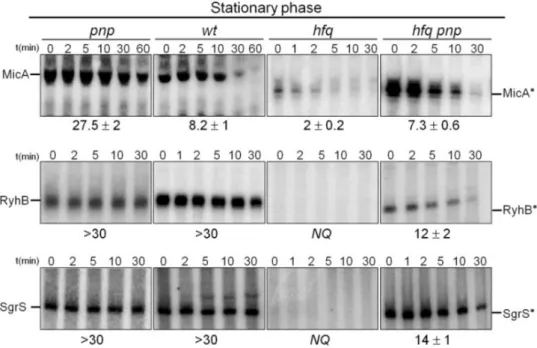
the degradation pathways of sRNAs in the absence of Hfq. We have found that
PNPase is the main ribonuclease involved in the rapid degradation of sRNAs,
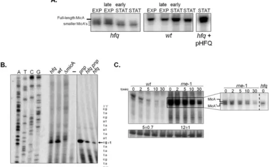
that in the absence of Hfq all sRNAs are trimmed at their 3’-end resulting in slightly shorter transcripts than their full-length species.
The turnover of Hfq-free sRNAs is growth-phase regulated and PNPase
activity is particularly important in stationary phase. In fact, the contribution of
PNPase to degradation of sRNAs is far greater than RNase E, which was commonly
believed to be the main enzyme in the initiation of decay of sRNAs. The lack of
poly(A) polymerase I (PAP I) also affects the degradation of Hfq-free sRNAs,
although to a minor extent.
Small RNAs are not “innocent” molecules waiting to be degraded. The
sequence and structural features of the small RNAs influence their degradation. In
the second part of this work, we characterised the RNA determinants involved in
the stability of the sRNA MicA and further analysed how this may influence the
regulation of its targets. Based on MicA sequence and secondary structure we
predicted the following MicA domains: a linear 5’ end sequence; a structured module harbouring two stem loops, an internal A/U-rich sequence that is the
predicted Hfq binding site and a transcriptional terminator with a U-rich linear 3’ end. Mutations were introduced and designed to affect certain domains, but not
the global secondary structure of the MicA.
Our results showed that besides the 5´domain of MicA, the stem loops and
the 3´poly(U) tail are also important in target binding. In vivo and in vitro
experiments showed that not only the AU-rich sequence but also the
transcriptional terminator are critical for stability and Hfq-binding. The different
MicA modules confer different stabilities and once again, PNPase was shown to
be the most important exoribonuclease involved in MicA degradation. The specific
MicA modules differentially affect the expression of the targets. Disruption of the
mRNAs while STEM1 was critical for regulation of tsx mRNA levels. Disruption of
the 3´U-rich sequence greatly affects all the targets analysed. In conclusion, we
found that MicA RNA can use different modules to regulate its targets.
In the third part of this work, we analysed the entire RNA content of the
cell. To investigate the roles of the three main exoribonucleases we used a whole
transcriptome sequencing approach (RNA-seq). We used cufflinks algorithm to
determine the relative abundance of the transcripts and cuffdiff algorithm to find
significant changes in transcript expression when comparing two samples. After
this step, we clustered the differentially expressed transcripts into different
functional categories using the program GeneCodis to retrieve gene ontology
terms and integrate the diverse biological information.
We started by comparing the transcriptome changes that occur when cells
go from exponential to stationary phase. We identified more than 1000
transcripts that were significantly different between the exponential and
stationary wild-type samples. Most of these transcripts are somehow connected
to the E. coli membrane and transport. We found that the three exoribonucleases
have different roles depending on the growth phase. However, there is some
overlap between PNPase, RNase II and RNase R functions in both exponential and
stationary phases.
In exponential phase, RNase II significantly affected 187 transcripts. The
majority of these transcripts belong to flagellar assembly and motility functional
categories suggesting that RNase II mutant may present defects in motility. On the
other hand, RNase R affected 202 transcripts of which the most interesting ones
seems to link RNase R to anaerobic respiration. PNPase was the exoribonuclease
might have a very important role in their metabolism.
Regarding stationary phase, RNase R seems to be the most important
enzyme in RNA degradation. In a ∆rnr mutant there are almost 700 transcripts
that are differentially expressed, while ∆rnb and ∆pnp mutants only significantly
affect 117 and 228 transcripts, respectively. On the other hand PNPase seems to
be the most important exoribonuclease involved in the degradation of sRNAs. In
the ∆pnp mutant 41% of the E. coli sRNAs are up-regulated.
In summary, the work on this dissertation contributed to expand our
knowledge not only on the small RNA degradation and mRNA targets control but
Resumo
Pequenos RNAs não codificantes (sRNAs) são moléculas reguladoras que normalmente não são traduzidas em proteínas. Estas moléculas são na sua maioria muito estruturadas, muito estáveis e podem afectar múltiplas vias genéticas em todos os domínios da vida. Os pequenos RNAs reguladores (sRNAs) bacterianos são similares aos microRNAs na sua capacidade de controlar múltiplos alvos. Pequenos RNAs pode ligar-se a proteínas ou ao mRNA alvo. Os sRNAs que atuam por um mecanismo de antisense pode ter complementaridade completa (transcrito na mesma região mas em sentido contrário – cis) ou parcial (transcrito noutra região da sequência mas produzido em sentido contrário – trans) com os seus alvos. A maioria dos sRNAs trans-codificados estudados até agora em Escherichia coli ligam-se ao chaperone de RNA Hfq. A extremidade 5' dos sRNAs é
geralmente considerada crítica para a interacção com os alvos, e geralmente inibem a tradução e promovem a degradação do mRNA. RNases são elementos chave no controle dos níveis de RNA na célula e também são fundamentais na regulação dos sRNAs. Em E. coli, há três 3'-5'exoribonucleases que realizam a maior parte da actividade degradativa do RNA: Ribonuclease II (RNase II), Ribonuclease R (RNase R) e “polynucleotide phosphorylase” (PNPase).
O objetivo principal deste trabalho de Doutoramento foi estudar as vias de degradação de sRNAs. Já se sabia que a degradação exonucleolítica 3'-5 ' era uma importante via reguladora no controlo dos níveis do pequeno RNA não-codificante MicA, um importante regulador da expressão das proteínas da membrana externa. Além das ribonucleases há outros fatores envolvidos na degradação de sRNAs. Neste trabalho abordamos alguns desses fatores e suas funções na degradação de sRNAs.
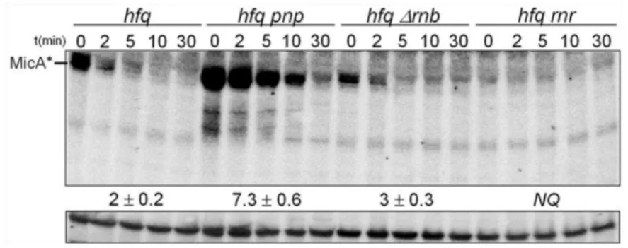
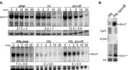
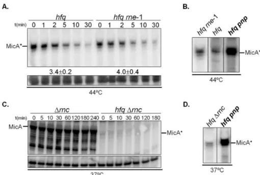
sRNAs livres da ligação ao Hfq faz parte do ciclo de vida normal e dinâmico de um sRNA. Na primeira parte deste trabalho, foram estudados os processos de degradação dos sRNAs na ausência de Hfq. Nós descobrimos que a PNPase é a principal ribonuclease envolvida na rápida degradação de sRNAs, especialmente aqueles que não estão ligados ao Hfq. Em células sem Hfq a inactivação da PNPase conduz ao aumento dos níveis dos sRNAs MicA, GlmY, RyhB e SgrS. Também descobrimos que na ausência de Hfq todos os sRNAs são cortados na sua extremidade 3', resultando em transcritos ligeiramente mais curtos do que os transcritos de comprimento normal.
A degradação dos sRNAs não ligados ao Hfq é regulada de acordo com a fase de crescimento e a actividade da PNPase é particularmente importante na fase estacionária. De facto, a contribuição da PNPase na degradação de sRNAs é muito maior do que a da RNase E, a qual foi geralmente reconhecida como a principal enzima involvida na degradação de sRNAs. A deleção da “poly(A) polimerase I” (PAP I) afecta também a degradação de sRNAs não ligados ao Hfq, mas em menor grau.
Os nossos resultados mostraram que, além do domínio 5' do MicA, os stem loops e a cauda 3'poly (U) também são importantes na ligação aos alvos. As experiências in vivo e in vitro mostraram que não só a sequência rica em A/Us, mas também o terminador de transcrição são críticos para a estabilidade e ligação com o Hfq. Os diferentes módulos do MicA conferem diferentes estabilidades e, mais uma vez, a PNPase mostrou ser a exoribonuclease mais importante envolvida na degradação do MicA. Os módulos específicos do MicA afetam diferencialmente a expressão dos alvos. Perturbação da região 5' do MicA não afecta significativamente o alvo lamB mRNA, no entanto os níveis de ompA e ecnB são dramaticamente aumentados. Em contraste, mutações nos stem loops aumentam fortemente os níveis do mRNA lamB, mas quase não afetam os mRNAs ompA e ecnB. Apenas interrupção da seqüência 3' rica em Us afeta muito todos os
alvos analisados.
Na terceira parte deste trabalho, analisamos todo o RNA da célula. Para investigar os papéis das três principais exoribonucleases usamos uma abordagem de sequenciamento de todo o transcriptoma (RNA-seq). Usou-se o algoritmo cufflinks para determinar a abundância relativa dos transcritos e o algoritmo cuffdiff para encontrar mudanças significativas na expressão dos transcritos ao comparar duas amostras. Após este passo, agruparam-se os transcritos diferencialmente expressos em diferentes categorias funcionais, utilizando o programa GeneCodis para obter os termos da ontologia de genes e integrar a diversificada informação biológica.
Nós descobrimos que as três exoribonucleases têm papéis diferentes, dependendo da fase de crescimento. No entanto, existe alguma sobreposição entre os papéis da PNPase, da RNase II e da RNase R em ambas as fases exponencial e estacionária.
Na fase exponencial, a RNase II alterava significativamente 187 transcritos. A maioria destes transcritos pertencem ás categorias funcionais de montagem de flagelos e mobilidade sugerindo que o mutante da RNase II pode apresentar defeitos na mobilidade. Por outro lado, a RNase R alterava 202 transcritos, dos quais os mais interessantes parece ligar a RNase R com a respiração anaeróbica. A PNPase foi a exoribonuclease cuja mutação alterou mais transcritos, num total de 226. Muitos desses transcritos são RNAs estáveis (rRNAs, tRNAs e sRNAs) sugerindo que a PNPase pode ter um papel muito importante no seu metabolismo.
No que diz respeito fase estacionária, a RNase R parece ser a enzima mais importante na degradação de RNA. No mutante Δrnr há quase 700 transcritos que são diferencialmente expressos, enquanto que os mutantes Δrnb e Δpnp apenas afectam significativamente 117 e 228 transcritos, respectivamente. Por outro lado, a PNPase parece ser a mais importante exoribonuclease envolvida na degradação de sRNAs. No mutante Δpnp os níveis de 41% dos sRNAs de E. coli estão aumentados.
The work presented in this Dissertation contributed to the following publications:
Papers in international scientific periodicals with referees:
Andrade J.M., Pobre V., Silva I.J., Domingues S., and Arraiano C.M. (2009). The
role of 3´-5´exonucleases in RNA degradation. Progress in Nucl. Acids Res. and
Molecular Biology Review, 85:187-229.
Arraiano C.M., Andrade J.M., Domingues S., Guinote I.B., Malecki M., Matos R.G.,
Moreira R.N., Pobre V., Reis F.P., Saramago M., Silva I.J. and Viegas S.C. (2010).
The critical role of RNA processing and degradation in the control of gene
expression. FEMS Microbiol Rev., Review, 34(5): 883-923.
Andrade J.M., Pobre V., Matos A.M. and Arraiano C.M. (2012). The crucial role of
PNPase in the degradation of small RNAs that are not associated with Hfq. RNA
18: 844-855.
Matos RG, Bárria C, Pobre V, Andrade JM and Arraiano CM (2012).
Exoribonucleases as modulators of virulence in pathogenic bacteria. Front. Cell.
Inf. Microbio. 2:65. doi: 10.3389/fcimb.2012.00065
Pobre V, Andrade J.M. And Arraiano C.M. (2012). Small RNA modules confer
different stabilities and selectively choose among different targets. Accepted with
modifications.
Reis F, Pobre V, Silva IJ, Malecki M, Arraiano CM (2012). The RNB Family of
Chapters in books:
Matos R.G., Pobre V., Reis F.P., Malecki M., Andrade J.M. and Arraiano C.M.
(2011) Structure and Degradation Mechanisms of 3′ to 5′ Exoribonucleases,
Chapter 8 in Ribonucleases, Nucleic Acids and Molecular Biology Series. Allen W.
Thesis outline
This dissertation is divided into five main chapters
Chapter 1 is a general introduction relating RNA degradation mechanisms and
some factors involved in RNA. This introduction focuses mainly on the degradative
ribonucleases, small RNAs, Hfq and their importance for RNA degradation.
The results of this Doctoral work are presented in the in the chapters 2, 3 and 4.
Each of these chapters has its own Introduction, Results, Discussion, Materials and
Methods and References sections.
Chapter 2 explores the degradation pathways of small RNAs in the absence of the
RNA chaperone Hfq. It is shown that PNPase has a crucial role in the degradation
of small RNAs especially if they are not associated with Hfq. It was demonstrated
that under these conditions the PNPase contribution for sRNAs decay is even
higher than RNase E, which had been considered the main ribonuclease involved
in sRNA decay.
Chapter 3 analyses the role of the small RNA MicA sequence and structure in the
stability of this sRNA and their function in target selectivity. It is shown that the
different MicA modules confer different stabilities and that PNPase is the main
exoribonuclease involved in the degradation of MicA. The 5´domain of MicA, the
stem loops and the 3´poly(U) tail are also important in target-binding and the
Chapter 4 studies the global roles of the 3’-5’exoribonucleases in the
transcriptome of Escherichia coli by RNA-Seq. This advanced technology was used to identify the transcripts affected by RNase II, RNase R and PNPase in exponential
and stationary cells. The three exoribonucleases have different roles depending
on the growth phase. Some of their functions overlap. A deletion of both RNase II
and RNase R is somehow compensated by the cell. PNPase is the main enzyme
involved in small RNA degradation.
Chapter 5 is the final discussion based on the results from the previous chapters
and connects the main results from this Dissertation. This chapter also includes
Chapter 1
Introduction
This chapter was based on:
Arraiano CM, Andrade JM, Domingues S, Guinote IB, Malecki M, Matos RG,
Moreira RN, Pobre V, Reis FP, Saramago M, Silva IJ, Viegas SC. 2010. The critical role of RNA processing and degradation in the control of gene expression. FEMS
Microbiol Rev 34:883-923.
Table of Contents
1 General Introduction ... 5 2 RNA Degradation pathways in E. coli ... 5 3 RNase E ... 8 3.1 RNase E structure and function ... 9
3.2 Control of RNase E expression ...11
3.3 Relating RNase E and RNase G ...12
3.4 RNase E in other organisms ...13
4 Ribonuclease III ...13 4.1 RNase III family of enzymes ...14
4.2 RNase III structure and substrate recognition ...14
4.3 RNase III activity and function ...16
5 RNase II ...17 5.1 Control of RNase II expression ...18
5.2 RNase II activity and RNA degradation ...18
5.3 RNase II structure and function ...19
6 RNase R ...22 6.1 RNase R modular organization and function ...22
6.2 RNase R role in RNA and protein quality control and mRNA decay ...24
6.3 RNase R is a stress induced protein ...24
6.4 RNase R in other organisms and its role in virulence ...25
7.1 Control of PNPase expression ... 27
7.2 PNPase activities and RNA degradation ... 28
7.3 PNPase structure and function ... 30
7.4 PNPase role in virulence ... 32
8 Role of small RNAs and Hfq in RNA decay ... 33 8.1 Small RNAs mode of action ... 34
8.2 Hfq roles in the cell ... 36
8.3 Hfq structure and function ... 38
8.4 Hfq complexes ... 39
1
General Introduction
The RNA levels in the cell depend on the efficiency of the transcription,
translation and the rate of degradation. Although transcription and translation are
important to determine RNA steady state levels, the processing and degradation
of RNA are also key factors in the regulation of gene expression. During this
Dissertation the main focus will be on RNA degradation. Ribonucleases (RNases)
are the enzymes that are able to process and degrade RNA. RNases are present in
all domains of life, and play a central role in the control of gene expression by
determining the levels of functional RNAs in the cell (Régnier & Arraiano, 2000;
Arraiano & Maquat, 2003; Parker & Song, 2004). Many of the RNases in the cell
are essential and others have overlapping functions (Régnier & Arraiano, 2000).
They are also involved in the quality control of all types of RNAs, allowing the
recycling of the ribonucleotides in the cell (Li et al., 2002; Silva et al., 2011). This
introduction will focus on the ribonucleases involved in RNA degradation in
Escherichia coli, which was the model organism throughout this Dissertation.
2
RNA Degradation pathways in
E. coli
In order to degrade RNAs, ribonucleases can act alone or they can be part
of RNA degradation complexes. Ribonucleases can be divided into
endoribonucleases (which cleave the RNA molecules internally) and
exoribonucleases (which degrade the RNA by removing terminal nucleotides from
the 3’ end of the RNA molecules). Exoribonucleases can act hydrolytically,
releasing nucleotide monophosphates, or phosphorolytically, if they use inorganic
phosphate to cleave the molecules releasing nucleotide diphosphates (Zuo &
can compete for access to the same substrate. In E. coli there are two
endoribonucleases (RNase E and RNase III) and four exoribonucleases (RNase II,
RNase R, PNPase and oligoribonuclease) involved in mRNA degradation (Table 1).
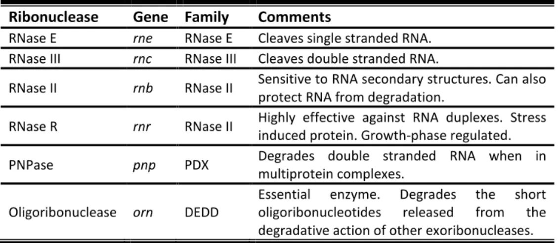
Table 1. Ribonuclease involved in RNA degradation in E. coli
Ribonuclease Gene Family Comments
RNase E rne RNase E Cleaves single stranded RNA. RNase III rnc RNase III Cleaves double stranded RNA.
RNase II rnb RNase II Sensitive to RNA secondary structures. Can also
protect RNA from degradation.
RNase R rnr RNase II Highly effective against RNA duplexes. Stress induced protein. Growth-phase regulated.
PNPase pnp PDX Degrades double stranded RNA when in multiprotein complexes.
Oligoribonuclease orn DEDD
Essential enzyme. Degrades the short oligoribonucleotides released from the degradative action of other exoribonucleases.
The RNA degradation pathways are not universal (Grunberg-Manago,
1999), and there are different mechanisms in bacteria (positive versus
gram-negative) and eukaryotes (Arraiano et al., 2010). However in all systems the
intrinsic characteristics of both available enzymes and RNA seem to control the
degradation of individual RNAs. Still, some common characteristics arise from the
analysis of different RNA degradation pathways. Perturbation of RNA structural
features may also work as an efficient degradation signal. Relaxation of secondary
structures may result in an easier accessibility of RNases, namely exposing the 3’
RNA end to exoribonucleolytic attack.
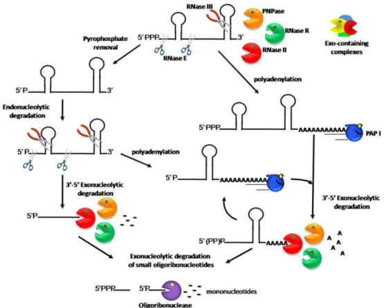
In E. coli the decay of the majority of transcripts starts with an
endoribonucleolytic cleavage by RNase E (Figure 1). This endoribonuclease prefers
a monophosphorylated 5’ end but not in a strict way, and several RNAs that do
not follow this rule have been described (Kime et al., 2010). RNase III is another
RNAs. However, unlike RNase E (that only cleaves single-stranded RNAs) RNase III
cleaves double-stranded RNAs.
Figure 1. Model of RNA degradation pathways in E. coli.
After endoribonucleolytic cleavages, the linear transcripts are rapidly
degraded by the 3’–5’ degradative exoribonucleases, RNase II, RNase R, and
PNPase. RNase II and PNPase are sensitive to secondary structures while RNase R
is the only exoribonuclease efficient against highly structured RNAs. However,
PNPase can associate with other proteins, namely RNA helicases, to unwind RNA
duplexes and consequently degrade structured RNAs (Andrade et al., 2009b). A
smaller pathway in the cell does not require the initial endoribonucleolytic
cleavage, instead polyadenylation emerges as important factor controlling the
exoribonucleolytic activity (Dreyfus & Régnier, 2002). It extends the 3’ linear
region providing a toehold that exoribonucleases can use to bind and initiate
degradation. Interestingly, this mechanism was conserved through evolution and
destabilizing poly(A) tails also promote exonucleolytic RNA degradation in
eukaryotes (LaCava et al., 2005; Vanácová et al., 2005; Wyers et al., 2005).
3
RNase E
RNase E, encoded by rne gene, was first identified by a temperature-sensitive
mutation (rne-3071) (Apirion & Lassar, 1978) and was initially described as an
activity required for the processing of E. coli 9S rRNA (Ghora & Apirion, 1978). The
ams (altered mRNA stability) locus was identified by a temperature-sensitive
mutation (ams-1) (Ono & Kuwano, 1980) and was shown to have an important
role in E. coli turnover (Ono & Kuwano, 1979). Later it was shown that these two
previously identified genes, rne and ams were actually different mutant alleles of
the same gene encoding RNase E (Mudd et al., 1990; Babitzke & Kushner, 1991;
Melefors & von Gabain, 1991; Taraseviciene et al., 1991). This important
endoribonuclease is essential for cell growth, and inactivation of
(Apirion & Lassar, 1978; Ono & Kuwano, 1979; Arraiano et al., 1988; Mudd et al.,
1990; Babitzke & Kushner, 1991; Melefors & von Gabain, 1991; Taraseviciene et
al., 1991). It has been described that RNase E plays a central role in the processing
of precursors of 5S ribosomal RNA (Apirion & Lassar, 1978; Misra & Apirion, 1979),
16S ribosomal RNA (Li et al., 1999), tRNAs (Ow & Kushner, 2002), tmRNA
(Lin-Chao et al., 1999) and the M1 RNA component of the RNase P ribozyme (Lundberg
& Altman, 1995; Ko et al., 2008). Homologous of RNase E have been identified in
more than 50 bacteria, archaea, and plants (Lee & Cohen, 2003).
3.1
RNase E structure and function
E. coli RNase E is a 1061 residue enzyme composed of two distinct
functional regions. The amino-terminal half forms the catalytic domain (residues
1–529) and is relatively conserved among prokaryotes (Marcaida et al., 2006). The
carboxy-terminal half of RNase E (residues 530-1061) is a non-catalytic region,
largely unstructured and poorly conserved (Callaghan et al., 2004). Segment-A is
located between residues 565 and 582 and is responsible for binding of the full
length RNase E to the inner cytoplasmic membrane (Khemici et al., 2008).
However, segment-A is not necessary for membrane interaction of the
catalytically active segment (Murashko et al., 2012). Residues 601–700 form an
arginine-rich segment that binds RNA in vitro and that is believed to enhance the
activity of RNase E in mRNA degradation in vivo (Lopez et al., 1999; Ow et al.,
2000). Residues 701– 1061 form a scaffold for interactions between RNase E and
the other major components of the degradosome, a protein complex involved in
mRNA decay (Kaberdin et al., 1998; Vanzo et al., 1998).
The first crystal structure for a member of the RNase E family has been
homotetramer with a molecular mass of roughly 260 kDa, organized as a dimer of
dimers (Callaghan et al., 2005). Each protomer is composed of two globular
portions, the large and small domains. The large domain consists of several
subdomains including the 5′-sensor as well as subdomains structurally similar to
protein folds found in S1, DNase I, and RNase H. In isolation each protomer
appears elongated, with a large domain comprising the subdomains (S1, 5’-sensor,
RNase H and DNase I), an elongated linker region (Zn-link) and then the small
domain. The dimer-dimer interface is formed by the small domains. At the
junction point there is a zinc binding site (Callaghan et al., 2005). The arrangement
of the domains within each dimer resembles the blades and handles of an open
pair of scissors. The positively charged surface within RNase H, 5’ sensor and
DNase I subdomains mediate the interaction of the catalytic domain of RNase E
with the membrane (Murashko et al., 2012).
E. coli RNase E is a single-stranded, nonspecific endoribonuclease with a
preference for cleaving A/U-rich sequences (Mackie, 1992; McDowall et al., 1995).
In vitro experiments have shown that purified E. coli RNase E prefers to cleave
RNAs that are monophosphorylated at the 5’ end (Mackie, 1998). It was shown
that RppH (RNA pyrophosphohydrolase) converts the 5’ terminus of primary
transcripts from a triphosphate to a monophosphate (Celesnik et al., 2007; Deana
et al., 2008). However, some structured substrates can be cleaved independently
of its state of phosphorylation by RNase E even if the 5’ end forms a secondary
structure (Baker & Mackie, 2003; Hankins et al., 2007). This indicates that while
5’-monophosphate-dependent pathway makes a significant contribution to mRNA
degradation (Mackie, 1998, 2000), there is another pathway of initial substrate
recognition by RNase E termed ‘bypass’ or ‘internal entry’ (Baker & Mackie, 2003;
Kime et al., 2010). The requirements for this pathway seems to be only the
existence of multiple single stranded segments in a conformation that allows
The crystal structure explains some features of the protein and suggests a
mechanism of RNA recognition and cleavage. A pocket is formed between the 5’
sensor and the RNase H subdomains and can bind a monophosphate group at a 5’
end (Callaghan et al., 2005). The catalytic site is physically separated of the 5’
sensing site. It contains conserved residues on the surface of the DNase I
subdomain of RNase E and coordinate a magnesium ion implicated in catalysis. A
‘mouse-trap’ model for communication between the 5’ sensing pocket and the
site of catalysis has been suggested. S1 and 5’ sensing domains move together as
one body to clamp down the substrate (Koslover et al., 2008).This conformational
change suggests a mechanism of RNA recognition and catalysis that explains the
enzyme’s preference for substrates with a monophosphate over a
5’-triphosphate and 5’-hydroxy RNA. It was also observed substantial flexibility at
one of the dimer-dimer interfaces, a deformation that may be essential to
accommodate structured RNA for processing by internal entry.
3.2
Control of RNase E expression
The cellular level and activity of RNase E are subject to complex regulation.
First, the enzyme concentration in the cell is regulated by a feedback loop in
which RNase E modulates decay of its own mRNA maintaining the level of the
enzyme within a narrow range (Mudd & Higgins, 1993; Jain & Belasco, 1995; Diwa
et al., 2000; Sousa et al., 2001; Ow et al., 2002). Recently it was shown that the 5’
sensor domain is essential for efficient autoregulation of RNase E (Garrey &
Mackie, 2011). Second, the efficiency of RNase E cleavage depends on the
structure of the substrates and the accessibility of putative cleavage sites. A 5’
monophosphate in substrate RNAs serves as an allosteric activator of RNase E
activity (Mackie, 1998; Jiang & Belasco, 2004). Third, interactions of mRNA targets
mRNAs by RNase E (Wagner et al., 2002). Fourth, the activity of RNase E is globally
affected by protein inhibitors, namely L4 ribosomal protein, RraA and RraB
(regulator of ribonuclease activity A and B, respectively) that interact with RNase
E and inhibit RNase E endonucleolytic cleavages of a selective group of transcripts
(Lee et al., 2003; Gao et al., 2006). Fifth, the membrane localization of RNase E
and its association with the bacterial cytoskeleton may affect its function through
various mechanisms (Liou et al., 2001; Khemici et al., 2008; Taghbalout &
Rothfield, 2008).
3.3
Relating RNase E and RNase G
RNase G is a paralogue of RNase E (McDowall et al., 1993), belonging to the
RNase E/G family, and is also involved in the degradation and processing of RNA
(Carpousis et al., 2009). E. coli RNase G was initially identified by its role in
chromosome segregation and cell division (Okada et al., 1994). RNase G was
subsequently shown to exhibit endoribonuclease activity both in vivo (Li et al.,
1999; Wachi et al., 1999; Umitsuki et al., 2001) and in vitro (Jiang et al., 2000; Tock
et al., 2000). A strong resemblance has been identified between RNase G and the
amino-terminal portion of E. coli RNase E, sharing a high level of sequence identity
(35%) and similarity (50%) (McDowall et al., 1993). Purified RNase G has in vitro
properties similar to RNase E and both enzymes are required for a two-step
sequential reaction of 5’ maturation of the 16S rRNA gene (Li et al., 1999; Wachi
et al., 1999). Residues of RNase E that can contact a 5’-monophosphorylated end
and coordinate the catalytic magnesium ion are conserved in RNase G (McDowall
et al., 1993; Callaghan et al., 2005). The precise cleavage sites of RNase E and
RNase G are not strictly conserved (Li et al., 1999; Tock et al., 2000). The 5’-
monophosphate end, which stimulates RNase G, is generated by RppH (Deana et
that RNase G interaction with a single-stranded segment, linked physically to a
5’-monophosphorylated-end, is an important determinant of the overall affinity of
RNA binding (Jourdan et al., 2010). Moreover, it was demonstrated that the
sequence of a site bound by RNase G can moderate the maximal cleavage rate
(Jourdan et al., 2010). RNase G is a paralogue of RNase E but up to now most of
the research on RNA degradation has been focusing on RNase E.
3.4
RNase E in other organisms
Some variants of RNase E can be found in α-Proteobacteria, Synechocystis sp.
and in the high G+C Gram-positive bacteria (Condon & Putzer, 2002). In
Rhodobacter capsulatus, RNase E is the responsible enzyme for the majority of
the endonucleolytic cleavages. In this organism RNase E has 118 KDa with a
conserved N-terminal region (Jager et al., 2001) and a C-terminal portion,
probably involved in the scaffold of degradosome assembly. It was purified in two
different complexes, one where it is associated with a helicase and an unidentified
protein, while in the other one was coupled with a helicase, the transcription
terminator Rho and an unidentified protein (Jager et al., 2001). Moreover, in R.
capsulatus, this enzyme is involved in the endonucleolytic process and
stabilization of cspA mRNA (Jager et al., 2004). Similarly to R. capsulatus,
Pseudomonas syringae, a psychrophilic bacterium, has also an RNase E which is
associated with RNase R and the DEAD-box helicase RhlE in a degradosome
(Purusharth et al., 2005).
Ribonuclease III (RNase III) was originally identified by Robertson and
co-workers in extracts of E. coli as the first specific double-stranded RNA (dsRNAs)
endoribonuclease (Robertson et al., 1968). Members of RNase III family are widely
distributed among prokaryotic and eukaryotic organisms, sharing structural and
functional features (Lamontagne et al., 2001). However, until now homologues of
RNase III have not been found in the genomes of archaea (Condon & Putzer,
2002).
4.1
RNase III family of enzymes
The RNase III family comprises four classes, according to their polypeptide
structure. The class I members of the RNase III family are ubiquitously found in
bacteria, bacteriophages and some fungi (MacRae & Doudna, 2007).
The Class II is exemplified by the eukaryotic Drosha protein while the class
III is represented by the eukaryotic Dicer (MacRae & Doudna, 2007). The
nucleases Drosha and Dicer have very important roles in RNA interference. Finally,
the Class IV is represented by the Mini-RNase III of Bacillus subtilis (Redko et al.,
2008). Taken together, the functional and evolutionary conservation of RNase III
family in bacteria and higher organisms is indicative of their biological relevance in
RNA maturation and degradation. Despite the fact that RNase E is considered the
major ribonuclease that catalyses the initial rate-determining cleavage of several
transcripts, RNase III family of enzymes has emerged as one of the most
important group of endoribonucleases in the control of RNA stability (Jaskiewicz &
Filipowicz, 2008).
E. coli RNase III has served as the prototypical member of the family. In this
model microorganism, RNase III is encoded by the rnc gene, and is active as a 52
kDa homodimer (Li & Nicholson, 1996). Each monomer contains a C-terminal
dsRBD, located in the last 74 amino acids, which is responsible for substrate
recognition and adopts a tertiary fold with the characteristic α1-β1-β2-β3-α 2-structure that is conserved throughout the RNase III family (Blaszczyk et al., 2001).
Additionally, each monomer is also composed by an N-terminal NucD. When the
two monomers are combined (RNase III homodimer), they form a single
processing center in the subunit interface, in which each monomer contributes to
the hydrolysis of one RNA strand of the duplex substrate. Ji and collaborators
(Blaszczyk et al., 2004; Gan et al., 2006) solved the structure of the
hyperthermophilic bacteria Aquifex aeolicus RNase III and the data has revealed
two functional forms of dsRNA binding by RNase III: a catalytic form, functioning
as a dsRNA-processing enzyme, cleaving both natural and synthetic dsRNA; and a
non-catalytic form, in which RNase III has a role of dsRNA binding protein (without
cleaving). The later activity is in agreement with previous studies in which this
enzyme binds certain substrates in order to influence gene expression, affecting
RNA structures (Calin-Jageman & Nicholson, 2003), (Court, 1993; Oppenheim et
al., 1993; Dasgupta et al., 1998). Magnesium (Mg2+) is the preferred co-factor.
Recent data are indicative that each active site contains two divalent cations
during substrate hydrolysis (Meng & Nicholson, 2008).
The RNase III substrate selection consists in a combination of structural
determinants and sequence elements referred as reactivity epitopes, such as the
helix length, the strength of base-pairing or the occurrence of specific nucleotide
pairs (termed proximal and distal boxes) located at defined positions related to
the cleavage site. In addition, there are also two classes of double-helical
recognition of this endoribonuclease or suppress the cleavage (without affecting
recognition) (Zhang & Nicholson, 1997; Pertzev & Nicholson, 2006).
4.3
RNase III activity and function
All enzymes of this family are hydrolytic and have specificity for dsRNAs,
generating 5’ monophosphate and 3’ hydroxyl termini with a two base overhang
at the 3’ end (Meng & Nicholson, 2008). RNase III in E. coli is not essential,
however it was observed that mutants for this endoribonuclease have a
slow-growth phenotype (Nicholson, 1999). This enzyme was initially identified due to
its role in the maturation of tRNA precursors and rRNA. Regarding maturation of
rRNA, RNase III is involved in the processing of 16S and 23S from a 30S rRNA
precursor (Babitzke et al. 1993). In Salmonella and other members of α
-proteobacteria, RNase III is also responsible for the cleavage of intervening
sequences (IVS) found in their 23S rRNA (Evguenieva-Hackenberg & Klug, 2000).
RNase III is also involved in the decay of several mRNA species (Condon & Putzer,
2002; Calin-Jageman & Nicholson, 2003). For example, in E. coli, this enzyme
participates in the first step of the decay of pnp mRNA (Régnier & Portier, 1986),
the gene encoding Polynucleotide Phosphorylase (PNPase), downregulating its
synthesis (Régnier & Grunberg-Manago, 1990; Robert-Le Meur & Portier, 1992;
Jarrige et al., 2001). Interestingly, this endoribonuclease has also the ability to
regulate its own synthesis with a specific cleavage near the 5’ end of its own
mRNA that removes a stem-loop, which acts as a degradation barrier (Bardwell et
al., 1989; Matsunaga et al., 1996; Lioliou et al., 2012). A recent work show that in
Staphylococcus aureus RNase III is involved in rRNA and tRNA maturation and
regulates the turnover of mRNAs and non-coding RNAs (Lasa et al., 2011; Lioliou
RNase III has been seen to work as a stress response modulator, controlling
the steady state levels of genes involved in cellular adaptation to stress (Santos et
al., 1997; Freire et al., 2006; Sim et al., 2010). It was seen in Salmonella
typhimurium that RNase III regulates the levels of the small RNA (sRNA) MicA
(Viegas et al., 2007), a main regulator of the abundant outer membrane protein
OmpA that has an important structural role in the cell and is involved in
pathogenesis (Guillier et al., 2006). The enzyme is also involved in the decay of
sRNA/mRNA complexes upon translational silencing (Vogel et al., 2004;
Afonyushkin et al., 2005), (Huntzinger et al., 2005; Kaberdin & Blasi, 2006). In this
way, cleavage by RNase III within the sRNA/mRNA duplex and the resulting
subsequent decay of the mRNA intermediate by the E. coli RNA decay machinery
could resemble the RNA interference (RNAi) in the eukaryotic cells (Agrawal et al.,
2003). RNAi is an evolutionary conserved phenomenon that functions as a
safeguard for the maintenance of genomic integrity. This phenomenon permits
the selective post-transcriptional downregulation of target genes in the cells, in
which RNase III-like enzymes dictate the degradation of dsRNA molecules
(Jagannath & Wood, 2007; Ma et al., 2007; Jinek & Doudna, 2009). Accordingly,
RNase III family has been associated with gene expression regulation, potential
antivirus agent, and tumor suppressor (Lamontagne et al., 2001).
5
RNase II
E. coli RNase II is a 3’-5’ exoribonucleases and is the prototype of the RNase II
family of enzymes (Mian, 1997; Mitchell et al., 1997; Zuo & Deutscher, 2001;
Grossman & van Hoof, 2006). RNase II-like proteins are widespread among the
three domains of life and in eukaryotes they are the catalytic component of the
multi-protein complex involved in RNA degradation in eukaryotes. In Saccharomyces
cerevisiae Rrp44/Dis3 is the RNase II family member. Rrp44 has some extra
domains at the N-terminal (CR3 and Pin) and can work also as an
endoribonuclease (Schaeffer et al., 2009). In Schizosaccharomyces pombe and
mammals there are several important members of this family such as Dis3, Dis3L
and Dis3L2 (Tomecki et al., 2010; Malecki et al., 2012).
5.1
Control of RNase II expression
RNase II is encoded by the rnb gene that can be transcribed from two
promoters P1 and P2 and terminates in a Rho-independent terminator 10
nucleotides downstream of rnb stop codon (Zilhão et al., 1993; Zilhão et al.,
1995a; Zilhão et al., 1996). PNPase regulates RNase II expression by degrading the
rnb mRNA (Zilhão et al., 1996). RNase III and RNase E endoribonucleases are also
involved in the control of RNase II expression at the post-transcriptional level.
RNase III does not affect rnb mRNA directly, but affects PNPase levels and RNase E
is directly involved in the rnb mRNA degradation (Zilhão et al., 1995b).
RNase II is also post-translationally regulated at level of protein stability and
its levels are also adjusted according to growth conditions. gmr (Gene Modulating
RNase II) is located downstream of rnb and is involved in the modulation of
stability of RNase II (Cairrão et al., 2001). Gmr has a PAS domain which can act as
an environmental sensor detecting changes in growth conditions.
5.2
RNase II activity and RNA degradation
E. coli RNase II is a sequence-independent hydrolytic exoribonuclease that
monophosphates. However, the processive degradation of an RNA molecule by
RNase II is easily blocked by secondary structures, and the enzyme is known to
stall around seven nucleotides before it reaches a double-stranded region
(Cannistraro & Kennell, 1999; Spickler & Mackie, 2000). In E. coli RNase II is the
major hydrolytic enzyme and participates in the terminal stages of mRNA
degradation (Deutscher & Reuven, 1991). However the enzyme is not essential for
E. coli growth unless PNPase is also missing (Donovan & Kushner, 1986; Zilhão et
al., 1995a). Although RNase II degrading activity is sequence-independent, its
favourite substrate is the homopolymer poly(A). Since the presence of a poly(A)
tail is often needed for the RNA degradative process, the rapid degradation of
polyadenylated stretches by RNase II can paradoxically protect some RNAs by
impairing the access of other exoribonucleases (Hajnsdorf et al., 1994; Pepe et al.,
1994; Coburn & Mackie, 1996; Marujo et al., 2000; Mohanty & Kushner, 2000;
Folichon et al., 2005b). Indeed, in the absence of RNase II a large number (31%) of
E. coli mRNAs are decreased, especially ribosomal protein genes, suggesting a
major function for this enzyme in the protection of specific mRNAs through
poly(A) tail removal (Mohanty & Kushner, 2003).
5.3
RNase II structure and function
The structure of E. coli RNase II and its RNA-bound complex was
determined (Frazão et al., 2006). This was the first structure of an
exoribonuclease from the RNase II family that has been solved (Frazão et al.,
2006). The overall X-ray crystallographic structure of the wild-type enzyme
(Frazão et al., 2006; Zuo et al., 2006) revealed four domains, as previously
predicted by Amblar et al. (Amblar et al., 2006). Three RNA binding domains have
been identified: two cold shock domains (CSD1 and CSD2) in the N-terminal region
central RNB domain, whose structure has shown an unprecedented fold
characteristic of this family. This domain contains four highly conserved sequence
motifs (I-IV) with some invariant carboxylate residues (Mian, 1997). The
RNA-binding domains (CSD1, CSD2 and S1) are grouped together on one side of the
structure, while the active site is on the other side of the molecule (Frazão et al.,
2006).
Elimination of the N-terminal CSD1 resulted in an increase of the
RNA-binding affinity of the enzyme for poly(A), suggesting that this domain may have a
role in controlling the movement of the enzyme on the poly(A) chain (Amblar et
al., 2006; Arraiano et al., 2008). Interestingly, without all the RNA-binding
domains the enzyme is still able to degrade RNA, although with much less
efficiency than the wild-type enzyme (Matos et al., 2009; Vincent & Deutscher,
2009).
The structure of the RNA-bound enzyme revealed that the RNA fragment
interacts with the protein at two non-contiguous regions, the “anchor” and
catalytic regions (Cannistraro & Kennell, 1994; Frazão et al., 2006). Nucleotides
1-5, at the 5’-end of the RNA fragment, are located in the “anchor” region in a deep
cleft between the two CSDs and the S1 domain. The final nucleotides 9-13 are
located in a cavity deep within the RNB domain, stacked and “clamped” between
the conserved residues Phe358 and Tyr253. A 10-nucleotide fragment is the
shortest RNA able to retain contacts with both anchor and catalytic regions. This
fact explains why RNase II is processive on long RNA molecules but becomes
distributive on substrates shorter than 10-15 nucleotides. When the RNA
molecule is shorter than five nucleotides, the required packing of the bases can no
longer occur, preventing the translocation of the RNA and a final end product of
four nucleotides is released (Frazão et al., 2006). Tyr-253 has been identified as
was shown to change the smallest end product of degradation from 4 to 10
nucleotides (Barbas et al., 2008). This mutation has been proposed to cause
loosening of the RNA substrate at the catalytic site and, as a consequence, binding
at the anchor region would be essential to keep the RNA attached to the protein
and allow cleavage. Molecules shorter than 10 nucleotides are too small to be
simultaneously bound at both sites meaning that they would have to be degraded
distributivety (Barbas et al., 2008).
The access to the catalytic pocket is restricted to single-stranded RNA by
steric hindrance, which explains the inability of RNase II to degrade
double-stranded RNA. DNA is not a substrate because there is a specific interaction
between the protein and the ribose rings of nucleotides that directly contact the
enzyme (Frazão et al., 2006). Residues Tyr-313 and Glu-390 have been
demonstrated to be responsible for the discrimination of cleavage of RNA versus
DNA (Barbas et al., 2009).
Several residues in the catalytic region are important for catalysis (Amblar &
Arraiano, 2005; Frazão et al., 2006). Asp-201 and Asp-210 substitution led to a
significant loss of RNase II activity and Arg-500 has also been shown to be crucial
for RNA cleavage (Frazão et al., 2006; Barbas et al., 2008, 2009). However,
Asp-209 is the only essential residue for RNA degradation (Amblar & Arraiano, 2005;
Barbas et al., 2008). The conserved residue Glu-542 has been proposed to
facilitate the elimination of the leaving nucleotide upon phosphodiester cleavage
(Frazão et al., 2006). Interestingly its substitution by alanine rendered the mutant
RNase II much more active than the wild-type and significantly increased the
RNA-binding ability. 3D modelling of the mutant enzyme indicated that the substitution
induced a subtle conformational change in the RNB domain. This resulted in a
so-called “super-enzyme”, an enzyme with extraordinary catalysis and binding
abilities (Barbas et al., 2009).
6
RNase R
RNase R encoded by the rnr gene (previously vacB) is a 3’-5’ hydrolytic
exoribonuclease from the RNase II family of exoribonucleases (Cheng &
Deutscher, 2002; Vincent & Deutscher, 2006). The rnr gene is second in an operon
together with nsrR (a transcriptional regulator), rlmB (rRNA methyltransferase),
and yjfI (unknown function). Transcription is driven from a possible σ70 promoter
upstream of nsrR (Cairrão et al., 2003). rnr mRNAs are post-transcriptional
regulated by RNase E, although RNase G may also participate (Cairrão & Arraiano,
2006). RNase R is a processive and sequence independent enzyme, with a wide
impact in RNA metabolism (Cairrão et al., 2003; Cheng & Deutscher, 2005;
Oussenko et al., 2005; Andrade et al., 2006; Purusharth et al., 2007; Andrade et
al., 2009a). It is unique amongst the RNA degradative exoribonucleases present in
E. coli as it can easily degrade highly structured RNAs (Cheng & Deutscher, 2002,
2003; Awano et al., 2010). RNase R is able to degrade a RNA duplex provided
there is a single stranded 3’ overhang (Cheng & Deutscher, 2002; Vincent &
Deutscher, 2006). In fact, RNase R was shown to be a key enzyme involved in the
degradation of polyadenylated RNA (Andrade et al., 2009a)
.
6.1
RNase R modular organization and function
RNase R shows a modular organization of RNA binding domains (CSD1 and
catalytic RNB domain, typically found on RNase II-family members. A
three-dimensional model of RNase R has been proposed based on the structure of its
paralogue RNase II (Barbas et al., 2008). Mutational analysis identified important
residues located in the active centre: D272, D278 and D280 (Matos et al., 2009;
Vincent & Deutscher, 2009). A D280N mutant showed no exonucleolytic activity,
analogous to what was reported with the D209N mutant in RNase II (Amblar &
Arraiano, 2005; Matos et al., 2009; Awano et al., 2010). RNase R degradation is
processive and unlike RNase II, the final end-product of digestion is a dinucleotide.
Tyrosine Y324 was found to be responsible for setting the final end-product of
RNase R (Matos et al., 2009).
RNase R was shown to bind RNA more tightly within its catalytic channel
than does RNase II. Surprisingly, a mutant expressing only the nuclease domain
(RNB) is able to degrade a perfect double stranded RNA (Matos et al., 2009;
Vincent & Deutscher, 2009). Probably the RNA binding domains “block” the
entrance of dsRNA into the catalytic channel. Accordingly, it was proposed that
RNA binding domains actually discriminate the substrates that can be targeted by
RNase R, favouring the selection of RNA molecules harbouring a 3’ linear tail. It
has been suggested that RNase R can function both as an exoribonuclease as well
as an RNA “helicase” (Awano et al., 2010). RNase R intrinsic “helicase” unwinding
activity is dependent on RNA-binding regions (S1, CDS1 and most importantly
CDS2). The double stranded RNA must have a 3’ linear overhang in order to
become a suitable substrate to RNase R helicase activity. Altogether, RNA binding
domains of RNase R seem to be responsible for the selection of RNA substrates
harbouring a 3’ linear region, which can be provided by polyadenylation (Andrade
et al., 2009a). Clearly, only the resolution of RNase R structure will allow the fully
6.2
RNase R role in RNA and protein quality control and mRNA
decay
RNase R is critical in RNA quality control, namely in degradation of defective
tRNAs (Vincent & Deutscher, 2006; Awano et al., 2010) and rRNA (Cheng &
Deutscher, 2003). Together with PNPase, RNase R eliminates aberrant fragments
of 16S and 23S rRNA whose accumulation potentially affects ribosome maturation
and assembly. Furthermore, the importance of RNase R in the accuracy of gene
expression is broadening with its role in protein quality control. In the absence of
RNase R, the small stable SsrA/tmRNA is not properly processed, leading to
defects in trans-translation and significant errors in protein tagging for proteolysis
(Cairrão et al., 2003). RNase R has also emerged as an important novel contributor
to mRNA degradation. The absence of both RNase R and PNPase results in the
strong accumulation of REP-containing mRNA sequences (Cheng & Deutscher,
2005). However, the presence of only one of these exoribonucleases is sufficient
to remove such transcripts, revealing again a functional overlap between these
two enzymes. Remarkably, RNase R was also shown to degrade the ompA
transcript in a growth-phase specific manner (Andrade et al., 2006). In the
stationary phase of growth, the single inactivation of RNase R results in the
accumulation of ompA mRNA and this correlated with increasing intracellular
levels of OmpA protein. This work revealed a role for RNase R in control of gene
expression that could not be replaced by any of the other exoribonucleases.
6.3
RNase R is a stress induced protein
The activity of RNase R is modulated according to the growth conditions of
the cell and responds to environmental stimuli. RNase R is a general
namely in cold-shock, and stationary-phase of growth (Cairrão et al., 2003;
Andrade et al., 2006). RNase R is a highly unstable protein in exponential phase,
however this protein is stabilized in stationary phase and other stress conditions,
leading to its relative increase (Chen & Deutscher, 2010). tmRNA and SmpB
binding to the C-terminal region of RNase R is responsible for the unstability of
RNase R in exponential phase (Liang & Deutscher, 2010). This binding is regulated
by acetylation of RNase R by the Pka acetylating enzyme that is absent from the
cell under stress conditions such as stationary phase and cold-shock (Liang &
Deutscher, 2012).
6.4
RNase R in other organisms and its role in virulence
RNase R-like enzymes are widespread in most sequenced genomes.
Although most of the knowledge on this protein came from work in E. coli, many
RNase R from other bacterial species have been identified. Notably, RNase R has
also been implicated in the establishment of virulence in a growing number of
pathogens.
In Shigella flexneri RNase R was shown to be required for the expression of
the invasion factors IpaB, IpaC, IpaD and VirG (Tobe et al., 1992). The disruption of
VacB gene in other Shigella spp. and enteroinvasive Escherichia coli resulted in
reduced expression of virulence phenotypes (Tobe et al., 1992). In Legionella
pneumophila RNase R is the only hydrolytic exoribonuclease present. This protein
is not essential for growth at optimal temperature, however, it is important for
growth and viability at low temperatures and induces the competence
development (Charpentier et al., 2008). To the date, only one exoribonuclease,
RNase R (MgR), was identified in Mycoplasma genitalium, where is an essential
R and RNase II and can carry out a broad range of RNA processing and degradative
functions (Lalonde et al., 2007). Similarly to what happens in E. coli, RNase R from
Aeromonas hydrophila is also a cold-shock protein essential for the viability at
lower temperatures and it absence leads to a reduction in A. hydrophila motility
(Erova et al., 2008). The infection of mouse cells with ∆rnr strains shows that the
virulence is attenuated, confirming the role of this enzyme in the pathogenesis of
this organism (Erova et al., 2008). In Streptococcus pneumoniae there is a unique
homolog of RNase II family of enzymes which was shown to be a RNase R-like
protein (Domingues et al., 2009). Proteins isolated from different strains regarding
their virulence ability (virulent vs. non-virulent) are different regarding their
activity and RNA affinity (Domingues et al., 2009). Further studies are still
necessary to confirm if the differences observed in RNase R protein are
responsible for the virulence of these strains.
In Pseudomonas syringae, RNase R is the exoribonuclease present in the
degradosome as opposed to most other systems where PNPase is part of such
complexes (Purusharth et al., 2005). Like in E. coli, RNase R is also particularly
important at low temperatures, since inactivation of the rnr gene inhibits growth
of both P. putida (Reva et al., 2006) and P. syringae (Purusharth et al., 2007) at
4°C. In P. syringae RNase R is involved in 3’-end maturation of 16S and 5S rRNA,
and in tmRNA turnover (Purusharth et al., 2007).
Overall, RNase R-deficient bacteria have been shown to be less virulent than
the wild-type parental strains. However, how this is achieved is still not
completely clear. This is probably related to critical RNA degradation pathways.
The fact that RNase R was found to be essential in the degradation of small RNAs,
namely the virulence regulator SsrA/tmRNA, opens the way to broaden its role in
be attractive as a potential therapeutic agent but clearly more studies are
required.
7
PNPase
PNPase is a 3’-5’ phosphorolytic exoribonuclease that belongs to the PDX
family of exoribonucleases, which also includes RNase PH from bacteria, and the
core of the exosome in archaea and eukaryotes (Mian, 1997; Zuo & Deutscher,
2001; Pruijn, 2005). The enzyme is involved in global mRNA decay, being widely
conserved from bacteria to plants and metazoans (Zuo & Deutscher, 2001;
Bermúdez-Cruz et al., 2005).
7.1
Control of PNPase expression
PNPase is encoded by the pnp gene and is transcribed from two promoters
(Portier & Régnier, 1984). pnp expression is negatively autoregulated at the
post-transcriptional level by the concerted action of PNPase and RNase III (Portier et
al., 1987; Robert-Le Meur & Portier, 1992, 1994; Jarrige et al., 2001; Carzaniga et
al., 2009). This autoregulation can be disrupted by Ribosomal protein S1 that
binds to the pnp mRNA 5’-UTR (Briani et al., 2008). In an RNase III deficient strain
there is a 10-fold increase of the PNPase levels (Portier, et al., 1987). PNPase
levels are also affected by polyadenylation. It is likely that polyadenylated
transcripts titrate out the amount of PNPase available to carry out normal
autoregulation (Mohanty & Kushner, 2002). PNPase and RNase II are
cross-regulated (Zilhão et al., 1996). In the absence of RNase II, PNPase levels are
increased and PNPase overexpression leads to a decrease in RNase II activity