Transdisciplinary treatment of Class III
malocclusion using conventional implant-supported
anchorage: 10-year posttreatment follow-up
Mariana Roennau Lemos Rinaldi1, Susana Maria Deon Rizzatto2, Luciane Macedo de Menezes3, Waldemar Daudt Polido3, Eduardo Martinelli Santayanna de Lima4
How to cite this article: Rinaldi MRL, Rizzatto SMD, Menezes LM, Poli-do WD, Lima EMS. Transdisciplinary treatment of Class III malocclusion us-ing conventional implant-supported anchorage: 10-year posttreatment follow-up. Dental Press J Orthod. 2015 May-June;20(3):69-79.
DOI: http://dx.doi.org/10.1590/2176-9451.20.3.069-079.oar
Submitted: July 02, 2014 - Revised and accepted: November 25, 2014 » Patients displayed in this article previously approved the use of their facial and intraoral photographs.
Contact address: Eduardo Martinelli S. de Lima
Av. Ipiranga, 6681 - Prédio 6 (Faculdade de Odontologia), Porto Alegre - RS, CEP 91530-000 - Brazil.
E-mail: [email protected]
1 PhD resident in Orthodontics, Pontifícia Universidade Católica do Rio Grande
do Sul (PUCRS), Porto Alegre, Rio Grande do Sul, Brazil.
2 Professor of Orthodontics, Pontifícia Universidade Católica do Rio Grande do
Sul (PUCRS), Porto Alegre, Rio Grande do Sul, Brazil.
3 PhD in Oral and Maxillofacial Surgery, Pontifícia Universidade Católica do
Rio Grande do Sul (PUCRS), Porto Alegre, Rio Grande do Sul, Brazil.
4 Adjunct professor of Orthodontics, Pontifícia Universidade Católica do Rio
Grande do Sul (PUCRS), Porto Alegre, Rio Grande do Sul, Brazil.
» The authors report no commercial, proprietary or financial interest in the prod-ucts or companies described in this article.
Introduction: Combined treatment offers advantages for partially edentulous patients. Conventional implants, used as orthodontic anchorage, enable previous orthodontic movement, which provides appropriate space gain for crown insertion. Objective: This case report describes the treatment of a 61-year and 10-month-old patient with negative overjet which made ideal prosthetic rehabilitation impossible, thereby hindering dental and facial esthetics. Case report:
After a diagnostic setup, conventional implants were placed in the upper arch to anchor intrusion and retract anterior teeth. Space gain for lateral incisors was achieved in the lower arch by means of an orthodontic appliance. Conclusions:
Integrated planning combining Orthodontics and Implantology provided successful treatment by means of conventional implant-supported anchorage. The resulting occlusal relationship proved stable after 10 years.
Keywords:Orthodontic anchorage procedures. Dental implants. Angle Class III malocclusion. Tooth loss. DOI: http://dx.doi.org/10.1590/2176-9451.20.3.069-079.oar
Introdução: tratamentos combinados podem oferecer vantagens em casos de edentulismo parcial. O uso de implantes convencionais como ancoragem ortodôntica permite a movimentação ortodôntica prévia, fornecendo os espaços apro-priados para inserção das coroas. Objetivo: este relato de caso descreve o tratamento de uma paciente, com 61 anos e 10 meses de idade, que possuía um overjet negativo que tornava impossível uma reabilitação protética ideal, comprometendo a estética dentária e facial. Relato do caso: após um setup diagnóstico, os implantes convencionais foram colocados na mandíbula para ancorar a intrusão e retração dos dentes anteriores. Espaços para os incisivos laterais foram abertos na maxila, usando-se aparelho ortodôntico. Conclusões: o planejamento integrado entre Ortodontia e Implantodontia propiciou um tratamento bem-sucedido, usando implantes convencionais como ancoragem. A relação oclusal obtida apresenta estabilidade 10 anos pós-tratamento.
INTRODUCTION
Transdisciplinarity is a trend in Dentistry as well as in other areas of the health sciences. This is because the in-teraction established among diferent specialties provides patients with a comprehensive treatment plan.1,2,3
Os-seointegration has opened up new possibilities not only for Prosthodontics, but also for Orthodontics. Proper anchorage has always been fundamental for orthodontic treatment eiciency, as it allows the desired orthodon-tic movements to be performed and reduces poten-tial adverse efects. The use of conventional implants and temporary anchorage devices (TAD) has improved anchorage control and provided absolute resistance units against movement. Absolute anchorage allows space clo-sure, intrusion, extrusion, protraction, retraction of teeth and stabilization of periodontally compromised teeth.4-8
Conventional implants for prosthetic restoration can also be used for orthodontic anchorage.9 Implant
se-lection and insertion site should be appropriate for the dual function of implants: rehabilitation and anchor-age. The anatomical aspects of the case, the intended orthodontic movement and the ideal position for inal rehabilitation should be planned ahead of time.5,10,11
Combined treatment ofers advantages for partially edentulous patients, and so does previous orthodontic movement, as it provides appropriate space gain for im-plant insertion.12-15
The aim of this case report is to demonstrate the transdisciplinary treatment of a Class III malocclusion patient with multiple missing teeth. Conventional im-plants were used as anchorage to retract lower teeth. This combined transdisciplinary plan intended to maxi-mize patient’s beneits, enhance dental esthetics and es-tablish a balanced occlusion associated with healthy tis-sues. This report illustrates a case of successful 10-year posttreatment stability.
CASE REPORT
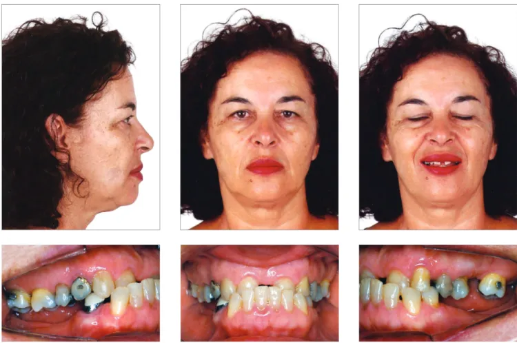
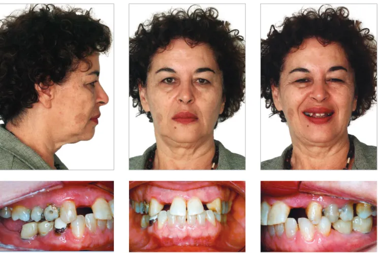
In 1998, a healthy female patient, aged 61 years and 10 months old, presented at the orthodontic service of the Brazilian Dental Association with anterior crossbite and multiple missing teeth. Her chief complaint was related to poor dental esthetics. Prosthetic rehabilita-tion was thought to be determined by the condirehabilita-tions of dental occlusion. There was premature contact between lower central incisors, and anterior crossbite was most-ly caused by functional sliding resulting from contact.
In occlusion, the patient had Class I canine relationship on both sides (Figs 1 and 2).
Patient’s face was symmetrical in frontal view, with a marked nasolabial fold. Facial profile was unbal-anced, with mild maxillary deficiency and protrusion of the lower lip which was positioned ahead of the upper lip. The nasolabial angle denoted the incorrect anterior-posterior position of the maxilla, which was confirmed by cephalometric findings. The mentola-bial sulcus was flat, most likely due to muscle adapta-tion to anterior crossbite.
Lower dentition was mutilated: molars and second premolars were absent on both sides. On the right side, there was a single-unit ceramic crown over the lower first premolar. In the upper arch, posterior teeth were extruded, left first molar was absent and right first premolar had a ceramic crown. A midline diastema of 5 mm was found in the upper arch and associated to the migration of central incisors and to the space of missing lateral incisors (Fig 3).
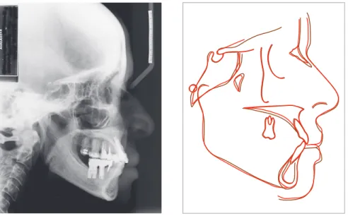
Periapical radiographs revealed generalized mild attachment loss, which suggested judicious periodontal control during orthodontic treatment. In spite of the edentulous regions, bone height was enough for conventional implant placement. Cepha-lometric evaluation revealed skeletal Class III maloc-clusion associated with retrusion of upper incisors and protrusion of lower incisors (Fig 4 and Table 1).
The objectives of treatment were: (1) correct ante-rior crossbite; (2) reestablish vertical dimensions in the posterior region, which would provide space gain for implant-retained prosthetic restorations in the region of lower premolars and molars; (3) close interincisal diaste-ma; (4) gain space for implants and prosthetic crowns in the region of upper lateral incisors; and (5) improve the relationship established between upper and lower lips.
Delay in rehabilitation treatment ater extraction of posterior teeth is expected to provoke alveolar bone atrophy; therefore, only basal bones of the maxilla and mandible remain intact. Lack of dental occlusion in the posterior region leads to a reduction in lower fa-cial height and changes in the position of remaining teeth. The mandible rotates anticlockwise, remod-eling the condyle process and the glenoid fossa.16,17
Figure 2 - Initial dental casts.
Pre-treatment Post-treatment 10-year follow-up
SNA (degrees) 82 81 81
SNB (degrees) 83 81 81.5
ANB (degrees) -1 0 -0.5
1.NA (degrees) 14 25 25.5
1-NA (mm) 4.0 9 9
1.NB (degrees) 24.5 23 23
1-NB (mm) 8.0 5.5 5.5
Pog-NB (mm) 1.5 0.5 0.6
1:1 (degrees) 142 132
SN:OP (degrees) 9 12 12.5
SN:GoGn (degrees) 31 32 32
S to upper lip (mm) -1 1 1
S to lower lip (mm) 2 1 3
FMA (degrees) 28 29.5 29.5
FMIA (degrees) 61 62.5 61.5
IMPA (degrees) 91 88 89
Angle of convexity (degrees) -4 -2 -2.5
Table 1 - Cephalometric data.
Figure 4 - Initial cephalometric tracing.
Presurgical Orthodontics may create space for im-plants to replace missing lateral incisors.
Surgery was considered a risky procedure for a 61-year-old patient. She presented favorable conditions for camoulage, since adequate anchorage could be pro-vided. Conventional dental implants can also be used for orthodontic anchorage. Upper incisors should be pro-clined so as to increase arch perimeter, which would help space gain for upper lateral incisors. Treatment plan was designed according to patient’s needs and expectations.
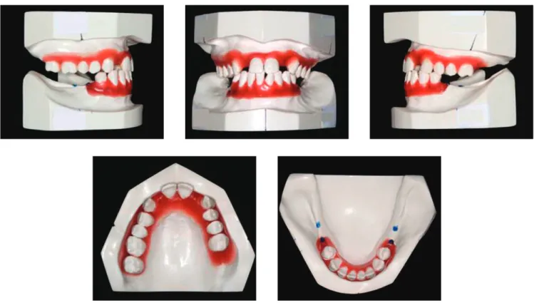
A diagnostic setup was performed according to cephalometric indings. Lower central incisors under-went retrusion of 4 mm and intrusion of 1.5 mm (Fig 5). Upper central incisors were subsequently positioned in contact, with an increase in buccal inclination so as to achieve a 2-mm overjet. Bilateral spaces of 6 mm were created to replace missing upper lateral incisors.
Implants should be inserted 2 mm distal to the
lower right first premolar and 3 mm distal to the
lower left first premolar
. Temporary acrylic crowns were adapted over conventional dental implants (Bråne-mark System, Nobel Biocare, Kloten, Switzerland: 11.5 x 5 mm in the region of molars and 4 x 11.5 mm in the region of premolars) on both sides of the lower arch. Orthodontic brackets were bonded ater six months. Absolute anchorage unit allowed distal movement oflower right canine and let premolar; in addition, it pro-vided retraction and intrusion of lower incisors.
Upper molars were banded and a full ixed orth-odontic appliance was placed (Standard Edgewise 0.022 x 0.028-in, 3M-Unitek, Monrovia, USA). Leveling and alignment followed the sequence of stainless steel archwires in increasing stifness (3M-Unitek, Monro-via, USA). Upper diastema closure and distal movement of lower teeth were performed by sliding mechanics with elastomeric chains.
Retraction of lower incisors and proclination of upper incisors occurred simultaneously. Tear drop loops were bent in 0.018 x 0.025-in stainless steel arch-wires halfway between lateral incisors and canines. Ideal 0.019 x 0.026-in stainless steel archwires allowed detailed angulation to be performed. Total treatment lasted 36 months.
Maxillary implants were inserted ater orthodontic space opening (Brånemark System, Nobel Biocare, Kloten, Switerland: 3.3 x 13 mm on the right side and 3.3 x 15 mm on the let side).
RESULTS
Figure 5 - Setup records.
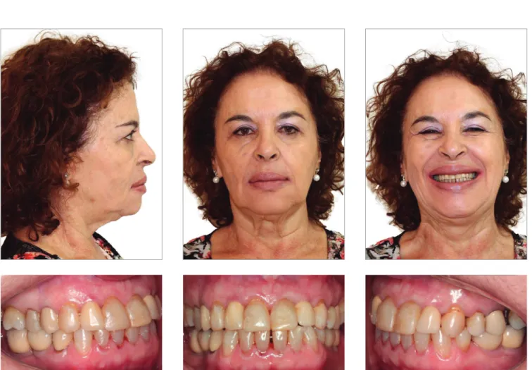
in the posterior region of the lower arch, was achieved (Figs 6 and 7). Midline upper diastema was closed, which favored prosthetic rehabilitation of lateral incisors. Ater bracket debonding, deinite crowns were placed over im-plants, central incisors were restored with resin veneers and other damaged restorations were replaced (Fig 8).
Superimposition of cephalometric tracings revealed that the mandible rotated clockwise (FMA from 28° to 29.5°). There was an increase in the occlusal plane angle (SN-OP from 9° to 12°) and a decrease in the incisor-mandible plane angle (IMPA from 91° to 88°), which relects intrusion and retrusion, respectively, of lower incisors. Proclination of upper incisors was high-lighted by an increase in the 1.NA angle (14° to 25°) (Figs 9 and 10, Table 1).
Regarding the facial proile, maxillary deiciency was camoulaged. Upper and lower lips were improved (upper lip to line, from -1 to 1 mm; lower lip to S-line, from 2 to 1 mm) with upper incisors support.
Ten years ater the completion of the case, the pa-tient showed occlusal stability, as well as integrity of dentition and prostheses. Resin veneers showed pig-mentation and discoloration, as expected. Periodontal structures remained healthy (Figs 11 and 12).
DISCUSSION
Anterior-posterior and transversal Class III mal-occlusion relationships tend to worsen with aging.18,19
Patient’s Class III skeletal pattern associated with loss of lower posterior teeth were limiting factors in the planning of this case. Without orthognathic surgery, conventional mechanics would not solve the patient’s problem. However, there are increased risks associated with surgery and, for this reason, the patient ultimately elected not to undergo surgery.
In this case, prognosis for camouflage was very favorable, considering mild maxillary deficiency and the possibility to procline upper central incisors. The need for oral rehabilitation led this case to be planned based on the use of implants and prosthe-ses. Dentistry restored key features of patient’s qual-ity of life: proper mastication as well as smile and fa-cial esthetics. In 1998, the life expectancy for women in Brazil was 72 years.20 Thus, we offered a reliable
treatment which promotes long-term oral health to our patient. Additionally, transdisciplinary treatment plan fulfilled patient’s needs and expectations.
Figure 6 - Post-treatment facial and intraoral photographs.
Figure 8 - Rehabilitation of the upper incisor region.
Figure 10 - Superimposition of cephalometric tracings at treatment onset (black) and after
treatment completion (red): A) Sella-nasion
plane at sella; B) Best-fit of the maxilla; C) Man-dibular plane at the internal symphysis cortical plate to assess tooth movement, intrusion and incisor repositioning.
Figure 11 - Facial and intraoral photographs 10 years after treatment completion.
A
B
Figure 12 - Radiograph 10 years after treatment completion. Superimposition of cephalomet-ric tracings at treatment completion (red) and 10 years after treatment (brown). Sella-nasion plane at sella.
the possibility of losing implants with the application of orthodontic movement and occlusal forces, leading to decreased alveolar width and height.
Orthodontic forces are small when compared to the complex system of intermittent and multidirectional forces acting on implants during mastication. Thus, biomechani-cal responses are within biologibiomechani-cal limits; for instance, an elastic chain used for canine retraction leads to a force of 1 N or less. The association between orthodontic forces and function stimulates responses of bone modeling and remodeling, which may lead to a new balance of forces.10,16
The approach presented herein took advantage of conventional implants which functioned as orthodontic anchorage before prosthetic procedures.8,21 Therefore,
implants placed in the region of lower molars provided anchorage necessary for intrusion and retraction of an-terior lower teeth. This was considered a worthwhile strategy: previous orthodontic treatment improved oc-clusion and created space necessary for crown place-ment (Fig 6).8,10,14,22 Implant selection and insertion
site must consider patient’s anatomical features, qual-ity and quantqual-ity of bone available (alveolar width and height), gingival conditions, ideal position for teeth re-placement and orthodontic movement.4,14,23,24
Whenever anterior teeth are missing, it is challenging to obtain a natural smile and achieve correct occlusion. Before implants were developed, alternative therapies
for these cases included the use of adhesive crowns and preparation of healthy teeth to function as pillars. Both treatment options have esthetics limitations.25,26
The esthetic objectives of implant therapy include cre-ating adequate gingival margins without abrupt changes in tissue height, maintaining the papilla intact and pre-serving alveolar crest convex contour. To this end, 1-mm space or more, between the implant and the adjacent tooth root, is required in addition to adequate space for crown placement.14 Whenever it is impossible to gain the
space required, space closure with mesial movement of posterior teeth is a reasonable option, especially if only one or two teeth are missing in the anterior region.13
No consensus has been reached regarding the best treatment option to replace missing lateral upper in-cisors.27 It is important to consider various aspects of
1. Agarwal S, Gupta S, Chugh VK, Jain E, Valiathan A, Nanda R. Interdisciplinary treatment of a periodontally compromised adult patient with multiple missing posterior teeth. Am J Orthod Dentofacial Orthop. 2014;145(2):238-48. 2. Pinho T, Neves M, Alves C. Multidisciplinary management including periodontics,
orthodontics, implants, and prosthetics for an adult. Am J Orthod Dentofacial Orthop. 2012;142(2):235-45.
3. Uribe F, Janakiraman N, Nanda R. Interdisciplinary approach for increasing the vertical dimension of occlusion in an adult patient with several missing teeth. Am J Orthod Dentofacial Orthop. 2013;143(6):867-76.
4. Shapiro PA, Kokich VG. Uses of implants in orthodontics. Dent Clin North Am. 1988;32(3):539-50.
5. Nanda R. Biomechanics and esthetic strategies in clinical orthodontics. 1st ed. Philadelphia: WB Saunders; 2005. 385 p.
6. Uribe F, Nanda R. Intramaxillary and intermaxillary absolute anchorage with an endosseous dental implant and rare-earth magnets. Am J Orthod Dentofacial Orthop. 2009;136(1):124-33.
7. Barros LAB, Almeida Cardoso M, de Avila ÉD, Molon RS, Siqueira DF, Mollo-Junior FA, et al. Six-year follow-up of maxillary anterior rehabilitation with forced orthodontic extrusion: achieving esthetic excellence with a multidisciplinary approach. Am J Orthod Dentofacial Orthop. 2013;144(4):607-15.
8. Kuroda S, Iwata M, Tamamura N, Ganzorig K, Hichijo N, Tomita Y, et al. Interdisciplinary treatment of a nonsyndromic oligodontia patient with implant-anchored orthodontics. Am J Orthod Dentofacial Orthop. 2014;145(4 Suppl):S136-47.
9. Alani A, Bishop K, Renton T, Djemal S. Update on guidelines for selecting appropriate patients to receive treatment with dental implants: priorities for the NHS–the position after 15 years. Br Dent J. 2014;217(4):189-90.
10. Favero L, Brollo P, Bressan E. Orthodontic anchorage with speciic ixtures: related study analysis. Am J Orthod Dentofacial Orthop. 2002;122(1):84-94. 11. Chang JZ-C, Liu P-H, Wang Y-T, Chen Y-J, Yao C-CJ, Lai EH-H.
Orthodontic-prosthetic implant anchorage in a partially edentulous patient. J Dent Sci. 2011;6(3):176-80.
12. Farret MM, Benitez Farret MM. Skeletal Class III malocclusion treated using a non-surgical approach supplemented with mini-implants: a case report. J Orthod. 2013;40(3):256-63.
13. Bilodeau JE. A “midline dilemma” in an adult mutilated dentition. Am J Orthod Dentofacial Orthop. 2014;146(3):364-70.
14. Rose TP, Jivraj S, Chee W. The role of orthodontics in implant dentistry. Br Dent J. 2006;201(12):753-64.
REFERENCES
15. Moslehifard E, Nikzad S, Geraminpanah F, Mahboub F. Full-mouth rehabilitation of a patient with severely worn dentition and uneven occlusal plane: a clinical report. J Prosthodont. 2012;21(1):56-64.
16. Higuchi KW. Orthodontic applications of osseointegrated implants. 1st ed. Hanover Park , IL: Quintessence; 2000. 218 p.
17. Melsen B, Lang NP. Biological reactions of alveolar bone to orthodontic loading of oral implants. Clin Oral Implants Res. 2001;12(2):144-52.
18. Berg RE, Espeland L, Stenvik A. A 57-year follow-up study of occlusion. Part 3: Oral health and attitudes to teeth among individuals with crossbite at the age of 8 years. J Orofac Orthop. 2008;69(6):463-83.
19. Miyajima K, McNamara JA, Sana M, Murata S. An estimation of craniofacial growth in the untreated Class III female with anterior crossbite. Am J Orthod Dentofacial Orthop. 1997;112(4):425-34.
20. IBGE. Coordenação de População e Indicadores Sociais. Gerência de estudos e análises da dinâmica demográica projeção da população do Brasil por sexo e idade para o período 1980-2050 revisão 2008. Available from: http://www.ibge.gov.br/home/estatistica/populacao/tabuadevida/evolucao_ da_mortalidade.shtm.
21. Martin W, Hefernan M, Ruskin J. Template fabrication for a midpalatal orthodontic implant: technical note. Int J Oral Maxillofac Implants. 2002;17(5):720-2.
22. Smalley WM. Implants for tooth movement: determining implant location and orientation. J Esthet Dent. 1995;7(2):62-72.
23. Odman J, Lekholm U, Jemt T, Branemark PI, Thilander B. Osseointegrated titanium implants—a new approach in orthodontic treatment. Eur J Orthod. 1988;10(2):98-105.
24. Thilander B, Odman J, Lekholm U. Orthodontic aspects of the use of oral implants in adolescents: a 10-year follow-up study. Eur J Orthod. 2001;23(6):715-31.
25. Zachrisson BU. Improving orthodontic results in cases with maxillary incisors missing. Am J Orthod. 1978;73(3):274-89.
26. Czochrowska EM, Skaare AB, Stenvik A, Zachrisson BU. Outcome of orthodontic space closure with a missing maxillary central incisor. Am J Orthod Dentofacial Orthop. 2003;123(6):597-603.
27. Andrade D, Loureiro C, Araújo V, Riera R, Atallah A. Treatment for agenesis of maxillary lateral incisors: a systematic review. Orthod Craniofac Res. 2013;16(3):129-36.
28. Southard TE, Buckley MJ, Spivey JD, Krizan KE, Casko JS. Intrusion anchorage potential of teeth versus rigid endosseous implants: a clinical and radiographic evaluation. Am J Orthod Dentofacial Orthop. 1995;107(2):115-20.
In addition to absolute anchorage provided by im-plants, biomechanics was similar to the conventional technique. This treatment option requires the under-standing of forces involved in the system and the abil-ity to control the magnitude of forces on implants. Implants are structures ixed to bone and which transfer the load to the teeth which, in turn, are connected by the appliance. It is important to consider the functional characteristics of occlusion with implants, which assures stability and success (Figs 11 and 12).17,28
CONCLUSION