Image
Severe Coronary Milking in Obstructive Hypertrophic Cardiomyopathy
Jorge Humberto Guardado, Hélder Pereira, Carlos Catarino, Hugo Vinhas, Jorge Marques, Manuel Carrageta
Hospital Garcia de Orta - Riachos, Portugal
Mailing Address: Jorge Humberto Guardado •
Serviço de Cardiologia – Hospital Garcia de Orta - 2800 Almada – Portugal E-mail: [email protected]
Manuscript received May 28, 2006; revised manuscript received June 12, 2006; accepted June12, 2006.
Key words
Cardiomyopathy, hypertrophic; coronary disease.
A 65 year old woman with recent onset angina was admitted for acute coronary syndrome without ST elevation. EKG show deep T wave inversion on the anterior and lateral leads.
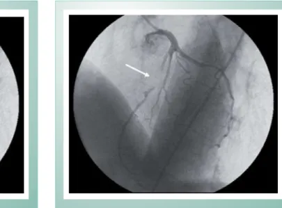
At angiography no coronary stenosis were found, but severe “milking” of the mid left anterior descending coronary artery, up to 100% systolic narrowing was observed (fig. 1, 2, 3 and 4).
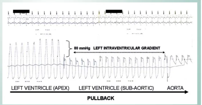
Intraventricular gradient could be elicited at rest by multipurpose catheter during left ventricle pullback (fig. 5).
Two dimensional (cross sectional) echocardiography disclosed asymmetric septal hypertrophy (anterior septum 22 mm, posterior wall 10 mm) with severe and diffuse involvement of the entire interventricular septum and anterolateral wall and left intraventricular gradient at rest was confirmed.
Fig. 1 -End diastolic left coronary angiogram (LAO 45; CRAN 25). The white arrowhead indicates a angiographic normal mid LAD segment.
Fig. 2 -End systolic left coronary angiogram (LAO 45; CRAN 25). The white arrowhead indicates 100% systolic narrowing of mid LAD segment by severe “milking”.
Image
Guardado et al
SEVERE CORONARY MILKING IN OBSTRUCTIVE HYPERTROPHIC CARDIOMYOPATHY
Arq Bras Cardiol 2007; 88(1) : e23-e24
Fig. 3 -End diastolic left coronary angiogram. (RAO 10; CRAN 40). The white arrowhead indicates a angiographic normal mid LAD segment.
Fig. 4 -End systolic left coronary angiogram. (RAO 10; CRAN 40). The white arrowhead indicates 100% systolic narrowing of mid LAD segment by severe “milking”.
Fig. 5 -Left ventricle wave pressure pullback. 80mmHg intraventricular gradient was registered by a multipurpose catheter.