Letters to the Editor
Radiol Bras. 2015 Set/Out;48(5):333–340
337
Primary intercavernous lymphoma of the central nervous system
Linfoma intercavernoso primário do sistema nervoso central
Dear. Editor,
A male, 63-year-old, HIV-negative patient was admitted to the hospital with intermittent frontal headache, right facial pain and diplopia for at least two months. Neurological examination revealed both cranial nerves VI paresis and facial pain in the area innervated by the branches V2 and V3 of the right trigeminal nerve. No alteration was observed in the other cranial nerves. The patients presented with normal gait and unaltered muscle strength and balance. Laboratory tests and cerebrospinal fluid puncture revealed normal results.
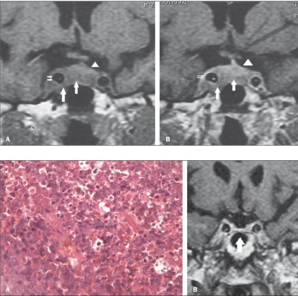
Cranial magnetic resonance imaging (MRI) demonstrated an expansile, intrasellar homogeneous, solid, well delimited lesion, with isosignal on T1- and T2-weighted sequences with a lower enhancement pattern in relation to the hypophysis. The lesion occupied both cavernous sinuses, particularly at right. The way in which the lesion extended suggested the involvement of the in-tercavernous sinus (Figure 1). The patient was submitted to sur-gical treatment, with an uneventful postoperative period. Anatomopathological analysis (Figure 2A) demonstrated diffuse non-Hodgkin’s B-cell lymphoma. The patient underwent comple-mentary radiotherapy, and successive follow-up with MRI did not demonstrate lesion recurrence in up to five years (Figure 2B).
Central nervous system (CNS) lymphoma is a quite rare neo-plasm, affecting most the supratentorial region. In the case of immunocompetent patients, CNS lymphoma manifests as a single, solid lesion, generally without necrosis, with hypo/isosignal on T2- and isosignal on T1-weighted sequences, and intense contrast-enhancement(1,2).
Anatomically, cavernous sinuses are irregularly-shaped, tra-beculated/compartmentalized venous sinuses located along the lateral aspect of the sella turcica(3). Cranial nerves III, IV, V1 and
V2 are located within the lateral dural wall, and not within the cav-ernous sinus(3). The two cavernous sinuses communicate with
each other via anterior and posterior intercavernous venous plex-uses(3). Such connections allow for extension of the
inflamma-tory or neoplastic process to the contralateral sinus(4).
In the case of cavernous sinus lymphoma, considering the proximity to several cranial pairs, cranial nerve lesions may be ob-served; however this is an uncommon finding(1).
CNS lymphomas are rarely found, particularly in immuno-competent individuals. In the present case, the unprecedented characteristic is the infra-hypophyseal involvement by dissemina-tion through the intercavernous sinus. Despite its rarity, particu-larly in immunocompetent individuals, the finding of a solid, ho-mogeneous lesion with isosignal on T1- and T2-weighted se-quences in the region of the cavernous sinuses should raise the hypothesis of lymphoma.
Figure 1. Pre-resection coronal magnetic resonance imaging T1-weighted sequence, before (A) and after (B) contrast agent injection: homogeneous, solid mass (large arrows) showing intermediate signal intensity in the right cavernous sinus, in-volving the right internal carotid artery (small arrows), with con-tralateral extension under the hypophysis. The gland (arrow-heads) is superiorly displaced by
the mass. A B
Figure 2. A: Histopathological analysis demonstrates the pres-ence of cells with large nuclei, sometimes with centrally lo-cated nucleoli and basophilic cytoplasm. B: Six-month post-operative and post-radiotherapy follow-up. Coronal magnetic resonance imaging T1-weighted sequence: no sign of the lesion is seen neither in the sellar cav-ity nor in the cavernous sinuses. The hypophysis is slightly
Letters to the Editor
Radiol Bras. 2015 Set/Out;48(5):333–340
338
http://dx.doi.org/10.1590/0100-3984.2014.0078
Chordoid glioma of the third ventricle
Glioma cordoide do terceiro ventrículo
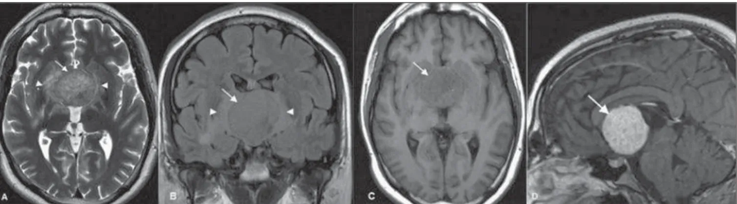
Figure 1. Axial MRI T2-weighted (A) and coronal FLAIR (B) sequences reveal a slightly hyperintense, well-defined hypothalamic/third ventricular tumor (arrows), with perilesional vasogenic edema (arrowheads). C: Axial MRI T1-weighted sequence reveals a predominantly isointense tumor (arrow). D: Gadolinium-enhanced sagittal MRI T1-weighted sequence reveals the tumor with uniform contrast enhancement (arrow).
REFERENCES
1. Reis F, Schwingel R, Nascimento FBP. Central nervous system lym-phoma: iconographic essay. Radiol Bras. 2013;46:110–6.
2. Barreira Junior AK, Moura FC, Monteiro MLR. Linfoma não-Hodgkin bilateral do seio cavernoso como manifestação inicial da síndrome de imunodeficiência adquirida: relato de caso. Arq Bras Oftalmol. 2011;74: 130–1.
3. Osborn AG. Encéfalo de Osborn. Imagem, patologia e anatomia. Porto Alegre, RS: Artmed; 2014.
4. Rocha AJ, Vedolin L, Mendonça RA. Encéfalo. Série CBR. São Paulo, SP: Elsevier; 2012.
Arthur Henrique de Aquino Dultra1, Fabio Noro2, Alessandro
Severo Alves de Melo3, José Alberto Landeiro4, Edson Marchiori4,
Marilene Filgueira do Nascimento5
1. Hospital Copa D’Or, Rio de Janeiro, RJ, Brazil. 2. Universidade Federal do Rio de Janeiro (UFRJ), Rede D’Or, Rio de Janeiro, RJ, Brazil. 3. Universidade Federal Fluminense (UFF), Niterói, RJ, Hospital Barra D’Or, Rio de Janeiro, RJ, Brazil. 4. Universidade Federal Fluminense (UFF), Niterói, RJ, Brazil. 5. Instituto Nacional de Câncer (INCA), Rio de Janeiro, RJ, Brazil. Mailing Address: Dr. Arthur Henrique de Aquino Dultra. Rua Real Grandeza, 281, Botafogo. Rio de Janeiro, RJ, Brazil, 22281-035. E-mail: [email protected].
Dear Editor,
A previously healthy 27-year-old man was referred with an 8-month history of headaches, memory loss, progressive weight gain (obesity), hyperphagia and behavior changes.
Computed tomography (CT) scans revealed the presence of a midline, solid, and homogeneously enhancing mass involving the anterior aspect of the third ventricle.
Brain magnetic resonance imaging (MRI) (Figure 1) showed a well-defined, rounded mass in the third ventricle, measuring about 4.0 cm in the craniocaudal axis. The tumor was slightly heterogeneous, predominantly isointense at T1- and T2-weighted MRI sequences, presenting with diffuse enhancement after ga-dolinium injection. Perilesional vasogenic edema, compression and subsequent displacement of midbrain and hypothalamic structures were observed.
A subtotal resection of the tumor was microsurgically per-formed by interhemispheric transcallosal approach to the third ventricle.
The tumor was histologically classified as a chordoid glioma. The mass showed nests of regular epithelioid cells with large nuclei, prominent nucleoli, and abundant eosinophilic cytoplasm, within a myxoid stroma. Sparse lymphocytic infiltrate was present. Im-munohistochemical studies demonstrated diffuse cytoplasmic expression for glial fibrillary acidic protein, vimentin, and CD34. The patient died three months after surgery as a conse-quence of massive hypothalamic invasion combined with pneu-monia.
Chordoid glioma is an unusual, noninvasive and slow-grow-ing tumor that arises from the anterior third ventricle, frequently adherent to the hypothalamus(1). There are reports in the
litera-ture about chordoid gliomas in other locations, such as the tem-poroparietal region, left thalamus and the corona radiata/thala-mus(2,3), most of them affecting children(2).
It is typically a well-circumscribed, round or oval-shaped tu-mor, with greatest diameter in the craniocaudal direction. The tumor is hyperdense to the gray matter at CT, isointense at MRI T1-weighted sequences, and isointense to slightly hyperintense at MRI long-TR, with strong, uniform enhancement after con-trast agent administration(1,2,4–6). Cystic changes and necrosis
may be present(2,5,7). Calcifications are usually rare(2,5,7). Usually,
bilateral and symmetric perilesional vasogenic edema may also be observed(3–5).
Given the tumor location, patients usually present with signs and symptoms related to obstructive hydrocephalus, such as nau-sea and headache, although endocrine imbalance, visual distur-bances, behavior disorders and autonomic dysfunction are also reported in the literature(1,4–6).
The histological and immunohistochemical features of these tumors are very typical and uniform, characterized by cords of oval to polygonal epithelioid cells with abundant eosinophilic cytoplasm and avid staining for glial fibrillary acidic protein and vimentin(1,2,4).
The differential diagnosis includes masses of suprasellar re-gion, such as pituitary macroadenoma, craniopharyngioma, optic and hypothalamic pilocytic astrocytoma, meningioma, ependy-moma and lymphoma(2,4).
Currently, the treatment of choice is complete surgical re-section of the tumor(1,4,6). Adjuvant radiotherapy has been used
following subtotal resection(2).
Despite being a low-grade tumor, the prognosis is usually poor because of its location and the difficulty in obtaining complete surgical resection without causing severe hypothalamic symp-toms(4). On the other hand, partial resection of the tumor is