Medullary breast carcinoma: anatomo-radiological
correlation*
Carcinoma medular da mama: correlação anátomo-radiológica
Valéria Soares Matheus1, Fabíola Procaci Kestelman2, Ellyete de Oliveira Canella2, Maria Célia Resende Djahjah3, Hilton Augusto Koch4
OBJECTIVE: To evaluate radiological findings in patients submitted to surgical treatment for medullary breast cancer at Instituto Nacional de Câncer (INCA), Rio de Janeiro, RJ, Brazil, correlating them with histological results. MATERIALS AND METHODS: A retrospective descriptive study was developed with patients submitted to surgery at INCA, in the period from January 1997 to December 2006, for identifying the presence of medullary breast carcinoma and analyzing radiological findings. RESULTS: Among 21,287 patients diagnosed with carcinoma, 76 (0.357%) had typical medullary breast carcinoma. The age range of these patients was 32–81 years (mean = 59.1 years). Mammography demonstrated lesions in 19 of these patients, 17 (89.5%) of them with masses, and 2 with focal asymmetry. Among the patients with masses, 15 (88.1%) presented with high density and 2 (11.9%) with isodensity. Twelve patients presented sonographic findings, 11 (91.6%) of them with hypoechoic masses, and one with an anechoic mass with areas of cystic degeneration. CONCLUSION: Nodular mass was the predominant radiological finding (89.5%), 88.1% of them corresponding to masses with high density and circumscribed margins. Despite the radiological characteristics of benignity, a solid, fast-growing, highly dense mass with circumscribed margins should be further investigated to confirm the diagnosis. Keywords: Medullary breast cancer; Radiological findings; Diagnosis.
OBJETIVO: Avaliar as características radiológicas do câncer de mama medular em pacientes submetidas a tratamento cirúrgico no Instituto Nacional de Câncer (INCA) – Ministério da Saúde, Rio de Janeiro, RJ, cor-relacionando os achados com estudo histopatológico. MATERIAIS E MÉTODOS: Foi realizado estudo descri-tivo retrospecdescri-tivo de mulheres submetidas a tratamento cirúrgico no INCA, no período de janeiro de 1997 a dezembro de 2006, para identificação das pacientes com carcinoma medular e análise dos achados radioló-gicos. RESULTADOS: Foram identificadas 21.287 pacientes com diagnóstico de carcinoma neste período, sendo 76 pacientes com diagnóstico de carcinoma medular típico (0,357%). Nessas pacientes seleciona-das, a idade média foi de 51,9 anos (32 a 81 anos). Dezenove pacientes apresentavam lesão na mamogra-fia, sendo 17 (89,5%) nódulos e 2 assimetrias focais (10,5%). Entre as pacientes com nódulo, 15 (88,1%) apresentavam alta densidade e 2 eram isodensos (11,9%). Doze pacientes apresentavam achados ultra-so-nográficos e, destas, 11 (91,6%) apresentavam nódulos hipoecóicos. Foi observada uma paciente com nódulo anecóico com áreas de degeneração cística. CONCLUSÃO: O nódulo foi o achado radiológico dominante (89,5%), dos quais 88,1% apresentaram nódulos com alta densidade e margens circunscritas. Apesar das características radiológicas de benignidade, um nódulo com alta densidade, sólido, margens circunscritas e crescimento rápido deve ser investigado para confirmar o diagnóstico.
Unitermos: Câncer de mama medular; Achados radiológicos; Diagnóstico. Abstract
Resumo
* Study developed at Instituto Nacional de Câncer (INCA), Rio de Janeiro, RJ, Brazil.
1. MD, Gynecologist and Mastologist at Hospital Naval Mar-cílio Dias, Rio de Janeiro, RJ, Brazil.
2. Masters, MDs, Radiologists, Instituto Nacional de Câncer (INCA), Rio de Janeiro, RJ, Brazil.
3. PhD, Associate Professor, Universidade Federal do Rio de Janeiro (UFRJ), MD, Radiologist, Instituto Nacional de Câncer (INCA), Rio de Janeiro, RJ, Brazil.
4. PhD, Titular Professor of Radiology, Universidade Federal do Rio de Janeiro (UFRJ) and Pontifícia Universidade Católica do Rio de Janeiro (PUC Rio), Rio de Janeiro, RJ, Brazil
Mailing address: Dra. Valéria Soares Matheus. Avenida Er-nani do Amaral Peixoto, 500, sala 901, Centro. Niterói, RJ, Brazil, 24020-077. E-mail: [email protected]
Received August 12, 2008. Accepted after revision October 13, 2008.
breast diseases, and is considered as the golden-standard for screening of minimal breast lesions(2,3). The utilization of
mam-mography as a screening method for women with > 40 years of age is defined in the United States of America(4–6). In Brazil, this
method is recommended for women aged between 50 and 60 years, according to the INCA consensus on breast cancer(1–7). In
Canada and United Kingdom mammogra-phy is recommended for asymptomatic women from 50 years of age(8–11).
.
Matheus VS, Kestelman FP, Canella EO, Djahjah MCR, Koch HA. Medullary breast carcinoma: anatomo-radiological corre-lation. Radiol Bras. 2008;41(6):379–383.
INTRODUCTION
In Brazil, breast cancer is the most fre-quent malignant neoplasm among women. According to Instituto Nacional de Câncer (INCA), the city of Rio de Janeiro presents the highest incidence, with Porto Alegre in the second place(1).
Reduction in the mortality rate depends on the early detection and an appropriate therapeutic planning(1). Mammography
Mammography is highly sensitive for detecting clinically occult breast cancer. A review of clinical studies evaluating the performance of this method has demon-strated that its sensitivity ranged between 71% and 98% for yearly screening mam-mography(1,10–16). However, many lesions
considered suspicious and indicating the necessity of histopathological study corre-spond to benign alterations. In the United States of America, the positive predictive value of biopsies performed because of mammographic findings, i.e., the propor-tion of malignant lesions diagnosed in a total of biopsies performed ranges between 15% and 40%. Both cost and morbidity of interventions for diagnosis of these lesions are taken into consideration for confirming the utilization of mammography as screen-ing for breast cancer(12,13,15–19).
Medullary breast carcinoma, currently considered as basal carcinoma, corre-sponds to a subgroup of malignant tumors most frequently detected as an “interval cancer” in comparison with tumors diag-nosed in the mammographic screening(20– 26). This tumor represents 2% to 7% of
breast cancers and is most frequently found in young women(27–30).
One of the difficulties observed in the analysis of the images is the absence of pathognomonic features in a great number of diagnosed lesions. Knutzen and Gis-vold(9) have developed a study about the
likelihood of malignancy in several catego-ries of non-palpable lesions detected by mammography, with the objective of defin-ing the necessity of follow-up or biopsy for these lesions. The authors have observed that if morphological criteria were taken into consideration in the evaluation of these lesions, the rate of malignant lesions sub-mitted to biopsy could reach 40%. Few studies in the literature have evaluated this type of breast cancer.
The relevance of the investigation of these lesions as well as their imaginological features increases, considering that many times medullary breast cancer presents morphologically like a benign lesion, and that this histological type of lesion is asso-ciated with high-risk patients. The present study was aimed at evaluating mammo-graphic and sonomammo-graphic findings of med-ullary breast carcinoma.
MATERIALS AND METHODS
In the period between January 1997 and December 2006, 21,287 patients were di-agnosed with breast carcinoma at INCA-III. Among these patients, 76 (0.357%) pre-sented a diagnosis of medullary carcinoma. Out of these 76 patients, those who had not a pure medullary carcinoma were excluded (n = 4), as well as those whose previous
radiological studies could not be found (n
= 53). Among the patients without previous radiological studies (n = 53), 20 had radio-logical reports with no image, and 33 had neither radiological report nor image, like one patient with a large ulcerated blastoma referred for investigation with no previous radiological study of the involved breast.
Seventy-six records and radiological studies were retrospectively evaluated, and 19 patients diagnosed with typical medul-lary carcinoma and with preoperative ra-diological study were included in the present study. Radiological findings, BI-RADS® category, tumor size at diagnosis
and patient’s age were analyzed.
The present study was approved by the INCA Council of Ethics in Research under the number 041/08.
RESULTS
Nineteen patients were included be-cause they had preoperative radiological imaging studies. The mean age of these selected patients was 51.9 years (32 to 81 years). All of them had their lesions de-tected by mammography with the follow-ing findfollow-ings: 17 masses (89.5%) and two focal asymmetries (10.5%). Among the masses, 15 (88.2%) presented high density and two (11.9%) were isodense. Twelve patients presented sonographic findings, 11 (91.6%) of them with hypoechoic masses. Anechoic mass with areas of cystic degen-eration was found in one patient.
Lobulated margin was most frequently observed in 11 patients (64.7%), and microlobulated margins in 9 (52.9%). Mean tumor size was 27.2 mm (14–50 mm). Most frequently, the tumors ware classified as BI-RADS category IV (52.7% of the patients).
The prevalent morphological aspect was mass, in 89.5% of the patients, with a
lobular shape in 11 (64.7%) and irregular shape in 6 (35.3%) patients. Microlobu-lated margin was found in 9 patients (52.9%), obscured margin in 4 (23.5%), speculate in 2 (11.8%) and indistinct in 2 patients (11.8%). High density was ob-served in 15 patients (88.1%), while iso-density was found in only 2 patients (11.8%). Architectural distortion and microcalcifications were not observed. Two patients presented “focal asymmetry” as a sole radiological alteration.
DISCUSSION
Breast cancer is a multifactorial disease, and in most of cases is considered as be-ing of epithelial origin, and is divided into an array of histological types whose behav-ior varies according to several factors such as degree of histological differentiation, subtype, etc. Some histological types of breast tumors such as medullary carci-noma, mucinous carcinoma and tubular carcinoma are associated with a better prognosis. Although rarely reported, other malignant tumors such as lymphomas, sar-comas and melanomas can also occur in the breast(22).
Among the malignant tumors of the breast, infiltrating ductal carcinoma is the most frequently found neoplasm. Progno-sis is unfavorable in cases of isolated le-sions, contrarily to the cases with associa-tion of other histological types of tumors such as tubular, invasive cribriform, cystic adenoid, mucinous and papillary, espe-cially in cases where these tumors consti-tute more than 90% of the mass.
Infiltrating ductal carcinoma and ductal carcinoma in situ represent 85% of malig-nant breast tumors(20). Typically, infiltrating
ductal carcinoma presents as a spiculate, irregular or focal asymmetrical mass, while ductal carcinoma in situ presents as pleomophic or linear microcalcifications(22).
Medullary carcinoma represents rare tu-mor of intermediate prognosis (20,24), with
an incidence ranging from 1% to 7% of all cases of breast cancer(22). In the present
reference center for treatment of breast cancer in Rio de Janeiro. In the general population the age range of patients diag-nosed with breast cancer is between 45 and 52 years. Mean age of patients in the present study was 51.9 years, similarly to data reported in the literature re-viewed(20,24,27–29). The World Health
Orga-nization describes this tumor as “a well-circumscribed carcinoma composed of poorly differentiated cells with scant
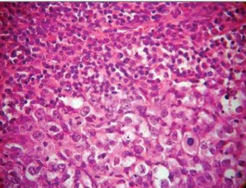
stroma and prominent lymphoid infiltra-tion”(23,25,29–32)(Figure 1).
It is a tumor with well-circumscribed margins with high histological and nuclear degree, composed of poorly differentiated cells, that does not present a tubular or glandular structure, but rather a prominent lymphoplasmocytary infiltrate with nuclear pleomorphism and high proliferation(32).
The tumor cells are large, with macronu-clei, syncytially arranged, with moderate,
abundant and diffuse inflammatory lymphoplasmocytary infiltrate among cell clusters. Clinically, the tumor is well-de-limited, and at mammography is typically well-circumscribed (Figures 2 and 3), and may be confused with a benign lesion(32–34).
Nodular, lobulated lesion with micro-lobulated margins was the radiological as-pect most frequently found in the present casuistic, likewise in several studies in the literature(20,23,24,28,29,32,33)(Figures 2 and 3).
In the present study, high density was a preponderant finding similarly to the litera-ture(35). The BI-RADS IV was the most
fre-quently observed category, in agreement with the worldwide literature, although category II has also been found in the present study(19,22,26,31). At ultrasonography,
the lesion is homogeneous and hypoechoic, or hypoechoic with intermediate heteroge-neity, according to the present casuis-tic(26,27)(Figures 2 and 3). Macroscopically,
well-delimited margins and soft consis-tency are observed. Hemorrhage and necro-sis are frequent findings. Fibrotic stroma is scarce. These tumors are frequently asso-ciated with the BRCA1 and BRCA2 genes. They are estrogen-receptors and pro-gestagen negative(36,37). Despite the
pres-ence of biological markers compatible with
A
B C
D
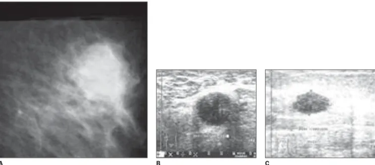
Figure 2. Mediolateral oblique mammogram dem-onstrating a lobulated mass with ill-defined mar-gins (A). BI-RADS category III. The mass is hypo-echoic at ultrasonography (B,C,D).
high aggressiveness, the pure medullary carcinoma presents a favorable prognosis. Despite the questioning of some authors(26, 35,36), the ten-year survival rate ranges
be-tween 50% and 90%. The differences in the diagnostic criteria can justify the character-istics of the tumor prognosis. Additionally, according to some authors, the incidence of this tumor is higher in younger women (35 years)(21). Clinically, these tumors are
char-acterized by fast growth and frequently manifest as a palpable mass(29).
According to Korsching et al.(28),
med-ullary carcinoma in included in a subgroup of invasive breast tumors called basal car-cinomas which have been focused by breast cancer research as a result of the introduction of the global gene expression analysis. This subgroup is characterized by a distinctive biology generating many ques-tions regarding pathogenesis, chemosensi-tivity and optimal clinical management of these tumors. Basal carcinoma is usually estrogen-receptor, progestagen- and HER2-negative, but they are not identical to the triple negative tumors(36,37)..
Bertucci et al.(37)refer to medullary
breast cancer as a rare but enigmatic tumor. Little is known about the molecular alter-ations associated with medullary carci-noma(21,37). When compared with
medul-lary carcinoma, basal-like ductal carcinoma overexpress genes involved in muscular differentiation, suggesting that medullary
carcinoma is a distinct subgroup of basal cancer with a limited myoepithelial differ-entiation(28,29,31,36). In addition, Bertucci et
al. report an overexpression of a series of genes located in the chromosomal regions 12p 13 and 6p 21, which are known by their pluripotential genes content(37). The
major-ity of patients are young and asymptomatic, and can be investigated by mammography, especially if associated with clinical exami-nation and ultrasonography.
Few studies have evaluated this type of breast cancer. Considering that, many times, medullary carcinoma presents a morphological aspect of benign lesion and that this histological type of tumor is asso-ciated with high-risk women, the radiologi-cal evaluation plays a relevant role in the diagnosis of this type of lesion.
Considering the increase in the number of cases in young women, the insidious on-set and fast-growth of the disease, the in-creasing significance of the investigation of basal cancer, dubious prognosis, and the radiological aspect of medullary cancer similar to that of a benign lesion (false-negative), thus delaying the early diagno-sis, imaginological findings are essential in the determination of the diagnosis. This study demonstrated that the findings in the sample evaluated were nodular lesion and asymmetry. In the nodular lesions, the ra-diological characteristics most frequently found were increased density, lobular
shape, circumscribed margins and size up to 20 mm. The relevance of this study is in the increasing correlation observed be-tween the presence of medullary carcinoma and BRCA1, and less frequently, BRCA2 gene mutation(26) and moreover, because of
the radiological aspect of these lesions which may be interpreted as benign tumors and therefore, false-negative(25–28,34–37).
The present study was limited by rarity of this tumor and, above all, because of the radiological data loss.
CONCLUSION
In spite of being a fairly rare type of tu-mor, medullary breast carcinoma is found in high-risk women. Morphological char-acteristics on imaginological studies should be taken into consideration and the hypoth-esis of medullary carcinoma should not be ruled out, considering that these lesions should be investigated by histopathologi-cal study and not by radiologihistopathologi-cal follow-up.
Acknowledgements
To the colleagues of the Division of Ra-diology, especially Dr. Fátima Maria Car-doso Garcia; Division of Pathological Anatomy, especially Dr. Paulo Farias; and Division of Epidemiology of Instituto Nacional de Câncer, especially Dr. Luiz Cláudio Thuler, whose support was essen-tial to bring the present study into reality.
Figure 3. Mammography demonstrating an irregular mass with obscured margins (A). BI-RADS category IV. The mass is hypoechoic at ultrasonography (B,C).
REFERENCES
1. Brasil. Ministério da Saúde. Secretaria de Aten-ção à Saúde. Instituto Nacional de Câncer. Coor-denação de Prevenção e Vigilância. Parâmetros técnicos para programação de ações de detecção precoce do câncer de mama. Recomendações para gestores estaduais e municipais. Rio de Ja-neiro: Ministério da Saúde – INCA; 2006.
2. Melhado VC, Alvares BR, Almeida OJ. Correla-ção radiológica e histológica de lesões mamárias não-palpáveis em pacientes submetidas a marca-ção pré-cirúrgica, utilizando-se o sistema BI-RADS. Radiol Bras. 2007;40:9–11.
3. Roveda Jr DR, Piato S, Oliveira VM, et al. Valo-res preditivos das categorias 3, 4 e 5 do sistema BI-RADS em lesões mamárias nodulares não-palpáveis avaliadas por mamografia, ultra-sono-grafia e ressonância magnética. Radiol Bras. 2007;40:93–8.
4. Ball CG, Butchart M, MacFarlane JK. Effect on biopsy technique of the breast imaging reporting and data system (BI-RADS) for nonpalpable mammographic abnormalities. Can J Surg. 2002; 45:259–63.
5. Baségio DL. Métodos de diagnóstico do câncer de mama – uma contribuição às Bases para um Programa de Detecção Precoce do Câncer de Mama [tese de doutorado]. Rio de Janeiro: Uni-versidade Federal do Rio de Janeiro; 1999. 6. Bérubé M, Curpen B, Ugolini P, et al. Level of
suspicion of a mammographic lesion: use of fea-tures defined by BI-RADS lexicon and correla-tion with large-core breast biopsy. Can Assoc Radiol J. 1988;49:223–8.
7. Brasil. Ministério da Saúde. Conselho Nacional de Saúde. Resolução nº 196/96 sobre pesquisa envolvendo seres humanos. Bioética. 1996;4 Supl:15–25.
8. Heywang-Köbrunner SH, Dershaw DD, Schreer I. Diagnostic breast imaging. Mammography, sonography, magnetic resonance imaging, and interventional procedures. 2nd ed. New York: Thieme; 2001.
9. Knutzen AM, Gisvold JJ. Likelihood of malignant disease for various categories of mammographic-ally detected, nonpalpable breast lesions. Mayo Clin Proc. 1993;68:454–60.
10. Morrison BJ. Screening for breast cancer. In: Ca-nadian Task Force on Preventive Health Care. [cited 2004 Nov 10]. Available from: http://www. ctfphc.org
11. Ciatto S, Cataliotti L, Distante V. Nonpalpable lesions detected with mammography: review of 512 consecutive cases. Radiology. 1987;165:99– 102.
12. Chala LF, Barros N. Avaliação das mamas como método de imagem. Radiol Bras. 2007;40(1):iv– vi.
13. Kestelman FP, Souza GA, Thuler LC, et al. Breast Imaging Reporting and Data System — BI-RADS®: valor preditivo positivo das categorias 3, 4 e 5. Revisão sistemática da literature. Radiol Bras. 2007;40:173–7.
14. Newman LA, Sabel M. Advances in breast can-cer detection and management. Med Clin North Am. 2003;87:997–1028.
15. Humphrey LL, Helfand M, Chan B, et al. Breast cancer screening: a summary of the evidence for the U.S. Preventive Services Task Force. Ann Intern Med. 2002;137(5 Part 1):347–60. 16. Thuler LC. Considerações sobre a prevenção do
câncer de mama feminino. Rev Bras Cancerol. 2003;49:227–38.
17. Liberman L, Menell JH. Breast imaging report-ing and data system (BI-RADS). Radiol Clin North Am. 2002;40:409–30.
18. Louveira MH. Avaliação da eficácia da ultra-so-nografia na diferenciação entre nódulos mamá-rios sólidos benignos e malignos baseada na as-sociação das características ultra-sonográficas de 176 nódulos com o resultado do estudo anatomo-patológico [tese de doutorado]. São Paulo: Uni-versidade Federal de São Paulo; 2003.
19. Calas MJG. Ultra-sonografia mamária: revisão e validação de uma proposta de classificação eco-gráfica [tese de mestrado]. Rio de Janeiro: Uni-versidade Federal do Rio de Janeiro; 2005.
20. Khomsi F, Ben Bachouche W, Bouzaiene H, et al. Typical medullary carcinoma of the breast: a ret-rospective study about 33 cases. Gynécol Obstét Fertil. 2007;35:1117–22.
21. Bertucci F, Finetti P, Cervera N, et al. Gene ex-pression profiling shows medullary breast cancer is a subgrup of basal breast cancers. Cancer Res. 2006;66:4636–44.
22. Schreer I, Lüttges J. Precursor lesions of invasive breast cancer. Eur J Radiol. 2005;54:62–71.
23. Liberman L, LaTrenta LR, Samli B, et al. Over-diagnosis of medullary carcinoma: a mammo-graphic-pathologic correlative study. Radiology. 1996;201:443–6.
24. Yilmaz E, Lebe B, Balci P, et al. Comparison of mammographic and sonographic findings in typi-cal and atypitypi-cal medullary carcinomas of the breast. Clin Radiol. 2002;57:640–5.
25. Eichhorn JH. Medullary carcinoma, provocative now as then. Semin Diagn Pathol. 2004;21:65– 73.
26. Meyer JE, Amin E, Lindfors KK, et al. Medullary carcinoma of the breast: mammographic and US appearance. Radiology. 1989;170(1 Pt 1):79–82.
27. Harvey JA. Unusual breast cancers: useful clues to expanding the differential diagnosis. Radiol-ogy. 2007;242:683–94.
28. Korsching E, Jeffrey SS, Meinerz W, et al. Basal carcinoma of the breast revisited: an old entity with new interpretations. J Clin Pathol. 2008;61: 553–60.
29. Cheung YC, Chen SC, Lee KF, et al. Sonographic and pathologic findings in typical and atypical medullary carcinomas of the breast. J Clin Ultra-sound. 2000;28:325–31.
30. Ashida A, Fukutomi T, Tsuda H, et al. Atypical medullary carcinoma of the breast with cartilagi-nous metaplasia in a patient with BRCA1 germ-line mutation. Jpn J Clin Oncol. 2000;30:30–2. 31. Schrading S, Kuhl CK. Mammographic, US, and MR imaging phenotypes of familial breast can-cer. Radiology. 2008;246:58–70.
32. Ribeiro-Silva A, Ramalho LNZ, Garcia SB, et al. Does the correlation between EBNA-1 and p63 expression in breast carcinomas provide a clue to tumorigenesis in Epstein-Barr virus-related breast malignancies? Braz J Med Biol Res. 2004;37:89– 95.
33. Jury O, Pizarro Z, Díaz A, et al. Presentación mamográfica y compromiso axilar en cáncer de mama específico. Rev Chil Cir. 2005;57:389–92. 34. Lorente Ramos RM, del Valle Sanz Y, Alcaraz Mexía MJ, et al. Carcinoma medular de mama: una lesion maligna que simula benignidad. Ra-diología. 2006;48:165–8.
35. Rakha EA, Reis-Filho JS, Ellis IO. Basal-like breast cancer: a critical review. J Clin Oncol. 2008;26:2568–81.