Value of thyroid echogenicity in the diagnosis of chronic
autoimmune thyroiditis*
Importância da ecogenicidade da tireóide no diagnóstico da tireoidite crônica auto-imune
Danilo Bianchini Höfling1, Giovanni Guido Cerri2, Adriana Gonçalves Juliano3, Suemi Marui4, Maria Cristina Chammas5
Chronic autoimmune thyroiditis is currently considered as the main cause for hypothyroidism and its diagnosis is based on clinical manifestations and laboratory tests results. The most significant laboratory marker for this disease is the presence of anti-thyroperoxidase and anti-thyroglobulin antibodies, the latter being the most sensitive one. Aspiration biopsy shows high sensitivity and specificity but, considering the invasiveness of this method it is reserved for cases of suspected nodules or fast growing goiter. Scintigraphy is unnecessary for the diagnosis, considering its low specificity and sensitivity. Both B-mode and duplex color Doppler ultrasonography have rapidly evolved, becoming a simple, noninvasive and reproducible method with high sensitivity for the diagnosis of chronic autoimmune thyroiditis. Echogenicity is a relevant parameter at B-mode ultrasonography because of the close correlation with cytopathological findings besides the high sensitivity in the diagnosis of the disease. Although this is not a specific parameter for chronic autoimmune thyroiditis, considering that it may also be found in Graves’ disease, postpartum thyroiditis and subacute thyroiditis, such disorders can be easily differentiated both by clinical-laboratory and duplex color Doppler findings. Thus, the present study is aimed at evaluating the significance of thyroid echogenicity in the diagnosis of chronic autoimmune thyroiditis.
Keywords: Ultrasonography; Echogenicity; Thyroiditis; Chronic; Autoimmune.
A tireoidite crônica auto-imune é, atualmente, a principal causa de hipotireoidismo e seu diagnóstico baseia-se nas manifestações clínico-laboratoriais. O marcador laboratorial mais importante é a prebaseia-sença de anticor-pos antitireoglobulina e antiperoxidase, sendo este último o teste mais sensível. A biópsia aspirativa apre-senta alta sensibilidade e especificidade, porém, é um método invasivo e, por isso, reservado para quando há presença de nódulo ou bócio de crescimento rápido. A cintilografia é desnecessária para o diagnóstico, já que apresenta baixa sensibilidade e especificidade. A ultra-sonografia, tanto ao modo B como ao dúplex-Doppler colorido, evoluiu de forma muito rápida e tornou-se um método simples, não-invasivo, reprodutível e com alta sensibilidade para o diagnóstico da tireoidite crônica auto-imune. Ao modo B, a ecogenicidade é um parâmetro de extrema importância, já que, além de apresentar alta correlação com o quadro citopatoló-gico, também apresenta alta sensibilidade para o diagnóstico da tireoidite crônica auto-imune. Embora este parâmetro não seja específico da tireoidite crônica auto-imune, pois também pode estar presente na doença de Graves, na tireoidite pós-parto e na tireoidite subaguda, tais desordens podem ser facilmente diferencia-das tanto pelo quadro clínico-laboratorial quanto pelo dúplex-Doppler colorido. Assim, este artigo tem o objetivo de revisar a importância do estudo da ecogenicidade no diagnóstico da tireoidite crônica auto-imune. Unitermos: Ultra-sonografia; Ecogenicidade; Tireoidite; Crônica; Auto-imune.
Abstract
Resumo
* Study developed in the Instituto de Radiologia do Hospital das Clínicas da Faculdade de Medicina da Universidade de São Paulo (InRad/HC-FMUSP), São Paulo, SP, Brazil.
1. MD, Fellow PhD degree, Instituto de Radiologia do Hospi-tal das Clínicas da Faculdade de Medicina da Universidade de São Paulo (InRad/HC-FMUSP), São Paulo, SP, Brazil.
2. Full Professor, Division of Radiology at Hospital das Clíni-cas da Faculdade de Medicina da Universidade de São Paulo (HC-FMUSP), São Paulo, SP, Brazil.
3. Assistant Physician at the Unit of Ultrasonography, Instituto de Radiologia do Hospital das Clínicas da Faculdade de Medicina da Universidade de São Paulo (InRad/HC-FMUSP), São Paulo, SP, Brazil.
4. PhD, Assistant Physician at the Unit of Endocrinology and Metabology, Hospital das Clínicas da Faculdade de Medicina da Universidade de São Paulo (HC-FMUSP), São Paulo, SP, Brazil. 5. PhD, Director for the Unit of Ultrasonography, Instituto de
cytic infiltrate at variable intensities in the thyroid parenchyma and production of anti-thyroid antibodies(1). Amongst such
disor-ders, chronic autoimmune lymphocytic thyroiditis stands out as the most frequent cause of hypothyroidism(2) and one of the
most frequent specific autoimmune dis-eases affecting humans(3). The prevalence
of chronic autoimmune thyroiditis is higher after 50 years of age(4,5), affecting 5%-15%
of women and 1%-5% of men, according to diagnostic criteria and geographic
local-Höfling DB, Cerri GG, Juliano AG, Marui S, Chammas MC. Value of thyroid echogenicity in the diagnosis of chronic auto-immune thyroiditis. Radiol Bras. 2008;41(6):409–417.
Radiologia do Hospital das Clínicas da Faculdade de Medicina da Universidade de São Paulo (InRad/HC-FMUSP), São Paulo, SP, Brazil.
Mailing address: Dr. Danilo Bianchini Höfling. Rua Tabapuã, 111, 9º andar, Itaim Bibi. São Paulo, SP, Brazil, 04533-010. E-mail: [email protected]
Received April 14, 2008. Accepted after revision June 5, 2008.
INTRODUCTION
lympho-ization(6), with a potential incidence nine
times higher in women than in men(7).
Under the clinical point of view, chronic autoimmune thyroiditis may present under two forms, namely: atrophic thyroiditis and goiter or Hashimotos’s thyroiditis de-scribed by this author in 1912(8,9). Both
forms are characterized by the presence of lymphocytic thyroiditis, thyroid anti-bodies in the blood, and different degrees of thyroid dysfunction(6). Grave’s disease
is one of the forms of autoimmune diseases of the thyroid gland that may be associated with the presence of antithyroid antibod-ies(6). Equally, silent thyroiditis (painless)
and postpartum thyroiditis constitute self-limited autoimmune disorders which present lymphocytic thyroid infiltration, both supposedly attributed to manifesta-tions of chronic autoimmune thyroiditis(4).
Focal thyroiditis — another presentation of autoimmune disease of the thyroid gland —, may simulate a thyroid neoplasm(1,10).
Chronic autoimmune thyroiditis with goiter is characterized by a diffuse lympho-cytic thyroid infiltration, with occasional germinative centers, thyroid follicles with scarce colloid, fibrosis, and oxyphilic alter-ations, such as Hürthle or Askanazy cells. In atrophic chronic autoimmune thyroidi-tis, the thyroid gland is small, with lympho-cytic infiltrate and fibrosis replacing the glandular parenchyma(6). In both variants,
lymphocytic infiltrate is the finding that characterizes the presence of thyroid gland autoimmunity(11).
CLINICAL, LABORATORY AND CYTOLOGICAL DIAGNOSIS
Diagnostic criteria for chronic autoim-mune thyroiditis are based both on clinical signs and subsidiary studies(6). The patients
may present with symptoms of hypothy-roidism, and clinical examination may demonstrate signs of this condition, but typically only a diffuse goiter is found, with increased consistency, lobulated surface and rarely large enough to cause compres-sive symptoms. Typically, patients with the atrophic presentation of chronic autoim-mune thyroiditis do not present goiter(6), on
the contrary, they present with a decreased glandular volume at the moment of the di-agnosis. This form of disease may present
in the absence of a pre-existing goiter, or may result from a slow and gradual pro-gression of the disease with goiter(12).
The main laboratory marker for chronic autoimmune thyroiditis is the presence of anti-thyroid serum antibodies. Antithyreo-globulin antibodies (TgAb) were present in 60% of patients, while anti-thyroid mi-crosomal antibodies are present in 95% of patients(6)
. Most of times, anti-thyroid mi-crosomal antibodies concentrations are higher than TgAb concentrations, and a positive result for thyroid peroxidase anti-body (TPOAb) is a subtly more sensitive marker for chronic autoimmune thyroidi-tis than a positive test for anti-thyroid mi-crosomal antibodies(6). Thyroid
scintig-raphy is unnecessary to confirm the diag-nosis of chronic autoimmune thyroidi-tis(4,6). In the clinical suspicion of this
dis-ease, high anti-thyroid antibodies concen-tration and a laboratory test determining TSH levels are sufficient to confirm the diagnosis(4,6).
Fine-needle aspiration biopsy should be considered for cases where, despite clini-cal manifestations of chronic autoimmune thyroiditis and high anti-thyroid antibodies concentrations, a thyroid nodule is sus-pected, and also in the presence of a dif-fuse, fast-growing goiter, especially in pa-tients submitted to thyroxin therapy, to rule-out the diagnosis of a thyroid gland lym-phoma(1,13)
.
SONOGRAPHIC DIAGNOSIS
Although in the presence of clinical manifestations of chronic autoimmune thy-roiditis a positive test for anti-thyroid an-tibodies may be considered as sufficient for the diagnosis of this disease(6), this method
may show negative results in the early phase of the disease, as well as in some patients with subclinical hypothyroidism(6).
With the development of more sensitive TPOAb tests, relatively low concentrations may be detected both in individuals with other thyroid diseases (multinodular goiter and thyroid neoplasm)(6) and healthy
indi-viduals (false-positive)(14). Additionally,
although the presence of thyroid anti-bodies indicates the presence of lympho-cytic infiltration in the thyroid gland (auto-immunity), these tests do not contribute to
the differentiation among the several pre-sentations of autoimmune diseases of the thyroid gland, such as focal thyroiditis, diffuse thyroiditis and Grave’s disease(1)
. Therefore, a simple, fast, non-invasive, reproducible, highly sensitive and specific method like ultrasonography (US) could be extremely useful in the early diagnosis of chronic autoimmune thyroiditis.
US has been rapidly developing in the last years and has played an increasingly significant role in the diagnosis of thyroid gland diseases. Several sonographic pa-rameters contribute to the diagnosis of chronic autoimmune thyroiditis, both in the evaluation by B-mode ultrasonography (glandular volume, texture and echoge-nicity of the thyroid parenchyma) and du-plex/color Doppler. In the clinical practice, thyroid volume and parenchyma texture are described practically in every sonographic reports but echogenicity is frequently ab-sent, despite being highly correlated with the presence of chronic autoimmune thy-roiditis. So, the present study is aimed at reviewing the relevance of the sonographic echogenicity of the thyroid parenchyma in the diagnosis of chronic autoimmune thy-roiditis, recommending a systematic de-scription of this variable.
The thyroid parenchyma echogenicity is subjectively analyzed as compared with the echogenicity of prethyroid muscles and submandibular gland and classified as isoechogenic, hyperechogenic or hypo-echogenic in relation to other structures. Typically, a normal thyroid gland presents a higher echogenicity as compared with prethyroid muscles, and a similar or slightly higher echogenicity as compared with sub-mandibular glands. The thyroid paren-chyma is considered as hypoechogenic in the presence of fewer internal echoes than the submandibular glands, or similar to the ones from prethyroid muscles(15,16).
Correlation between echogenicity and cytopathology in chronic autoimmune thyroiditis
lymphocytic infiltrate and fibrosis with a marked reduction in the thyroid echoge-nicity(15).
Additionally, the echogenicity level was correlated with the mean follicular lumen diameter, that is to say, a normal echoge-nicity was observed in patients with 67 µm mean follicular lumen diameter, and hypoechogenicity in those with 25 µm on average. Therefore, the normal echoge-nicity represented normofollicular or macrofollicular tissue structure, while a hypoechogenicity pattern represented a microfollicular or solid structure in patients with chronic autoimmune thyroiditis(16)
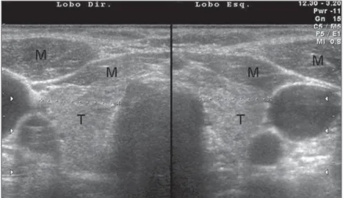
. The decrease in echogenicity was probably caused by the reduction in the colloid/cel-lular interface predominant in the thyroid gland, representing a critical factor in the sonographic reflection. So, in a normo-follicular (normal thyroid gland) or macro-follicular structure (colloid goiter), a great amount of ultrasonic waves interact with the colloidal/cellular portion and are re-flected to the transducer, resulting in a nor-mal echogenic pattern (Figure 1).
In the setting of reduction or absence of follicles, there is a decrease in the sono-graphic interface, causing an intense scat-tering and absorption of sonographic waves with a poor sonographic reflection, resulting in a hypoechogenic pattern (Fig-ure 2)(16). Therefore, it is clear that the
echogenicity of the thyroid parenchyma is inversely correlated to the presence of lym-phocytic infiltration and disaggregation of the follicular structure. In Grave’s disease, the presence of hypoechogenicity mani-fests by a mechanism different from that found at chronic autoimmune thyroiditis, where hypoechogenicity is a result of hypervascularization and hypercellu-larity(17)
.
Echogenicity in the diagnosis of chronic autoimmune thyroiditis: predictive value, sensitivity, specificity, accuracy and comparison with other methods
The predictive value of hypoechoge-nicity in the diagnosis of autoimmune dis-eases of the thyroid gland (chronic autoim-mune thyroiditis and Grave’s disease) was based on the evaluation of a sample of pa-tients who presented thyroid
hypoecho-Figure 1. Transversal view of left and right thyroid lobes. B-mode US demonstrates typical aspect of thyroid parenchyma echogenicity. The echogenicity level of the thyroid gland is higher than that of the adjacent musculature.
Figure 2. Longitudinal view of left thyroid lobe. B-mode US demonstrates micronodular texture and hypoechoic aspect of the thyroid parenchyma. The echogenicity level of the thyroid gland is lower than that of the adjacent musculature.
genicity. Fine-needle aspiration biopsy re-sults were compatible with chronic autoim-mune thyroiditis in 352 patients, and Grave’s disease in 47 patients. Positive and negative predictive values for hypoechoge-nicity as indicators for autoimmune dis-eases of the thyroid gland were respectively 88.3% and 93%, demonstrating that a de-creased echogenicity indicates a high prob-ability of diagnosis of these diseases. Isolatedly, positive and negative predictive values for chronic autoimmune thyroiditis were, respectively, 59% and 93%, clearly demonstrating that the absence of hypo-echogenicity corresponds to a low prob-ability of diagnosis of this disease(9).
The evaluation of patients diagnosed with chronic autoimmune thyroiditis by
means of fine-needle aspiration biopsy has shown that No clinical manifestation was found in 29.3% of patients, antithyroid antibodies were not found in 13%, and thy-roid-stimulating hormone (TSH) levels were normal in 41%. While a cytological study has been isolatedly effective in the diagnosis for 91.3% of patients, US has demonstrated 94.6% sensitivity in the di-agnosis of chronic autoimmune thyroiditis (presence of hypoechogenicity)(18). In cases
where the isoechogenic pattern is found in a general population, the specificity of this parameter to rule out chronic autoimmune thyroiditis exceeds 95%(19). In a selected
sample of patients in an ambulatory of thy-roid diseases, absence of hypoechogenicity was effective for ruling out the diagnosis of chronic autoimmune thyroiditis in 84% of patients(8).So, US is highly specific for
autoimmune disease even in a selected population(19). Additionally, duplex color
Doppler can aid in the differentiation be-tween chronic autoimmune thyroiditis and Grave’s disease(17).
Approximately 10% of individuals in the general population present some degree of hypothyroidism whose primary cause is chronic autoimmune thyroiditis(20). The presence of non-diagnosed hypothyroidism may represent severe problems, both in children because of the risk for growth re-tardation(21), and in pregnant women
be-cause of the risk for miscarriage, perinatal complications and impaired neurological development of the fetus(22). Hence the
in-terest in evaluating the most accurate diag-nostic methods.
Aiming at comparing the diagnostic accuracy of the sonographic hypoecho-genicity in association with TPOAb con-centrations in the diagnosis of chronic au-toimmune thyroiditis, Raber et al. have evaluated 451 patients with unknown thy-roid status in an ambulatory with high fre-quency of thyroid diseases. The thyroid parenchyma echogenicity was rated as fol-lows: grade 1 – normal, echogenicity simi-lar to the one of the prethyroid musculature; grade 2 – hypoechoic in relation to the sub-mandibular gland and hyperechoic in rela-tion to the prethyroid musculature; grade 3 – iso- or hypoechoic in relation to the prethyroid musculature. In cases where grade 3 was observed, the positive predic-tive value was 95% and the sensitivity was 56%. On the other hand, the sensitivity, including grades 2 and 3 for the diagnosis of the disease was 84%, while TPOAb sen-sitivity was 58%. Grade 1 (normal) speci-ficity to rule out chronic autoimmune thy-roiditis was 96%. These authors have ob-served that 95% of patients with echoge-nicity grade 3 at US presented with high TPOAb concentrations(23). Corroborating
these results, another study where the thy-roid was classified according to four differ-ent sonographic patterns, has demonstrated that the patients with the highest pattern (grade 4 – thyroid gland with increased volume and diffuse and marked paren-chyma hypoechogenicity) have shown the highest anti-thyroid antibodies concentra-tions, lowest T4 levels and highest TSH levels(24)
.
In patients diagnosed with chronic au-toimmune thyroiditis confirmed by a cyto-pathological study, it was demonstrated that the presence of thyroid parenchyma hypoechogenicity is associated with a higher probability of hypothyroidism than in the presence of isoechogenicity(25)
. Sonographic hypoechogenicity also is more effective than anti-thyroid antibodies in the prediction of hypothyroidism along the follow-up(26).
It is important to note that, in 90% of cases, the hypoechogenicity is a result of autoimmune diseases, particularly thyroidi-tis or Grave’s disease. However, a typical presentation of marked hypoechogenicity in association with the presence of a hyperechoic fibrotic pattern crossing the parenchyma definitely confirms the diag-nosis of chronic autoimmune thyroiditis(27).
The atrophic presentation of chronic autoimmune thyroiditis may also corre-spond to a sonographic pattern of diffuse hypoechogenicity at ultrasonography. This condition shows a typical decrease in vol-ume and glandular echogenicity in 94.5% of patients(28). In the setting of postpartum thyroiditis, hypoechogenicity reached maximum levels in the postpartum period with a gradual normalization along the fol-low-up period. A persistent hypoechoge-nicity demonstrated a continuous process of destructive thyroiditis(29).
Focal thyroiditis may represent a milder or earlier form of chronic autoimmune
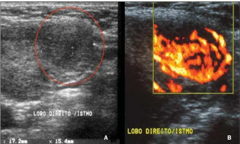
thy-roiditis, and usually is observed at US as a hypoechoic focal nodule (Figure 3)(7).
However, hyperechoic, isoechoic nodular aspects, or even a mixed echogenicity may be present. In this case, fine-needle aspira-tion biopsy is required to confirm the dis-ease and ruling out the presence of a neo-plasm(10).
So, several studies have reported a pat-tern characterized by a diffusely decreased echogenicity in patients with chronic au-toimmune thyroiditis. This is a relevant parameter both for the diagnosis of the dis-ease(8,9,15,16,18,19,24,25,30) and for predicting
the thyroid gland dysfunction along the patients follow-up(25,26). It is important to note that, besides the heterogeneous texture
(25,30)
or a finely micronodular pattern(31), the classic sonographic appearance of the thyroid gland in patients with chronic au-toimmune thyroiditis shows the presence of hypoechogenicity (Figures 2, 4, 5 and 6). The thyroid volume is increased in the majority of patients (Figure 7), decreased in patients with atrophic thyroiditis (Figure 8), but may also be normal in some patients. The presence of lymph nodes with a nor-mal aspect in the cervical chain VI rein-forces the diagnosis of chronic autoim-mune thyroiditis(32).
Sonographic diagnosis by means of com-puted histogram
In the above mentioned studies, the evaluation and classification of the thyroid
Figure 3. Focal chronic autoimmune thyroiditis represented by a hypoechoic, partially defined nodule (A) demonstrating hypervascularization at amplitude color Doppler (B).
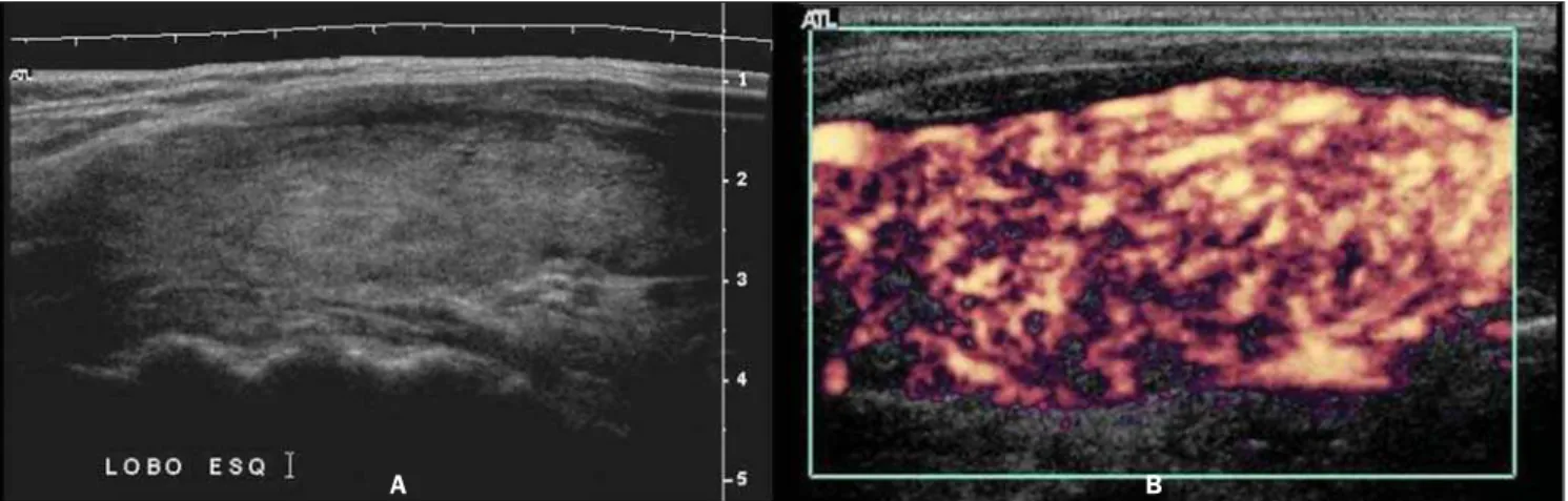
Figure 4. Longitudinal views of thyroid lobe. Mode-B US demonstrates a diffusely heterogeneous texture of the thyroid parenchyma with no nodule (A). Am-plitude color Doppler demonstrates a diffusely increased vascularization of the gland (B).
A B
Figure 5. Longitudinal views of thyroid lobe. Mode-B US demonstrates a diffusely heterogeneous texture of the thyroid parenchyma intermingled with areas of lower echogenicity (pseudonodules), where true nodules are not characterized (A). Amplitude color Doppler demonstrates increased vascularization of the thyroid gland; and nodules are not delimited (B).
A B
Figure 6. Longitudinal views of thyroid lobe. Mode-B US demonstrates a diffusely heterogeneous texture of the thyroid parenchyma intermingled with areas of lower echogenicity where true nodules are not characterized (A). Amplitude color Doppler demonstrates increased vascularization of the thyroid gland (B).
A B
echogenicity were accomplished by expe-rienced observers as compared with the echogenicity of the prethyroid musculature or submandibular glands; on the other hand, the interpretation and quantification
were subjectively accomplished. Although the standardization of procedures involv-ing the utilization of US equipment and classification criteria have been proposed by some authors, the method is still
(Graw-Wert-Einheiten – GWE). The higher the images resolution, the higher the number of gray tonalities identified. The black color corre-sponds to zero, and the white color, to the highest value that depends on the image resolution. Such technique allows the sup-pression of the method subjectivity as the echogenicity is numerically quantified, so this variable becomes objective, quantita-tive, highly sensitive and reproducible and, therefore, capable of providing more accu-rate statistical analyses. This method has been adopted by several authors for evalu-ating thyroid diseases as well as diseases related to other bodily structures.
Computed histogram was utilized in the evaluation of the transversal view of thy-roid lobes (steady brightness gain), and demonstrated a significantly decreased echogenicity in the patients with chronic autoimmune thyroiditis (GWE = 19.6) as compared with the control group (GWE = 25.6). Additionally, a marked hypoechoge-nicity was correlated with high TSH (in-cluding subclinical hypothyroidism) and f TPOAb levels, but with no correlation with
TgAb levels(33). In the experience of the
authors of the present review, computed histogram can also be performed utilizing a longitudinal view of the thyroid paren-chyma as compared with the adjacent mus-culature (Figure 9).
Considering that the thyroid echoge-nicity at computed histogram varies ac-cording to the equipment and also to the brightness gain adjustment, a comparison between values obtained in histograms based on transversal views of thyroid and prethyroid muscles to avoid such problems. This method is reproducible even when performed in different equipment and al-lows the observer to adjust the brightness gain for a better visualization of the paren-chyma, provided the same brightness gain is utilized in the evaluation of the muscu-lature. The thyroid was considered as hypoechoic in cases where mean values ± two standard deviations f the thyroid paren-chyma presented overlapping with those of the prethyroid musculature. All the patients with chronic autoimmune thyroiditis (100%) presented GWE values of the
thy-roid parenchyma overlapping those of the prethyroid muscles and all the patients with hypothyroidism presented hypoechoge-nicity involving more than 68% of the gland(34). Additionally, in patients with
chronic autoimmune thyroiditis, the histo-gram demonstrated a significantly de-creased echogenicity in the setting of hy-pothyroidism as compared with patients affected by euthyroidism(35). Results
ob-tained with computed histogram corrobo-rate the usefulness of the hypoechogenicity of the thyroid parenchyma in the diagno-sis of chronic autoimmune thyroiditis and also in the prediction of hypothyroidism.
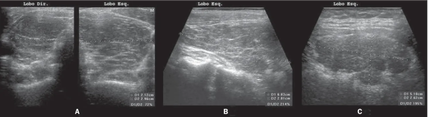
The above mentioned studies have evaluated the echogenicity by selecting regions of interest (ROI) on transversal views of the right and left lobes. Initially, the images were saved in JPEG files and transferred into a workstation for analysis, which limited the utilization of the method because of the work and time required for accomplishing this task. Presently, some ultrasonography systems are endowed with software allowing a real time images analy-Figure 7. B-mode US demonstrates chronic autoimmune thyroiditis with goiter: hypoechogenic and heterogeneous thyroid gland increase in volume (A,B,C). Presence of hyperechogenic pattern of fibrosis (A,B) and lobulated margins (C).
A B C
Figure 8. Atrophic presentation of chronic autoimmune thyroiditis with heterogeneous and hypoechogenic thyroid gland presenting with decrease in volume.
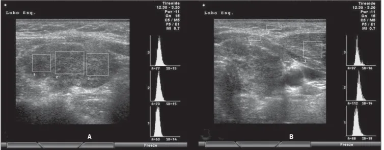
Figure 9. At left, computed histogram in a longitudinal view of the thyroid parenchyma, and at left, in a transverse view of the sternocleidomastoid muscle (1), prethyroid musculature (2) and subcutaneous fat tissue (3). One can observe that the mean value of the thyroid parenchyma echogenicity is lower than that of the adjacent muscles, configuring the hypoechogenic pattern of chronic autoimmune thyroiditis.
A B
Figure 10. At left, computed histogram in a transverse view of the left thyroid lobe, and at right, in a transverse view of the sternocleidomastoid muscle (1), prethyroid muscle (2) and subcutaneous fat tissue (3). One can observe that the mean value of the thyroid parenchyma echogenicity is lower than that of the adjacent muscles, configuring the hypoechogenic pattern of chronic autoimmune thyroiditis.
A B
sis, making the procedure easier to perform (Figures 10, 11 and 12).
The fast development of new softwares for evaluating echogenicity has resulted in several improvements. One of them allows the evaluation of echogenicity in a three-dimensional (3D) ROI(36); another allows
outlining a structure during the scanning, estimating the volume and volumetric mean values for the echogenicity of the whole tissue rather than only of a transver-sal or longitudinal view of the selected re-gion(37). This development will allow that
the region evaluated by the observer is rep-resentative of the actual mean echogenicity of the whole structure, resulting in ex-tremely high inter- and intraobserver agree-ment coefficients, besides making this pa-rameter even more sensitive and reproduc-ible.
CONCLUSION
Once the value of echogenicity in the diagnosis of chronic autoimmune thyroidi-tis is demonstrated, it is recommended that,
besides volume and texture data, a system-atic description of the thyroid parenchyma echogenicity, even if subjectively analyzed, should be included in thyroid US reports.
REFERENCES
1. Weetman AP. Autoimmune thyroid disease. Au-toimmunity. 2004;37:337–40.
2. Lindsay RS, Toft AD. Hypothyroidism. Lancet. 1997;349:413–7.
3. Rapoport B, McLachlan SM. Thyroid autoimmu-nity. J Clin Invest. 2001;108:1253–9. 4. Slatosky J, Shipton B, Wahba H. Thyroiditis:
5. Barbesino G, Chiovato L. The genetics of Hashi-moto’s disease. Endocrinol Metab Clin North Am. 2000;29:357–74.
6. Dayan CM, Daniels GH. Chronic autoimmune thyroiditis. N Engl J Med. 1996;335:99–107. 7. Solbiati L, Livraghi T, Ballarati E, et al. Thyroid
gland. In: Solbiati L, Rizzatto G, editors. Ultra-sound of superficial structures: high frequencies, Doppler and interventional procedures. New York: Churchill Livingstone; 1995. p. 73–6.
8. Nordmeyer JP, Shafeh TA, Heckmann C. Thyroid ultrasonography in autoimmune thyroiditis. A prospective study on 123 patients. Acta Endo-crinol (Copenh). 1990;122:391–5.
9. Pedersen OM, Aardal NP, Larssen TB, et al. The value of ultrasonography in predicting autoim-mune thyroid disease. Thyroid. 2000;10:251–9. 10. Langer JE, Khan A, Nisenbaum HL, et al. Sono-graphic appearance of focal thyroiditis. AJR Am J Roentgenol. 2001;176:751–4.
11. LiVolsi VA. The pathology of thyroid autoimmune disease: a review. Thyroid. 1994;4:333–9.
12. Buchanan WW, Harden RM. Primary hypothy-roidism and Hashimoto’s thyroiditis. A continu-ous spectrum. Arch Intern Med. 1965;115:411– 7.
13. Hegedüs L. Thyroid ultrasound. Endocrinol Metab Clin North Am. 2001;30:339–60.
14. Prummel MF, Wiersinga WM. Thyroid peroxi-dase autoantibodies in euthyroid subjects. Best Pract Res Clin Endocrinol Metab. 2005;19:1–15. 15. Yoshida A, Adachi T, Noguchi T, et al. Echo-graphic findings and histological feature of the thyroid: a reverse relationship between the level of echo-amplitude and lymphocytic infiltration. Endocrinol Jpn. 1985;32:681–90.
16. Müller HW, Schröder S, Schneider C, et al. Sono-graphic tissue characterization in thyroid gland diagnosis. A correlation between sonography and histology. Klin Wochenschr. 1985;63:706–10.
17. Vitti P, Rago T, Mazzeo S, et al. Thyroid blood flow evaluation by color-flow Doppler
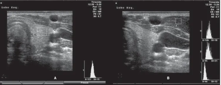
sono-Figure 11. At left, computed histogram in a transverse view of the left thyroid lobe, and at right, in a transverse view of the sternocleidomastoid muscle (1), prethyroid muscle (2) and subcutaneous fat tissue (3). One can observe that the mean value of the thyroid parenchyma ( ROI 1 at left) echogenicity is higher than that of the prethyroid and sternocleidomastoid muscles (ROI 1 and 2 at right), configuring the normal echogenicity pattern of the thyroid.
A B
graphy distinguishes Graves’ disease from Hashi-moto’s thyroiditis. J Endocrinol Invest. 1995;18: 857–62.
18. Gutekunst R, Hafermann W, Mansky T, et al. Ul-trasonography related to clinical and laboratory findings in lymphocytic thyroiditis. Acta Endo-crinol (Copenh). 1989;121:129–35.
19. Gutekunst R, Smolarek H, Hasenpusch U, et al. Goitre epidemiology: thyroid volume, iodine ex-cretion, thyroglobulin and thyrotropin in Ger-many and Sweden. Acta Endocrinol (Copenh). 1986;112:494–501.
20. Canaris GJ, Manowitz NR, Mayor G, et al. The Colorado thyroid disease prevalence study. Arch Intern Med. 2000;160:526–34.
21. Setian NS. Hypothyroidism in children: diagno-sis and treatment. J Pediatr (Rio J). 2007;83(5 Suppl):209–16.
22. Poppe K, Glinoer D. Thyroid autoimmunity and hypothyroidism before and during pregnancy. Hum Reprod Update. 2003;9:149–61. 23. Raber W, Gessl A, Nowotny P, et al. Thyroid
ul-trasound versus antithyroid peroxidase antibody determination: a cohort study of four hundred fifty-one subjects. Thyroid. 2002;12:725–31. 24. Sostre S, Reyes MM. Sonographic diagnosis and
grading of Hashimoto’s thyroiditis. J Endocrinol Invest. 1991;14:115–21.
25. Hayashi N, Tamaki N, Konishi J, et al. Sono-graphy of Hashimoto’s thyroiditis. J Clin Ultra-sound. 1986;14:123–6.
26. Rago T, Chiovato L, Grasso L, et al. Thyroid ul-trasonography as a tool for detecting thyroid au-toimmune diseases and predicting thyroid dis-function in apparently healthy subjects. J Endo-crinol Invest. 2001;24:763–9.
27. Chammas MC. Ultra-sonografia nas tireoidites. Radiol Bras. 2007;40(2):v–vi.
28. Vitti P, Lampis M, Piga M, et al. Diagnostic use-fulness of thyroid ultrasonography in atrophic thyroiditis. J Clin Ultrasound. 1994;22:375–9. 29. Premawardhana LD, Parkes AB, Ammari F, et al.
Postpartum thyroiditis and long-term thyroid sta-tus: prognostic influence of thyroid peroxidase antibodies and ultrasound echogenicity. J Clin Endocrinol Metab. 2000;85:71–5.
30. Aydin O, Apaydin FD, Bosdogan R, et al. Cyto-logical correlation in patients who have a pre-di-agnosis of thyroiditis ultrasonographically. Endocr Res. 2003;29:97–106.
31. Set PA, Oleszczuk-Raschke K, von Lengerke JH, et al. Sonographic features of Hashimoto thyroidi-tis in childhood. Clin Radiol. 1996;51:167–9.
32. Yamashiro I, Saito OC, Chammas MC, et al. Achados ultra-sonográficos na tireoidite. Radiol Bras. 2007;40:75–9.
33. Schiemann U, Avenhaus W, Konturek JW, et al. Relationship of clinical features and laboratory parameters to thyroid echogenicity measured by standardized grey scale ultrasonography in pa-tients with Hashimoto’s thyroiditis. Med Sci Monit. 2003;9:13–7.
34. Mazziotti G, Sorvillo F, Iorio S, et al. Grey-scale analysis allows a quantitative evaluation of thyroid echogenicity in the patients with Hashimoto’s thyroiditis. Clin Endocrinol. 2003;59:223–9. 35. Loy M, Cianchetti ME, Cardia F, et al.
Correla-tion of computerized gray-scale sonographic find-ings with thyroid function and thyroid autoim-mune activity in patients with Hashimoto’s thy-roiditis. J Clin Ultrasound. 2004;32:136–40.
36. Slapa RZ, Slowinska-Srzednicka J, Szopinski KT, et al. Grey-scale three-dimensional sonography of thyroid nodules: feasibility of the method and pre-liminary studies. Eur Radiol. 2006;16:428–36.