VII
Radiol Bras. 2008 Nov/Dez;41(6):VII–VIII
Laércio Castilho Lopes Neto1, Rodrigo Sampaio Chiarantano1, Marcelo Bordalo Rodrigues2
1. MDs, Residents at Instituto de Radiologia do Hospital das Clínicas da Faculdade de Medicina da Universidade de São Paulo (InRad/HC-FMUSP), São Paulo, SP, Brazil. 2. MD, Radiologist, Director for the Unit of Radiology at Instituto de Ortopedia e Traumatologia do Hospital das Clínicas da Faculdade de Medicina da Universidade de São Paulo (IOT/HC-FMUSP), São Paulo, SP, Brazil. Mailing address: Dr. Marcelo Bordalo Rodrigues. Avenida Doutor Enéas Carvalho de Aguiar, 255, Pinheiros. São Paulo, SP, Brazil, 05403-900. E-mail: [email protected]
0100-3984 © Colégio Brasileiro de Radiologia e Diagnóstico por Imagem
Which is your diagnosis?
•
Qual o seu diagnóstico?
Lopes Neto LC, Chiarantano RS, Bordalo-Rodrigues MB. Which is your diagnosis? Radiol Bras. 2008;41(6):VII–VIII.
A female, 34-year-old patient followed-up in the neurological care unit for a clinical picture of dis-tally initiated peripheral sensorimotor polyneuropa-thy. The patient attended the emergency department with sudden dyspnea, and was referred to the unit of imaging diagnosis with clinical suspicion of pul-monary thromboembolism. The occurrence of other previous significant medical conditions was denied by the patient.
Figure 4. Axial computed tomography image of the hip (bone window).
Figure 3. Thoracic spine computed tomography: sagittal re-formatting (bone window).
Figure 1. Chest computed tomography: axial image acquired after intravenous iodinated contrast injection (arterial phase).
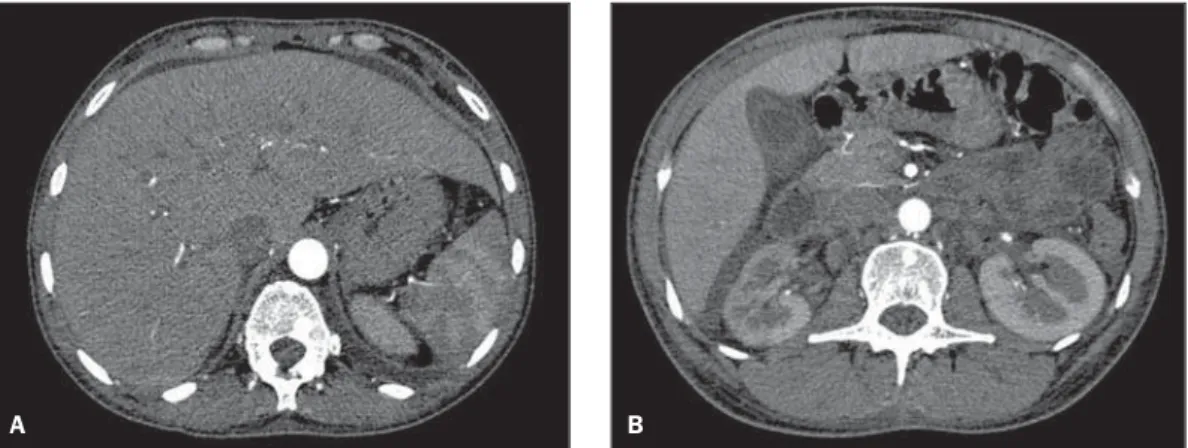
Figure 2. Upper abdominal computed tomography: axial images acquired after intravenous iodinated contrast injection (arte-rial phase).
VIII Radiol Bras. 2008 Nov/Dez;41(6):VII–VIII
Images description
Figure 1. Cardiomegaly and moderate bilateral pleural effusion are observed. There is no evidence of pulmonary throm-boembolism.
Figure 2. Presence of homogeneous hepatomegaly, free perihepatic and peri-vesicular fluid (ascites) and retroperitoneal lymphadenomegaly can be observed.
Figure 3. Multiple focal sclerotic le-sions sparsely scattered over the thoracic vertebral bodies, posterior elements and sternum. Also, a lytic lesion with irregular sclerotic margins on T10.
Figure 4. Multiple focal sclerotic bone lesions on the iliac bone and lumbar verte-bral body.
Diagnosis: POEMS syndrome.
COMMENTS
POEMS syndrome is a rare disorder that was firstly described by Bardwick et al.(1)
in 1980, and is characterized by a predomi-nantly sensorimotor polyneuropathy, organomegaly (hepatomegaly, lymph-adenomegaly), endocrinopathy (particu-larly hypothyroidism), monoclonal gam-mopathy and skin abnormalities.
The diagnosis is based on clinical find-ings, and the knowledge on radiological al-terations allows the radiologist to suggest this diagnostic hypothesis.
The patient presented monoclonal
gammopathy and subclinical hypothyroid-ism compatible with the diagnosis of PO-EMS syndrome.
POEMS syndrome is a rare disorder preferentially affecting men in the fifth or sixth decade of life. It is characterized by a range of clinical alterations and the patho-physiology is not well understood. Patients with POEMS syndrome essentially present plasma cell dyscrasia which may range from monoclonal gammopathy up to bones involvement (sclerotic myeloma)(2).
All the patients affected by this syn-drome present peripheral neuropathy and monoclonal gammopathy considered by some authors as the two major criteria re-quired for diagnosis of the disease. Minor criteria are the following: presence of scle-rotic bone lesions, organomegaly (hepa-tomegaly, splenomegaly, lymphadeno-megaly), edemas (pleural effusions, as-cites), endocrinopathy (involving adrenal glands, thyroid, parathyroid, gonads, hypo-physis and pancreas) and skin changes (hy-perpigmentation, hypertrichosis, heman-giomas)(3).
The main radiological findings in PO-EMS syndrome are the presence of organomegaly (particularly hepatosple-nomegaly and lymphadehepatosple-nomegaly) and bone lesions. The lesions are mainly scrotic and, less frequently, mixed. Lytic le-sions are rarely found. Preferentially, the axial skeleton is affected, with frequent
presence of lesions on elements of the pos-terior vertebral arch, which increases the difficulty in the differential diagnosis with secondary lesions. Additionally, abnormal bone proliferation may be observed, with lesions on intervertebral discs and costo-vertebral joints. In these cases, the sudden appearance of calcifications with irregular, spiculate borders is quite suggestive of this disease(4).
Generally, this syndrome progresses as a chronic disease, and the prognosis is di-rectly related to the presence of bone mar-row involvement and to the number and distribution of the bone lesions, hence the significant role played by the radiologist in the establishment of the prognosis for these patients(3).
REFERENCES
1. Bardwick PA, Zvaifler NJ, Gill GN, et al. Plasma cell dyscrasia with polyneuropathy, organomegaly, endocrinopathy, M protein, and skin changes: the POEMS syndrome. Report on two cases and a review of the literature. Medicine (Baltimore). 1980;59:311–22.
2. Aggarwal S, Goulatia RK, Sood A, et al. POEMS syndrome: a rare variety of plasma cell dyscrasia. AJR Am J Roentgenol. 1990;155:339–41. 3. Dispenzieri A, Kyle RA, Lacy MQ, et al. POEMS
syndrome: definitions and long-term outcome. Blood. 2003;101:2496–506.