Aid in the detection of myocardial perfusion abnormality
utilizing SPECT atlas and images registration: preliminary
results*
Auxílio à detecção de anormalidade perfusional miocárdica utilizando atlas de SPECT e registro de imagens: resultados preliminares
Rodrigo Donizete Santana de Pádua1, Lucas Ferrari de Oliveira2, Paulo Mazzoncini de Azevedo Marques3, Jean-Jacques Georges Soares de Groote4, Adelson Antonio de Castro5, Lauro Wichert Ana6, Marcus Vinicius Simões7
OBJECTIVE: To develop an atlas of myocardial perfusion scintigraphy and evaluating its applicability in computer-aided detection of myocardial perfusion defects in patients with ischemic heart disease. MATERIALS AND METHODS: The atlas was created with rest-stress myocardial perfusion scintigraphic images of 20 patients of both genders with low probability of coronary artery disease and considered as normal by two experienced observers. Techniques of image registration and mathematical operations on images were utilized for obtaining template images depicting mean myocardial uptake and standard deviation for each gender and physiological condition. RESULTS: Myocardial perfusion scintigraphy images of one male and one female patient were aligned with the corresponding atlas template image, and voxels with myocardial uptake rates two standard deviations below the mean voxel value of the respective region in the atlas template image were highlighted on the tomographic sections and confirmed as perfusion defects by both observers. CONCLUSION: The present study demonstrated the creation of an atlas of myocardial perfusion scintigraphy with promising results of this tool as an aid in the detection of myocardial perfusion defects. However, further prospective validation with a more representative sample is recommended.
Keywords: Scintigraphy; SPECT; Myocardium; Nuclear medicine; Computer-assisted image processing; Computer-assisted diagnosis.
OBJETIVO: Criar um atlas de cintilografia de perfusão miocárdica e verificar sua aplicabilidade no auxílio computadorizado à detecção de defeitos perfusionais miocárdicos em pacientes portadores de cardiopatia isquêmica. MATERIAIS E MÉTODOS: O atlas foi criado com imagens de cintilografia de perfusão miocárdica, em condições de repouso e estresse, de 20 pacientes de ambos os gêneros com baixa probabilidade de doença arterial coronariana e julgadas normais por dois observadores experientes. Técnicas de registro de imagens e operações matemáticas sobre imagens foram utilizadas para obtenção de modelos de média e desvio-padrão da captação miocárdica percentual de cada gênero e condição fisiológica. RESULTADOS: Imagens de um paciente masculino e um feminino foram alinhadas com os atlas correspondentes, e os voxels
apresentando valores de captação percentual dois desvios-padrão abaixo da média da respectiva região do atlas foram destacados nos cortes tomográficos e confirmados como defeitos de perfusão por dois observa-dores experientes. CONCLUSÃO: Demonstramos a criação de um atlas de cintilografia de perfusão miocár-dica e obtivemos resultados promissores na sua utilização para auxílio à detecção de defeitos perfusionais. Entretanto, uma validação prospectiva com um número mais representativo de casos é necessária. Unitermos: Cintilografia; SPECT; Miocárdio; Medicina nuclear; Processamento de imagem assistida por computador; Diagnóstico por computador.
Abstract
Resumo
* Study developed in the Division of Cardiology and Unit of Nuclear Medicine at Hospital das Clínicas da Faculdade de Medicina de Ribeirão Preto da Universidade de São Paulo (FMRP-USP), Ribeirão Preto, SP, Brazil.
1. Bachelor of Computational Science, Voluntary Researcher, Division of Cardiology and Unit of Nuclear Medicine at Hospital das Clínicas da Faculdade de Medicina de Ribeirão Preto da Universidade de São Paulo (FMRP-USP), Ribeirão Preto, SP, Brazil. 2. PhD, Professor, Department of Information Technology – Instituto de Física e Matemática da Universidade Federal de Pelotas (UFPel), Pelotas, RS, Brazil.
Pádua RDS, Oliveira LF, Azevedo-Marques PM, DeGroote JJ, Castro AA, Wichert-Ana L, Simões MV. Aid in the detection of myocardial perfusion abnormality utilizing SPECT atlas and images registration: preliminary results. Radiol Bras. 2008; 41(6):397–402.
3. PhD, Electronic Engineer, Professor, Centro de Ciências das Imagens e Física Médica da Faculdade de Medicina de Ribeirão Preto da Universidade de São Paulo (CCIFM/FMRP-USP), Ribei-rão Preto, SP, Brazil.
4. PhD, Physicist, Professor at Laboratory of Artificial Intelligence and Applications – Instituto de Ensino Superior COC, Ribeirão Preto, SP, Brazil.
5. Master, Physicist, Fellow PhD degree in Medicine, Facul-dade de Medicina de Ribeirão Preto da UniversiFacul-dade de São Paulo (FMRP-USP), Ribeirão Preto, SP, Brazil.
6. PhD, MD, Professor, Centro de Ciências das Imagens e
Física Médica da Faculdade de Medicina de Ribeirão Preto da Universidade de São Paulo (CCIFM/FMRP-USP), Ribeirão Preto, SP, Brazil.
7. PhD, MD, Professor at Division of Cardiology, Faculdade de Medicina de Ribeirão Preto da Universidade de São Paulo (FMRP-USP), Ribeirão Preto, SP, Brazil.
Mailing address: Rodrigo Donizete Santana de Pádua. Rua Eduardo Prado, 1356, Vila Tibério. Ribeirão Preto, SP, Brazil, 14050-480. E-mail: [email protected]
INTRODUCTION
Digital medical imaging techniques as-sociated with scientific and technological developments in the areas of images pro-cessing and graphic computation, have al-lowed the development of a range of com-puter-aided diagnosis – CAD systems. Besides improving the visualization and manipulation of medical images, such sys-tems allow the quantification of the abnor-malities detected(1,2).
Among the nuclear medicine imaging techniques applied to cardiology, myocar-dial perfusion scintigraphy (MPS) with single photon emission computed tomog-raphy (SPECT) allows the acquisition of digital images demonstrating the relative radiopharmaceutical uptake in the left ven-tricular myocardium for noninvasive detec-tion of ischemic or fibrotic areas resulting from ischemic cardiopathy. The study is performed with the patient under physical or pharmacological stress/rest conditions and the images acquisition is performed after a radiopharmaceutical injection into the patient’s blood current. Most fre-quently, thallium-201 (201Tl) or techne-tium-99m (99mTc)-sestamibi are utilized for this purpose. Then, the photons emitted by the radioactivity accumulated in the differ-ent segmdiffer-ents of the target-organs are de-tected by the scintillation chamber crystals, quantified and processed for images gen-eration(3,4).
A technical issue inherent to the myo-cardial perfusion images acquisition by SPECT is the frequent presence of attenu-ation artifacts or false uptake defects result-ing from the passage of photons through soft tissues. Dense breasts in women and subdiaphragmatic tissues in men are com-mon causes for attenuation artifacts. The presence of these artifacts results in a sig-nificantly decreased diagnostic specificity of this imaging method(3).
A technique called “transmission” uti-lizes an external radiation source for ob-taining maps of correction and minimiza-tion of photon attenuaminimiza-tion effects on the final image. However, the patients’ expo-sure to the effects and risks of higher lev-els of ionizing radiation should be taken into consideration as a significant disad-vantage of this technique(5).
An alternative approach aimed at im-proving the diagnostic accuracy of the method is the utilization of computational algorithms for comparison of patients’ im-ages with normality standards based on images of a group of healthy individuals of the same genre, which allows a mathemati-cal correction of attenuation artifacts. So, on the image of a determined patient, only those voxels with significantly lower up-take intensity as compared with the voxels on the template image (> two standard-deviations below the mean) strongly indi-cate the presence of regions with myocar-dial perfusion defects requiring further in-vestigation. Classically, this quantitative analysis is performed by means of polar mapping. Although this approach has al-ready been validated, differences between the visual representation of areas with perfusional defects by this technique and the traditional visualization of tomographic sections still represent a limitation to a more disseminated utilization of this method(6).
Aiming at preserving the tomographic sections visualization in the quantitative analysis and visualization of perfusional defects, an alternative approach utilizing 3D image registration techniques is sug-gested for obtention of normal template images for comparison and later identifica-tion of perfusional defects on tomographic sections(7,8).
Both the obtention of these template im-ages and their comparison with patients’ images require an intermediate processing phase corresponding to a spatial alignment between images that can be achieved with the utilization of medical images registra-tion techniques(7). Basically, images regis-tration is aimed at defining parameters for changing a “source” image in a way to achieve the best alignment possible with another image called “target image”(9–13).
The present study describes the utiliza-tion of image registrautiliza-tion and other tech-niques for digital images processing for the development of an atlas of myocardial per-fusion scintigraphy, and evaluates the pre-liminary results of the utilization of such atlas as an aid in the detection of myocar-dial perfusion defects through the compari-son between images. So, it is expected that the method applicability is evaluated for
later development of a computer-aided design tool for cardiology as well as pro-spective studies involving a higher number of patients aiming at validating this method. Despite the commercial availability in the market of similar already validated ap-plications, the local development of such tool can be useful for the author’s institu-tion in the reducinstitu-tion of costs with hardware and software licenses acquisition, as well as allowing a better integration with already implemented hospital and radiological in-formation systems. Furthermore, the future implementation of refinements and cus-tomization of this tool is feasible, consid-ering the possibility of reutilization of the source-code that will be already available, so encouraging a culture of research & development in information technology in the field of medical images.
MATERIALS AND METHODS
Tomographic images acquisition and processing
Aiming at obtaining the template im-ages for the atlas, the authors selected stress/rest images of myocardial perfusion scintigraphy with 99mTc-sestamibi which were visually analyzed and considered as normal by two experienced observers based on the pattern of relative radiophar-maceutical accumulation in myocardial segments. The images were divided into categories according to the patients’ genre and physiological condition as follows: male-rest, male-stress, female-rest and fe-male stress categories.
fil-ter (5th order, 0.25 cy/mm cutoff fre-quency) was utilized for construction of image sets. Subsequently, tomographic sec-tions on classical orthogonal planes were automatically generated by a commercially available software. The images were con-verted into DICOM 3.0 format and trans-mitted to the workstation for processing.
Images registration and obtention of template images for the atlas
All the images were aligned in relation to a single coordinate space with the soft-ware pack VTK CISG Registration Toolkit (vtkCisg), developed by Hartkens et al.(14) and made freely available by the Compu-tational Imaging Science Group of Kings College of London. The vtkCisg performs rigid and non-rigid images registration through voxels similarity, allowing the user the option for cross correlation – C), mu-tual information – MI, normalized mumu-tual information – NMI measurements, among others. The rigid registration method was adopted to avoid anatomical deformation on the images, utilizing only scale, rotation and translation transformations, although there are other alternative non-rigid regis-tration techniques(9,10,13). Figure 1 demon-strates the equation for measurement of voxels similarity by means of cross corre-lation utilized for images alignment.
In this equation, N corresponds to the number of voxels on each of the images, and A(i) and B(i) to the overlapping voxels intensity i respectively on the images A and B. It is important to note that, the higher the
C value, the more an image overlaps the other, and two identical images should cor-respond to the value 1 (one). So, the image registration algorithm will generate param-eters for spatial transformation of images performing several interactions until the C
value achieves the nearest possible to 1(9). Once the images were spatially regis-tered, the next step was the obtention of template images of the mean myocardial uptake rate for each gender/physiological condition. For this purpose, a software based on the Visualization Toolkit (VTK)
library(15,16), called vtkImageAVG was de-veloped. Through this software, n 3D medical images are entered and normalized according to a 100-level gray-scale, and a final image corresponding to a median image of all the normalized images is gen-erated. The definition of the images nor-malization in a 100-level gray-scale is aimed at allowing a direct quantitative evaluation of the rate of myocardial perfu-sion defect based on the comparison of the patients’ images with the respective values in the template images of the atlas. The gray-scale values indicate a maximum up-take at 100, with the normalization calcu-lation corresponding to the division of the original image voxels value by the highest voxel, and subsequent multiplication of this result by 100.
The statistical analysis of the myocar-dial perfusion cannot be performed only by comparison between the patient’s image and the mean image of normal individuals. It is necessary to have template images re-flecting the intensity variability found on images of normal individuals. A software called vtkImageSD was developed for this purpose. Based on n input images, this soft-ware generates template images reflecting the standard deviation of the individual’s myocardial perfusion uptake rate. In the present study, four mean images and four standard deviation images were generated - one for each gender-physiological condi-tion category. All of the images are normal-ized for 100 gray levels, so they can be compared with the patient’s images nor-malized according to the same gray-scale.
Algorithm for detection of perfusion defects
The algorithm for statistical comparison between patients’ images and appropriate gender-physiological condition template images consists of four elementary steps: 1) patient’s image alignment with the cor-responding template image with the mean myocardial perfusion uptake rate based on C; 2) patient’s image normalization accord-ing to a 100-level gray-scale; 3) selection of patient’s image voxels with myocardial uptake rates two standard deviations below the mean voxel value of the respective re-gion; 4) construction of the myocardial perfusion defect image.
Figura 1. Equação para obtenção da medida de correlação cruzada (C).
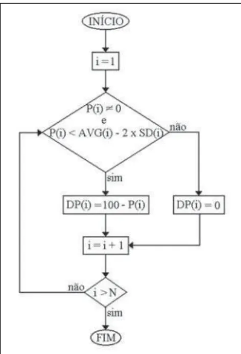
Figure 2. Diagram demonstrating the algorithm for selection of voxels with uptake rates two standard deviations below the mean voxel value of the re-spective region on the atlas template image indi-cating the presence of myocardial perfusion defect.
Figure 2 illustrates the step in the algo-rithm where the selection of voxels on the perfusion defect images is performed. Note that P(i), AVG(i), SD(i) and DP(i) store respectively the overlapping voxels inten-sity on the patient’s image, mean gender-physiological condition, standard-devia-tion for gender/physiological condistandard-devia-tion and perfusional defect images, with i ranging from 1 up to the total number of voxels N. If the P(i) value is different from de 0 (zero) and lower than the AVG(i) value less 2 x
SD(i), then DP(i) corresponds to the perfusional defect rate; otherwise it corre-sponds to 0. Considering that the images are normalized according to a 100-level gray-scale, subtracting the P(i) value from 100 will be enough to have the perfusion defect rate.
RESULTS
Images registration
process for alignment of patients’ images with the atlas template images, and genera-tion of perfusion defect images for each genre-physiological condition took ap-proximately 45-60 seconds in a standard PC (AMD Athlon XP-2000 processor, 1 Gb RAM, running operational system Ubuntu GNU/Linux version 8.04 LTS).
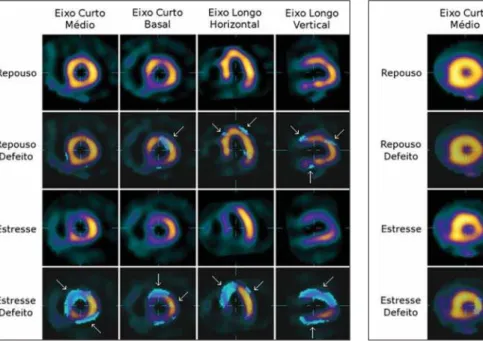
The first and third range of images on Figure 6 demonstrate three orthogonal axis of, respectively, rest/stress cardiac SPECT projections of the male patient already aligned with the template images of mean perfusion uptake for male individuals (Fig-ure 4). The second range demonstrates the same projections overlapped by the perfu-sion defect image where arrows indicate the areas with myocardial perfusion abnor-mality. In the same way, the fourth range shows areas of perfusion abnormality with the patient under stress condition. Accord-ingly with the visual analysis performed by two experienced observers, the results in-dicate a reversible, extensive perfusional defect on the anterior, septal, apical and postero-lateral walls of the myocardium.
Like Figure 6, Figure 7 demonstrates the female patient’s images already aligned with the corresponding template images (Figure 5), besides presenting the areas with myocardial perfusion defect indicated by the arrows. Also, in complete agreement with the visual analysis results, the
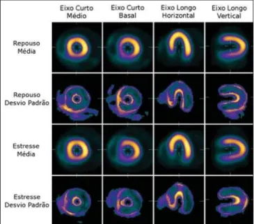
auto-Figure 4. Tomographic sections illustrating the atlas of male myocardial per-fusion SPECT scintigraphy demonstrating template images of mean and stan-dard-deviation under physiological stress and rest conditions.
Figure 5. Tomographic sections illustrating the atlas of female myocardial perfusion SPECT scintigraphy demonstrating template images of mean and standard-deviation under physiological stress and rest conditions.
Figure 3. Tomographic sections of SPECT of myocardial perfusion im-ages illustrating the patient’s images before and after alignment with the target images.
long axis and horizontal long axis): on the first range, views of the target-image; on the second, views of the patient’s image before the alignment; and on the third range, views of the patient’s image after alignment. By observing and comparing the landmarks at the center of each view, one can note that after the alignment, the patient’s images are in the same coordinate space of the target image, differently from the images before the alignment.
Construction of the normality atlas
Through registration and addition of rest/stress images of ten female and ten male normal individuals, the authors ob-tained template images normalized accord-ing to a 100-level gray-scale representaccord-ing
the mean uptake and standard deviation for each voxel on the respective images.
Figures 4 and 5 show SPECT images of the atlas of myocardial perfusion, with mean myocardial perfusion uptake rates and standard deviation. Images of mean uptake are similar to those of any male or female individual with low probability of myocardial ischemia. On the other hand, images of standard deviation present higher (clear) value on the borders where struc-tural differences can be more clearly ob-served in the myocardium.
Perfusion defect detection
matic detection technique indicates a versible perfusional defect in the apical re-gion and postero-lateral wall.
DISCUSSION
Images registration has played an essen-tial role in the development of the present study, both in the construction of template images and in their statistical comparison with patients’ images. This process is nec-essary because of the tomographic images reconstruction where the human operator cannot accurately define the sections posi-tioning in a way that the final images can be viewed and analyzed in the same coor-dinate system.
Image registration is classified as being “rigid” or “non-rigid”. The rigid technique allows only a global change on the image, whereas the non-rigid technique allows also local changes, without necessarily changing the image as a whole(9–13). The results de-scribed in the present study are associated with the utilization of rigid image registra-tion, only by a geometry-based approach without deformation. The reliability of the anatomical identification tends to improve with the utilization of the rigid technique,
but significant limitations may be observed in situations where the patient presents a marked anatomical variation such as, for example, presence of artifacts on images of patients with a marked ventricular dilata-tion.
The most recent techniques based on voxels similarity measurements utilize comparison between voxels intensity on source- and target-images to define trans-formation parameters with the advantage of being completely automated, differently from the previous techniques based on objects recognition which depended on preliminary marking and/or segmentation steps. Some measurements can be utilized by registration algorithms for calculation of image voxels similarity, namely, cross-cor-relation measurements and information theory measurements. The cross-correla-tion measurement technique presents bet-ter results in cases where there is a linear relationship between intensities on the images to be aligned, and most frequently is utilized for intramodality registration. On the other hand, for intermodality registra-tion (images acquired through different techniques) where the relationship between voxels intensities is non-linear, information
theory measurements, particularly mutual information and normalized mutual infor-mation, are most frequently utilized(9–13).
Image registration techniques based on voxels similarity through correlation mea-surements are considered as the ideal tech-niques for template images construction and other activities requiring alignment of images from a same modality(9–13). In the present study, the authors utilized the cross correlation measurement, a normalized correlation coefficient version for images registration.
Considering that all myocardial perfu-sion scintigraphy images present a same spatial disposition, voxel-by-voxel math-ematical operations can be performed, re-sulting in reliable images reflecting such operations. So, with images normalized according to a 100-level gray-scale for ten individuals of each gender with low prob-ability of coronary artery disease and con-sidered as normal by two experienced ob-servers, the authors obtained template im-ages corresponding to mean and standard deviation for perfusion uptake for a myo-cardium considered as normal for both male and female patients under physiologi-cal stress/rest conditions. Images from one
Figure 6. Tomographic sections representative of myocardial perfusion SPECT of a male patient demonstrating a reversible perfusional defect on the septal, anterior, infero-posterior and apical walls. The two upper series represent the initial gross images, and the two lower series show the fusion of the initial images with the perfusional defects projections automatically identified through statistical comparison with the corresponding template images of the atlas.
male and one female test-patients, also normalized according to a 100-level gray-scale and with reports of two specialists indicating ischemic cardiopathy, were sta-tistically compared with templates images created by means of a computational algo-rithm. The final results corresponded to images whose overlapping with test-pa-tients images indicate the presence of re-gions with a probable myocardial perfusion defect.
Also, it is important to note that the re-gions with perfusion defects are visualized on tomographic sections through the de-scribed algorithm, contrarily to the classi-cal visualization through polar maps, which makes the human observation pro-cess more intuitive.
A similar approach has been described by Slomka et al.(7), who have created nor-mality template images based on images of 23 men and 15 women, which were later utilized for quantifying perfusional abnor-malities in test-patients. For image registra-tion, these authors have developed and tested a hybrid image registration algorithm based on principal-axes transformation and minimization technique.
The present study is differentiated by the utilization of images registration based on correlation measurements classically ac-cepted as ideal for intramodality medical images registration and template images construction(9–13), besides the utilization of image templates, statistical measurements and images processing techniques for au-tomatic detection and definition of regions of probable perfusion defect on SPECT sections of patients with ischemic cardi-opathy aiming at offering the observer a “second diagnostic opinion”.
Although the initial results of the present study have demonstrated the poten-tial of the method proposed based on the agreement about the defect regions auto-matically detected by means of a visual
analysis by two experienced observers, it should be observed that, certainly, the cri-teria for selection of normal cases, as well their number and processing method uti-lized for creating the template images, are certainly not sufficient for validating the method. However, the proposal of the present study, in the current phase, was to prove the concept validity, so justifying further detailed investigation with a more representative number of cases, including comparison with a quantitative golden-standard to evaluate the sensitivity and specificity that could be achieved in actual clinical situations.
CONCLUSION
The techniques for images processing utilized in the present study have shown to be effective for the development of the at-las of SPECT myocardial perfusion scintig-raphy. However, further techniques should be evaluated, applied and compared for development of new template images, be-sides a more clear-sighted selection of nor-mal cases to enhance the reliability of the atlas.
As a proof of concept, the method pro-posed for comparing patients’ images with the atlas image templates has shown to be promising as an aid in the detection of myocardial perfusion defects on SPECT images. Future refinements in the algorithm and quantitative experiments with a higher number of patients, besides the inclusion of control cases, are required for evaluation of the sensitivity/specificity and validation of the method.
REFERENCES
1. Azevedo-Maques PM. Diagnóstico auxiliado por computador na radiologia. Radiol Bras. 2001;34: 285–93.
2. Doi K. Computer-aided diagnosis in medical im-aging: historical review, current status and future potential. Comput Med Imaging Graph. 2007;31: 198–211.
3. Cullom SJ. Principles of cardiac SPECT. In: DePuey EG, Garcia EV, Berman DS, editors. Car-diac SPECT imaging. 2nd ed. Philadelphia: Lippincott Williams & Wilkins; 2001. p. 3–14. 4. Yoo TS. Introduction. In: Yoo TS, editor. Insight
into images: principles and practice for segmen-tation, registration, and image analysis. Wellesey: AK Peters; 2004. p. 3–17.
5. Hendel RC, Corbett JR, Cullom SJ, et al. The value and practice of attenuation correction for myocardial perfusion SPECT imaging: a joint po-sition statement from the American Society of Nuclear Cardiology and the Society of Nuclear Medicine. J Nucl Med. 2002;43:273–80. 6. Van Train KF, Garcia EV, Cooke CD, et al.
Quan-titative analysis of SPECT myocardial perfusion. In: DePuey EG, Garcia EV, Berman DS, editors. Cardiac SPECT imaging. 2nd ed. Philadelphia: Lippincott Williams & Wilkins; 2001. p. 41–64. 7. Slomka PJ, Hurwitz GA, Stephenson J, et al. Automated alignment and sizing of myocardial stress and rest scans to three-dimensional normal templates using an image registration algorithm. J Nucl Med. 1995;36:1115–22.
8. Itti E, Klein G, Rosso J, et al. Assessment of myo-cardial reperfusion after myomyo-cardial infarction using automatic 3-dimensional quantification and template matching. J Nucl Med. 2004;45:1981–8. 9. Hajnal JV, Hill DLG, Hawkes DJ. Medical image
registration. Boca Raton: CRC Press; 2001. 10. Ng L, Ibanez L. Medical image registration:
con-cepts and implementation. In: Yoo TS, editor. Insight into images: principles and practice for segmentation, registration, and image analysis. Wellesey: AK Peters; 2004. p. 239–95. 11. Zitová B, Flusser J. Image registration methods:
a survey. Image and Vision Computing. 2003; 21:977–1000.
12. Oliveira LF, Azevedo-Marques PM, Wichert-Ana L, et al. Support software for clinical diagnosis in epilepsy: B.R.A.S.I.L. brain registration and subtraction: improved localization for SPECT analysis. Int J Comput Assist Radiol Surg. 2006; (1 Suppl 1):386–9.
13. Mäkelä T, Clarysse P, Sipilä O, et al. A review of cardiac image registration methods. IEEE Trans Med Imag. 2002;21:1011–21.
14. Hartkens T, Rueckert D, Schnabel JA, et al. VTK CISG registration toolkit: an open source software package for affine and nonrigid registration of single- and multimodal 3D images. Bildverarbei-tung für die Medizin. 2002:409–12.
15. Schroeder W, Martin K, Lorensen B. The visual-ization toolkit: an object-oriented approach to 3D graphics. 4th ed. Clifton Park: Kitware; 2006. 16. Kitware, Inc. The VTK user’s guide: install, use