109 Portable device for vacuum-assisted biopsy
Radiol Bras. 2010 Mar/Abr;43(2):109–112 Original Article • Artigo Original
Evaluation of a portable device for vacuum-assisted
biopsy of breast microcalcifications*
Avaliação de um dispositivo portátil para biópsia vácuo-assistida de microcalcificações mamárias
Hélio Sebastião Amâncio de Camargo Júnior1, Márcia Martos Amâncio de Camargo2, Sandra Regina Campos Teixeira2, Juliana Azevedo2, Maurício de Souza Arruda3
OBJECTIVE: Vacuum-assisted biopsy is the percutaneous technique of breast biopsy with the lowest underestimation rate. However, the cost of such procedure is high and currently there is a considerable interest in developing less expensive techniques. The present study was aimed at testing a less expensive device for vacuum-assisted biopsy of breast microcalcifications. MATERIALS AND METHODS: Thirty-five patients with clustered microcalcifications classified as BI-RADS® 4 or 5 were submitted to biopsy. Collected specimen appropriateness, difficulties in the reinsertion of the cannula and number of biopsy passes were evaluated. RESULTS: Successful specimens collection was achieved in all of the patients. Histo-radiological disagreement, difficulties in the cannula reinsertion or severe complications were not observed. CONCLUSION: The authors conclude that the method is effective in terms of specimens appropriateness and cost-benefit ratio as compared with of biopsy techniques for breast microcalcifications. Such findings are compatible with data reported in the literature.
Keywords: Breast biopsy; Vacuum-assisted biopsy; Mammotomy; Mammography; Microcalcifications; Vacora.
OBJETIVO: A biópsia vácuo-assistida é a forma percutânea de biópsia de microcalcificações que obtém a menor taxa de subestimação, porém, seu custo é alto, havendo interesse em se conseguir formas mais baratas de biópsia vácuo-assistida. O objetivo deste trabalho foi testar um dispositivo portátil de biópsia vácuo-as-sistida que apresenta custo menor. MATERIAIS E MÉTODOS: Foram biopsiadas 35 pacientes que apresen-tavam agrupamentos de microcalcificações BI-RADS® 4 ou 5. Foram testados a representatividade dos frag-mentos colhidos, as dificuldades na reintrodução da cânula e o número de ciclos de colheita. RESULTADOS: Houve obtenção de calcificações representativas em todas as pacientes. Não houve discordância anatomor-radiológica, dificuldade na reintrodução da cânula ou complicações graves. CONCLUSÃO: Os dados permi-tem concluir que o sispermi-tema apresenta boa eficácia na obtenção das amostras e com relação de custo-bene-fício favorável em relação a outros sistemas para a biópsia de microcalcificações, achados em concordância com outras publicações da literatura.
Unitermos: Biópsia mamária; Biópsia vácuo-assistida; Mamotomia; Mamografia; Microcalcificações; Vacora. Abstract
Resumo
* Study developed at CDE Diagnóstico por Imagem, Campi-nas, SP, Brazil.
1. Title of Specialist in Radiodiagnosis and Mastology, Direc-tor for CDE Diagnóstico por Imagem, Campinas, SP, Brazil.
2. MDs, Sonographers at CDE Diagnóstico por Imagem, Cam-pinas, SP, Brazil.
3. PhD, Sonographer at CDE Diagnóstico por Imagem, Cam-pinas, SP, Brazil.
Mailing address: Dr. Hélio Sebastião Amâncio de Camargo Júnior. CDE Diagnóstico por Imagem. Avenida Barão de Itapura, 933, Botafogo. Campinas, SP, Brazil, 13020-430. E-mail: [email protected]
Received October 20, 2009. Accepted after revision January 11, 2010.
Initially, there was a single sales repre-sentative of vacuum-assisted biopsy de-vices in Brazil, and the most common name attributed to the procedure in Portuguese,
mamotomia, is actually a reference to the
trademark of such device. Currently, other vacuum-assisted biopsy devices are avail-able in Brazil. One of them is a handheld device (Figure 1) that does not require cables or a separate vacuum generating unit. This system has a considerably lower cost (approximately 75% lower), and for this reason it may be an advantageous al-ternative when the economic reality of our country is considered. The potential disad-vantage of the method lies in the need of repeated probe insertions at each sample Camargo Júnior HSA, Camargo MMA, Teixeira SRC, Azevedo J, Arruda MS. Evaluation of a portable device for vacuum-assisted biopsy of breast microcalcifications. Radiol Bras. 2010;43(2):109–112.
dure. It can be performed under mammo-graphic guidance (in this case requiring the use of stereotactic resources), sonographic guidance or magnetic resonance imaging guidance. It is the type of percutaneous biopsy that obtains the largest sampling of tissue(1), and studies have demonstrated
that the adoption of such technique results in a lower underestimation rate in the di-agnosis of breast microcalcifications as compared with simple stereotactic core biopsy (that is, non-vacuum-assisted opsy), decreasing the need for surgical bi-opsies(2,3). The disadvantage of the
vacuum-assisted biopsy is related to cost, which is considerably higher than that of simple core biopsy.
0100-3984 © Colégio Brasileiro de Radiologia e Diagnóstico por Imagem INTRODUCTION
proce-110
Camargo Júnior HSA et al.
Radiol Bras. 2010 Mar/Abr;43(2):109–112 collection, which may increase the time
required for the procedure and affect the method accuracy. Additionally, one might question whether the vacuum produced by such portable system has enough pressure to produce samples with diagnostic qual-ity.
The present study was designed to test the performance of such equipment in microcalcifications biopsy concerning the appropriateness of tissue samples for his-topathological analysis, and the possible inconveniences caused by the need of re-peated probe insertions.
MATERIALS AND METHODS
Vacuum assisted biopsies were per-formed with a Vacora® device (Bard
Bi-opsy Systems; Temple, AZ, USA) in 35 consecutive patients presenting with BI-RADS® 4 or 5 microcalcifications between
January and October 2008. Mean patients’ age was 54 years. The exclusion criteria would be patients with allergy to anesthet-ics or those unable to remain still during the time required for the procedure, but no patient of the sample was excluded.
All the procedures were performed by one of two practitioners, both of them with more than seven-year experience in breast radiology.
Stereotactic guidance was utilized in all cases with a dedicated Giotto system (IMF; Bologne, Italy), with the patient positioned in ventral decubitus, the breast being ap-proached though an aperture on the exami-nation table (Figure 2). Local anesthesia (lidocaine), without vasoconstrictor on the skin, and with vasoconstrictor in the deep planes, was utilized. On average, 12 frag-ments were obtained in each collection cycle.
The device’s conventional technique was utilized. The cannula is always posi-tioned on the lesion central point, as deter-mined by stereotaxy. Successive collec-tions are then performed, in radial orienta-tion at 30° steps, until a complete circum-ference is completed around the cannula. The device is removed for tissue sample retrieval after each collection, and is then reinserted in the same position. The change in orientation of the collection window of the cannula is selected on the device itself (Figure 3). The presently tested system is not equipped with a post-collection he-matoma aspiration device.
The following parameters were tested: sample appropriateness, number of collec-tion cycles and technical difficulty in the repeated cannula insertion.
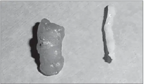
The retrieved fragments (Figure 4) were radiographed (Figure 5). In the presence of representative calcifications, a metal clip was placed, marking the biopsy site (Fig-ure 6), except in cases of very evident re-sidual lesions that could be utilized as a biological marker in the case of re-interven-tion. In the absence of representative cal-cifications, the procedure was repeated until they were obtained.
Representative calcifications were so considered when at least five clustered cal-cifications were obtained, and such calci-fications included some of the most suspi-cious for malignancy.
After the procedure was completed, two mammographic views were performed to confirm the calcifications extraction and the clips positioning.
111 Portable device for vacuum-assisted biopsy
Radiol Bras. 2010 Mar/Abr;43(2):109–112
In the cases diagnosed as benign, a six-month radiological follow-up was recom-mended.
RESULTS
All the biopsies obtained calcifications representative of the radiological lesions. Anatomopathological studies revealed be-nign findings in 23 cases (typical hyperpla-sia in 5, psammomatous calcifications in 6, simple adenosis in 1, dystrophic calcifica-tions in 2 and benign calcificacalcifica-tions not specified in the anatomopathological re-ports in 3), ductal carcinoma in situ in 8 cases, 4 of them high-grade, invasive duc-tal carcinoma in 2 cases, both of them high-grade, and high risk lesion (atypical ductal hyperplasia) in 2 cases. Among the 8 cases of ductal carcinoma in situ, definitive sur-gery revealed invasive ductal carcinoma in 1 case. In the 2 cases of high-risk lesions, the patient was submitted to surgical bi-opsy, and the diagnosis was confirmed as benign. In 6 cases two collection cycles were required, and in the remaining 29 cases a single collection cycle was per-formed. Technical difficulties were not observed with the repeated probe inser-tions, as it was demonstrated that the ster-eotactic guides directed the probe exactly to the skin nick previously made, since the patient’s breast remains fixed during the whole procedure.
Lesions were completely removed in 8 cases and partial sampling occurred in 27 cases. Metal clips were utilized in 31 cases. No error was observed in the marker clips placement.
Severe complications were not ob-served. Hematomas occurred in 15 cases (43%), 3 of them being large (9%). There was no need to drain such hematomas.
On average, each collection cycle took eight minutes to be completed.
With respect to pain, the procedure was well tolerated by all the patients. Occasion-ally, additional local anesthetic administra-tion was required because of pain com-plaint during the procedure. The tested device does not allow the application of an-esthetics directly through the biopsy can-nula. Therefore, whenever necessary, the additional anesthetic application was per-formed by means of a needle inserted be-Figure 3. Button for cannula radial orientation selection.
Figure 5. Radiographic image of one fragment retrieved with the device in study containing repre-sentative calcifications of the lesion.
112
Camargo Júnior HSA et al.
Radiol Bras. 2010 Mar/Abr;43(2):109–112 side the probe, or by removing the cannula
and inserting the needle in the same biopsy pathway.
DISCUSSION
This study was aimed at testing the per-formance of a hand-held vacuum assisted device and has found results similar to those achieved with non portable equipment.
The paradigm to be taken into consid-eration in the selection of a method for a given breast lesion biopsy should be the utilization of a less invasive and less expen-sive method capable of supplying sufficient material for analysis(4). In the case of
microcalcifications, vacuum-assisted bi-opsy is less invasive than surgery and pre-sents a lower smaller underestimation rate than simple core biopsy(1,5-7).
A problem related to vacuum-assisted biopsy is its cost, not only in set-up and de-vice acquisition but also the disposable consumables utilized the process. The cost of biopsies of lesions detected at screening programs may represent up to one-third of the program costs(8). The rational utilization
of resources may produce savings and al-low the application of such resources in other health-related activities, for example, extend the access to screening programs to more women(9). The tested system presents
a considerably lower set-up cost, although the cost of disposable consumables is simi-lar to that of other systems(10).
No difficulty was observed with the can-nula reinsertion for additional collections. Among the different vacuum-assisted devices currently available, some of them operate with a single cannula insertion, and others that operate with multiple insertions. In the first ones, there is a sampling cham-ber coupled to the cannula into where the collected fragment is moved, allowing its retrieval without removing the probe from the patients breast. In the presently tested device, the probe must be removed from the patient’s breast for the retrieval of the collected sample, and then be reinserted for collection of a new fragment. This caused some preoccupation that the system might cause additional trauma for the breast be-cause of the repeated probe insertions, and that the collection accuracy might be
af-fected as a result of the target lesion dis-placement caused by the probe movements, but such complications were not observed. On average, each collection cycle took eight minutes to be completed, longer than the time spent with single cannula insertion systems. Part of this time was spent with the repeated probe insertions. This longer collection time may potentially limit the method accuracy due to patients’ move-ments. The biopsy samples appropriateness suggests that accuracy was not compro-mised. It is important to remind that all biopsies performed in the present study were carried out on a dedicated examina-tion table, on which the patient’s immobi-lization tends to be more efficient. Hypo-thetically, when such procedure is per-formed with the patient on a sitting posi-tion, the probability of undesired move-ments is higher as the patient’s torso retrac-tion is not as well prevented by gravity force as it is with the dedicated prone ex-amination table. In fact, the authors have observed that, during stereotactic biopsies with the patient on a sitting position (not included in this study), movements are not rare (which is easily noticed when the skin nick loses its alignment with the direction of the biopsy guide). The remaining ques-tion is whether, in the case of biopsies per-formed with the patient on a sitting posi-tion, the longer time required by the collec-tion cycle with the portable device object of the present study, might interfere with the sampling accuracy.
One limitation of the present study is that the low number of cases does not al-low comparisons of rates of complications (among which the main one is the devel-opment of hematoma) with other vacuum-assisted biopsy methods. As the tested de-vice is not equipped with resources for post-collection wash out and hematomas aspiration, hypothetically its use might re-sult in a higher number of hematomas. A previous study has evaluated this matter and found that, with the handheld device, there was higher incidence of pain and lower incidence of early and late hemato-mas as compared with non-portable vacuum-assisted biopsy devices(11).
Another limitation of the present study that is also related to the low number of
cases is the fact that it did not compare the biopsies underestimation rate with that of non-portable devices.
Finally, the tested portable vacuum-as-sisted biopsy device obtained appropriate samples in all cases, and is a lower-cost alternative for the performance of such bi-opsies. This was the first test of the device in our country, and the results are compat-ible with other studies in the literature that likewise have demonstrated its good per-formance(12,13).
REFERENCES
1. Burbank F, Parker SH, Fogarty TJ. Stereotactic breast biopsy: improved tissue harvesting with the Mammotome. Am Surg. 1996;62:738–44.
2. Liberman L, Gougoutas CA, Zakowski MF, et al. Calcifications highly suggestive of malignancy: comparison of breast biopsy methods. AJR Am J Roentgenol. 2001;177:165–72.
3. Liberman L, Smolkin JH, Dershaw DD, et al. Cal-cification retrieval at stereotactic, 11-gauge, di-rectional, vacuum-assisted breast biopsy. Radiol-ogy. 1998;208:251–60.
4. Camargo Jr HSA. Diagnóstico por imagem da mama. Uma abordagem integrada. Rio de Ja-neiro: Revinter; 2008.
5. Fahrbach K, Sledge I, Cella C, et al. A compari-son of the accuracy of two minimally invasive breast biopsy methods: a systematic literature review and meta-analysis. Arch Gynecol Obstet. 2006;274:63–73.
6. Hoorntje LE, Peeters PH, Mali WP, et al. Vacuum-assisted breast biopsy: a critical review. Eur J Cancer. 2003;39:1676–83.
7. Plantade R, Hammou JC, Fighiera M, et al. Un-derestimation of breast carcinoma with 11-gauge stereotactically guided directional vacuum-as-sisted biopsy. J Radiol. 2004;85(4 Pt 1):391–401.
8. Lindfors KK, Rosenquist CJ. The cost-effective-ness of mammographic screening strategies. JAMA. 1995;274:881–4.
9. Camargo Jr HSA, Camargo MMA. Reflexões sobre os custos dos programas de rastreamento do câncer de mama. Diagn tratamento. 2003;8:193– 6.
10. Pistolese CA, Ciarrapico AM, Della Gatta F, et al. Cost-effectiveness analysis of two vacuum-as-sisted breast biopsy systems: Mammotome and Vacora. Radiol Med. 2009;114:743–56.
11. Salem C, Sakr R, Chopier J, et al. Pain and com-plications of directional vacuum-assisted stereo-tactic biopsy: comparison of the Mammotome and Vacora techniques. Eur J Radiol. 2009;72: 295–9.
12. Ghate SV, Rosen EL, Soo MS, et al. MRI-guided vacuum-assisted breast biopsy with a handheld portable biopsy system. AJR Am J Roentgenol. 2006;186:1733–6.