SYSTEMATICS, MORPHOLOGY AND PHYSIOLOGY
Morphometry and Morphology of the Antennae of
Panstrongylus megistus
Burmeister,
Rhodnius neglectus
Lent,
Rhodnius prolixus
Stal and
Triatoma
vitticeps
Stal (Hemiptera: Reduviidae)
J
OÃOA
DAR
OSA, S
ILVIAC M
DEF
REITAS, F
LÁVIOF M
ALARA, C
LÁUDIAS R
OCHALab de Parasitologia, Faculdade de Ciências Farmacêuticas/Unesp/Araraquara, Rod Araraquara-Jaú km 1, CP 502, 14801-902 Araraquara, SP, Brasil; [email protected]; [email protected]; [email protected];
[email protected] Edited by Eunice Galati – FSP/USP Neotropical Entomology 39(2):214-220 (2010)
ABSTRACT - The length of the four right antennal segments from nymphs and adults of Panstrongylus megistus Burmeister, Rhodnius neglectus Lent, Rhodnius prolixus Staland Triatoma vitticeps Stal were measured. The length of the antennal segments of the adults of all four species, 4th and 5th instars of P. megistus, and 5th instar of R. neglectus and R. prolixus followed the same pattern: 2nd>3rd>4th>1st. The pattern of 1st and2nd instars of P. megistus was: 4th>3rd>2nd>1st. For 3rd instars of P. megistus, 1st and 2nd instars of R. neglectusand R. prolixus they were: 3rd>4th>2nd>1st. Third and 4th instars of Rhodnius neglectus and R. prolixus had a pattern of: 3rd>2nd>4th>1st. Only T. vitticeps showed the same pattern (4th>3rd>2nd>1st) for all fi ve instars. The morphological study of the second antennal segment by scanning electron microscopy (SEM) disclosed that the fi rst instars of all four species exhibit type I bristles sensillae and one trichobothrium. Another type III bristle and basiconic, campaniform, coeloconic, trichoid sensillae and type I bristle and trichobothria were noted on their fourth instars and adults. Campaniform sensillae were noted only on T. vitticeps adults. Nodules were observed in the joint between 1st and 2nd antennal segments of adults of P. megistus and T. vitticeps, but not on R. neglectus and R. prolixus.
KEY WORDS:Triatominae, mensuration, electron microscopy, sensilla
The Chagas’ disease, which is caused by the fl agellate protozoan parasite Trypanosoma cruzi, is widespread in North and South America from Mexico to southern Argentina and Chile, and is still considered a major public health issue in Latin America (WHO 2005). Great advances have been made in the reduction of vectorial and transfusional transmission, with a resulting alleviation in the incidence of Chagas’ disease. Yet, it is estimated that a total of 18 million individuals are still infected in 17 Central and Latin American countries. Approximately 93 million people live in the endemic areas, and 200,000 new cases occur annually in these areas (WHO 2005).
Panstrongylus megistus Burmeister was fi rst recorded to transmit T. cruzi in 1909 (Chagas 1909), and is one of the six major species vectoring the Chagas’ disease (Silveira 1983, Dias 1993, Brazilian Health Ministry 2005). Rhodnius neglectus Lent plays a secondary role in transmitting the disease; however, it is widespread in the Brazilian states of Bahia, Goiás, Mato Grosso, Maranhão, Minas Gerais, Paraná, Pernambuco and São Paulo (Silveira 1983, Galvão et al 2003). Rhodnius prolixus Stal can be found in 16 Latin American countries and is an important vector of T. cruzi, especially in Venezuela, Colombia and French Guiana (Lent
& Wygodzinsky 1979, Galvão et al 2003). Even though Triatoma vitticeps Stal is considered the most important vector in Bahia, Espírito Santo, Minas Gerais and Rio de Janeiro, studies on this species are very scarce (Galvão et al 2003).
Despite the fact that immatures also play an important role in the transmission of the Chagas’ disease, there are very few information on their morphology. In an early article presented by Rangel (1979) some morphological aspects of the anatomy of the digestive apparatus of P. megistus nymphs are mentioned and distinct features of nymph instars are presented in other studies (Côrrea 1954, Ramírez-Pérez 1969, Carcavallo et al 1978, Lent & Wygodzinsky 1979,Jimenez & Fuentes 1981, Brewer et al 1981, Gonçalves et al, 1985, Rosa et al 1992a, 1992b, 1995, 1999, 2000, Rosa & Barata 1997, Galvão et al 2005).
The importance of the study of the morphological traits of triatomine vectors, beyond their physiological importance as reported by Wigglesworth & Gillett (1934), Chaika (1980) and McIver & Siemicki (1984, 1985), has already been emphasized by Lent & Wygodzinsky (1979), who showed that the length pattern of the four antennal segments can also be used in taxonomic analysis, a fact that justifi es the importance of this study.
We present a morphometric and morphological evaluation of the antennal segments of nymphs and adults of P. megistus, R. neglectus, R. prolixus and T. vitticeps, by means of stereomicroscopy, scanning electron microscopy, to provide new information for Triatominae taxonomic studies.
Material and Methods
Insects. A total of 120 specimens for each one of the four
above-mentioned species were used, 15 for each instar from 1st to 4th and 15 for each sex at the 5th instar and adult stage. Panstrongylus megistus (colony 139) was collected on February 11, 1985 in Santa Maria do Cambucá area, Pernambuco State, Brasil; R. neglectus (colony 16),collected on April 6, 1982 in Pitangueiras area, São Paulo State, Brasil; R. prolixus (colony 14), from Colombia and established in the insectary on March 6, 1982, and T. vitticeps (colony 41) from Minas Gerais State, Brasil, donated by the Faculty of Public Health, USP, on September 8, 1982. Species were identifi ed according to Lent & Wygodzinsky (1979). The insects were kept in the collection of the Triatominae Laboratory in the insectary of the Araraquara Special Health Service (SESA), São Paulo State. All specimens were blood-fed on ducks (Anas platyrhynchos) for a fortnight at 45 to 50 min intervals.
Insect preparation. Specimens were killed by exposure to
chloroform for 1-2 min, and had their right antennal segments excised for assessment of their size in dorsal position, as described by Rosa et al (2000).
Measurements. Antennal segments were measured at 800x
magnifi cation in a Leica MZ APO stereomicroscope coupled
with a CCD high performance COHU camera, and images were analyzed with the Leica Q-Win software. Trichobothria of adults of the four species (10 specimens each) were counted under the Leica MZ APO stereomicroscope.
Morphological procedures. Chloroform killed specimens
were sonicated (Thorton), dried at 50°C for 20 min. For the morphological study were used the second antennal segment of each specimen, which were fi xed horizontally to metallic supports. The antennae were sputter-coated with gold in a vaccum vaporizing metallizer (Edwards), using a pressure of 10-6. After the metallization, the antennal segments were observed and photographed under a scanning electron microscopy (SEM) Topcon – SM-300 Rosa et al (1992b).
Statistical analysis. Data on the size of the antennal segment were analyzed by using the GraphPad InStat version 3.06 package. Size differences among the four antennal segments were tested for signifi cance by ANOVA (One-way Variance Analysis) and analyzed by the post-test Tukey-Kramer multiple comparison method.
Results
Morphometric studies. Measurements of the antennal
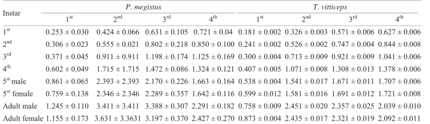
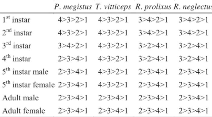
segments indicated the size of the segments varied among instars and the species studied, with the exception of T. vitticipes that showed the same pattern of size for all antennal segments throughout the immature development (Tables 1, 2 and 3). No differences were observed in the pattern of size of the antennal segments between sexes within the same species(Tables 1, 2, 3).
Multiple pairwise comparisons between species indicated that the length of all four antennal segments of each of the fi ve instars and adults differed between P. megistus and T. vitticeps, P. megistus and R. neglectus, P. megistus and R. prolixus, T. vitticeps and R. neglectus, R. neglectus and R. prolixus,exceptfor the third segment of the1stinstarof P. megistus and R. neglectus; there were no signifi cant differences between T. vitticeps and R. prolixus (Table 4).
Table 1 Length (mm) (mean + standard deviation) of the antennal segments of nymphs and adults of Panstrongylus megistus and Triatoma vitticeps.
Instar P. megistus T. vitticeps
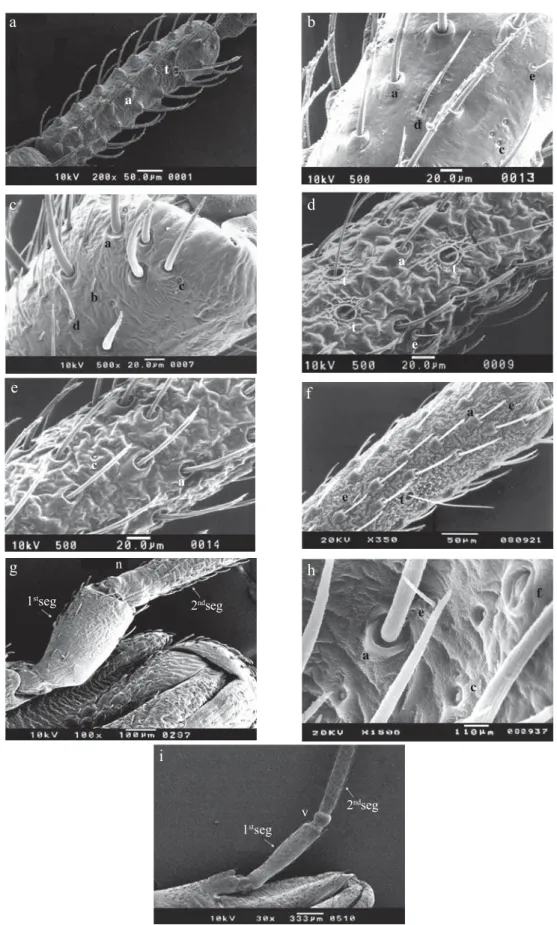
Morphological studies. The second antennal segments of all instars of P. megistus, R. neglectus, R. prolixus and T. vitticeps were examined by SEM, and differences and similarities in the number and type of sensillae were observed (Figs 1a-f).
The 1st instars of P. megistus, R. neglectus, R. prolixus and T. vitticeps displayed type I bristles and one trichobothrium located at the posterior third of the dorsal side of the second antennal segment (Fig 1a).
The 2nd and 3rd instars of P. megistus, 2nd, 3rd and 4th of R. neglectus and 2nd to 5th instars of R. prolixus and T. vitticeps displayed the same pattern of sensillae as the 1st instar, plus a few type III bristles (Fig 1f). The 4th and 5th instars of P. megistus showed types I and III bristles, coeloconic and trichoid sensillae and one trichobothrium (Fig 1b).
In adult male and females of P.megistus, the second antennal segment showed type I bristles, trichoid and coeloconic sensillae, basiconic sensillae and four to nine trichobothria (Fig 1c). Adult male and female of R. neglectus showed types I and III bristles, coeloconic sensillae and trichobothria (Fig 1d,e). Rhodnius prolixus showed types I and III bristles and trichobothria (Fig 1f). Triatoma vitticeps had types I and III bristles and campaniform and coeloconic sensillae and trichobothria (Fig 1h).
The number oftrichobothria on the right and left segments of the antenna of adults varied from two to nine in both sexes
for all studied species (Table 5).
Small nodules separating the fi rst and second antennal segments were observed in P. megistus and T. vitticeps, but were absent in R. neglectus and R. prolixus (Figs 1g, i).
Discussion
The origin of the Triatominae is controversial since they probably evolved from predatory Reduviidae. Triatominae are classifi ed as monophyletic according to Usinger (1944), Lent & Wygodzinsky (1979) and Hypsa et al (2002), or polyphyletic (Schofield 1988, Schofield & Dujardin 1999, Paula et al 2005). The combination of anatomical, physiological and ethological factors observed in this group calls for new research on the morphology of species, which could be useful for taxonomy and in the control of Chagas’ disease in endemic areas.
The occurrence of small nodules separating the antennal segments of Triatominae has been reported for several other species (Lent & Wygodzinsky 1979, Rosa et al 1999).
Although Lent & Wygodzinsky (1979) reported that in most species of Triatominae the length of the antennal segments decreased from the second to the fourth segment, the size of the segments varied according to the instar in Triatoma rubrovaria (Rosa et al 2000), as observed for all four species investigated in here (Tables 1, 2, 5). In the case of T. vitticeps, changes were observed only for the adult (Tables 1, 5).
Triatominae have hundreds of sensillae on their four antennal segments (Catalá 1994). Twelve different sensillae were identifi ed on the antennal segments of 10 out of the 16 species of Rhodnius studied from laboratory colonies (Catalá & Schofi eld 1994, Galvão et al 2003).
Trichobothria are a type of sensilla that is longer and thinner than the others and a varied length, with a diameter ranging from 60 μm to 210 μm. They are randomly distributed and may have mechano, thermo and chemoreceptor activity, and are very common to arthropods (Schuh 1975, Catalá & Schofi eld 1994). The number of trichobothria in Triatominae is also suggested to have a taxonomic value (Lent & Wygodzinsky 1979).
Table 2 Length (mm) (mean + standard deviation) of the antennal segment of nymphs and adults of Rhodnius neglectus and Rhodnius prolixus.
Instar R. neglectus R. prolixus
1st 2nd 3rd 4th 1st 2nd 3rd 4th 1st 0.137 ± 0.039 0.390 ± 0.061 0.665 ± 0.075 0.525 ± 0.075 0.119 ± 0.002 0.390 ± 0.003 0.711 ± 0.005 0.453 ± 0.008 2nd 0.219 ± 0.053 0.922 ± 0.048 1.142 ± 0.068 0.932 ± 0.048 0.144 ± 0.002 0.586 ± 0.108 0.803 ± 0.011 0.666 ± 0.008
3rd 0.328 ± 0.052 1.386 ± 0.074 1.489 ± 0.081 1.212 ± 0.114 0.185 ± 0.004 0.895 ± 0.007 1.094 ± 0.009 0.856 ± 0.009 4th 0.294 ± 0.046 1.388 ± 0.085 1.471 ± 0.061 1.142 ± 0.051 0.264 ± 0.009 1.401 ± 0.021 1.476 ± 0.027 1.068 ± 0.036 5st male 0.393 ± 0.037 2.086 ± 0.145 1.871 ± 0.096 1.319 ± 0.135 0.243 ± 0.003 1.341 ± 0.007 1.166 ± 0.006 0.848 ± 0.008 5st female 0.440 ± 0.028 2.207 ± 0.088 2.077 ± 0.365 1.399 ± 0.169 0.224 ± 0.004 1.381 ± 0.008 1.252 ± 0.009 0.865 ± 0.004 Adult male 0.404 ± 0.404 3.564 ± 0.385 2.264 ± 0.249 1.322 ± 0.096 0.281 ± 0.004 1.861 ± 0.009 1.371 ± 0.007 0.887 ± 0.008 Adult female 0.381 ± 0.055 3.388 ± 0.352 2.402 ± 0.280 1.324 ± 0.155 0.306 ± 0.006 1.904 ± 0.011 1.475 ± 0.011 0.933 ± 0.004
Table 3 Relative size difference among the absolute length of each antennal segments (antennal segment formula) of Panstrogylus megistus, Triatoma vitticeps, Rhodnius neglectus and Rhodnius prolixus.
P. megistus T. vitticeps R. prolixus R. neglectus
1stinstar 4>3>2>1 4>3>2>1 3>4>2>1 3>4>2>1 2nd instar 4>3>2>1 4>3>2>1 3>4>2>1 3>4>2>1 3rd instar 3>4>2>1 4>3>2>1 3>2>4>1 3>2>4>1 4th instar 2>3>4>1 4>3>2>1 3>2>4>1 3>2>4>1 5th instar male 2>3>4>1 4>3>2>1 2>3>4>1 2>3>4>1
Table 4 Statistical analysis of the 1st, 2nd, 3rd and 4th antennal segments of nymphs and adults of Panstrogylus megistus, Triatoma vitticeps, Rhodnius neglectus and Rhodnius prolixus.
***Extremely signifi cant ; *Less signifi cant; NS = not signifi cant
P
. meg
istu
s
x
T
. vitticeps
1st *** *** *** *** *** *** *** *** 2nd *** NS *** *** *** *** *** ***
3rd NS NS *** *** *** *** *** ***
4th NS *** NS NS NS NS *** ***
P
.
meg
istu
s
x
R
. pr
o
lix
us 1
st ** *** *** *** *** *** *** ***
2nd NS NS NS *** *** *** *** ***
3rd ** NS * NS *** *** *** ***
4th NS *** *** *** *** *** *** ***
T
. vitticeps
x
R
.
pr
o
lix
us 1
st
*** *** *** *** *** *** *** ***
2nd ** * *** *** *** *** *** **
3rd *** NS *** *** *** *** *** ***
4th NS NS ** *** *** *** *** ***
Antennal segment
Instars
1st 2nd 3rd 4th 5th male 5th female Adult male Adult female
1st *** *** ** *** *** *** *** ***
2nd NS *** *** *** *** *** NS ***
3rd NS *** *** NS *** NS *** ***
4th NS ** NS *** *** *** *** ***
T
.
vittic
eps
x
R
.
ne
g
le
ct
us 1st *** NS NS *** *** *** *** ***
2nd ** *** *** *** *** *** *** ***
3rd ** *** *** *** *** *** *** NS
4th NS *** ** *** *** *** *** ***
1st NS *** *** ** *** *** *** NS
2nd NS *** *** NS *** *** *** ***
3rd NS *** *** NS *** *** *** ***
4th NS *** *** * *** *** *** ***
P
.
m
egis
tus
x
R
.
ne
g
le
ct
us
R
.
ne
g
le
ct
us
x
R
.
pr
o
lix
us
Table 5 Number of trichobothria found on the second antennal segment of the right and left antenna on 10 adult males and females of Panstrogylus megistus, Triatoma vitticeps, Rhodnius neglectus and Rhodnius prolixus.
R = right antennal segment; L = left antennal segment
Specimens
P. megistus R. neglectus R. prolixus T. vitticeps
Male Female Male Female Male Female Male Female R L R L R L R L R L R L R L R L
1 5 6 5 6 4 3 3 4 5 5 6 5 7 6 6 5
2 5 5 6 8 5 4 5 3 8 7 9 8 5 3 6 4
3 4 6 7 5 5 4 6 5 6 5 5 9 6 8 6 6
4 8 6 8 8 5 5 4 6 5 4 6 5 5 5 6 9
5 6 4 5 6 3 4 3 4 4 5 4 7 3 6 4 5
6 7 7 5 7 6 3 5 5 9 6 4 6 7 8 7 5
7 4 6 6 5 7 5 4 5 8 7 5 5 6 5 4 6
8 5 5 8 6 4 4 7 5 6 6 4 6 6 8 6 4
9 5 6 7 4 4 6 5 5 7 6 5 7 7 5 7 7
Fig 1 Dorsal view of second antennal segment by SEM. a) nymph of 1st instar of P. megistus x500; b) nymph of 4th instar of P. megistus x500; c) adult of P. megistus x500; d and e) adult of R. neglectus x500; f) nymph of 3rd instar of R. prolixus x350; g) joint of the 1st and 2nd antennal segments of adult of R. prolixus x100; h) adult of T. vitticeps x1500; i) joint of the 1st and 2nd antennal segments of adults of T. vitticeps x100. a: type I bristle; b: basiconic sensilla; c: coeloconic sensilla; d: trichoid sensilla; e: type III bristle; f: campaniform sensilla; t: trichobothrium; v: nodule; n: nodule absent.
a
a
t
t
e a
t
d b
a
a
v c
n
a
e
c
f
1stseg
1stseg
2ndseg
2ndseg c
a
d
c e
t
g c
f b
h
i
d
e
a e
Our observations agree with those of Lent & Wygodzinsky (1979), which indicated the existence of a single trichobothrium situated subapically on the dorsal side of the second antennal segment. By contrast, adult triatomines display several (2-9) trichobothria on the second antennal segment (Table 4).
In adults, Lent & Wygodzinsky (1979) and Catalá & Schofield (1994) studied the number and location of trichobothria in species of Triatominae. The number of trichobothria described by them differs from those recorded in this study. Previous observation of many specimens of the four species studied in here showed that the number of trichobothria vary in the adults of P. megistus (four to eight), R. neglectus (two to seven), R. prolixus (four to nine) and T. vitticeps (three to nine) (Table 3), in contrast to the nymphs.
Another taxonomic contribution made by this study was the fi nding of campaniform sensillae on the posterior part of the second antennal segment of adults of T. vitticeps,but not found in P. megistus, R. neglectus or R. prolixus (Figs 1a-f). Triatoma vitticeps, differently from the other three species, presented only two patterns of antennal segment lengths, whereas P. megistus, R. neglectus and R. prolixus showed three patterns. These observations will be useful for phylogeny and, together with other studies, may contribute to determining whether the subfamily Triatominae is a polyphyletic or a monophyletic group.
Acknowledgments
We thank Prof Dr. José Maria Soares Barata, responsible for the Triatominae insectary of the School of Public Health (USP), located at the Special Health Service of Araraquara, who donated the specimens utilized. Thanks are also due to Prof Dr José Clóvis do Prado Jr for his revision and suggestions, Prof Dr Mario Cilense who consented to the utilization of SEM, João Luis Molina Gil and João Maurício Nóbrega Filho, who maintain the insect colonies and to Tim Roberts and João Eduardo da Rosa, who reviewed the English text. Financial support came from PIBIC/ CNPq.
References
Brewer M, Garay M, Gorla D, Murua F, Favarot R (1981) Caracterización de los estadios ninfales del genero Triatoma Laporte 1833. I Triatoma infestans, Klug, 1834 (Hemiptera, Reduviidae). Rev Soc Entomol Arg 40: 91-102.
Carcavallo R U, Justo N S, Martinez A (1978) Descripción de las ninfas de I, II y IV estadio de Alberprosenia goyovargasi Martinez & Carcavallo, 1977 (Hemiptera, Reduviidae, Triatominae). Observaciones con microscopia electrónica de barrido. Bol Mal San Amb 18: 132-138.
Catalá S (1994) The cave organ of Triatominae (Hemiptera, Reduviidae) under scanning electron microscopy. Mem Inst Oswaldo Cruz89: 275-277.
Catalá S, Schofield C (1994) Antennal sensilla of Rhodnius (Hemiptera, Reduviidae). J Morphol 219: 193-204.
Chagas C (1909) Nova tripanozomíase humana. Estudo sobre a morfologia e o ciclo evolutivo do Schizotrypanum cruzi, n. gen., n. sp., agente etiológico de nova entidade mórbida do homem. Mem Inst Oswaldo Cruz 1: 159-218.
Chaika S Y (1980) Ultra structure of the antennal sensilla of the bug Rhodnius prolixus (Hemiptera, Reduviidae). Parasitology 14: 486-492.
Corrêa R R (1954) Estudos sobre a morfologia externa do gênero Triatoma Laporte, 1833 (Hemiptera, Reduviidae). Fol Clin Biol 22: 23-50.
Dias J C P (1993) Aspectos clínicos, sociais e trabalhistas da doença de Chagas em área endêmica sob controle do estado de Minas Gerais, Brasil. Rev Soc Bras Med Trop 26: 93-99.
Galvão C, Carcavallo R, Rocha D S da, Jurberg J (2003) A checklist of the current valid species of the subfamily Triatominae Jeannel, 1919 (Hemiptera,Reduviidae) and their geographical distribution, with nomenclatural and taxonomic notes.Zootaxa202: 1-36.
Galvão C, Mcloon F M, Rocha D S, Schaefer C W, Patterson J S, Jurberg J (2005) Description of eggs and nymphs of Linshcosteus karupus Galvão, Patterson, Rocha, Jurberg, 2002 (Hemiptera: Reduviidae: Triatominae). Ann Entomol Soc Am 98: 861-872.
Gonçalves T C M, Jurberg J, Costa J M, Souza W (1985) Estudo morfológico comparativo de ovos e ninfas de Triatoma maculata (Erichson,1848) e Triatoma pseudomaculata Correa & Espínola, 1964 (Hemiptera, Reduviidae, Triatominae). Mem Inst Oswaldo Cruz 80: 276-280.
Hypsa V, Tietz D F, Zrzavy J, Rego R O M, Galvão C, Jurberg J (2002) Phylogeny and biogeography of Triatominae (Hemiptera: Reduviidae): molecular evidence of a New World origin of the Asiatic clade. Mol Phylogenet Evol 32: 447-457.
Jimenez O, Fuentes H O (1981) Triatoma fl avida Neiva, 1911 (Hemiptera, Reduviidae). I Estudio biometrico of larvas. Rev Cub Med Trop 33: 195-200.
Lent H, Wygodzinsky P (1979) Revision of the Triatominae (Hemiptera, Reduviidae) and their signifi cance as vectors of Chagas’ disease. Bull Am Mus Nat Hist163: 123-520.
McIver S, Siemick R (1984) Fine structure of antennal mechanosensilla of adult Rhodnius prolixus Stal (Hemiptera, Reduviidae). J Morphol180: 19-28.
McIver S, Siemick R (1985) Fine structure of antennal putative thermo/hygrosensilla of adult Rhodnius prolixus Stal (Hemiptera, Reduviidae). J Morphol183:15-23.
MS – Ministério da Saúde, Secretaria de Vigilância em Saúde (2005) Consenso brasileiro em doença de Chagas. Rev Soc Bras Med Trop 38 (Suppl III): 7-29.
Paula A S, Dioatiuti L, Schofi eld C J (2005) Testing the sister-group relationship of the Rhodniini and Triatomini (Insecta: Hemiptera, Reduviidae, Triatominae). Mol Phylogenet Evol 35: 712-718.
Ramírez-Pérez J (1969) Estudio sobre la anatomía de Rhodnius prolixus. Rev Venez Sanid Asist Soc 34: 10-98.
neglectus, Lent, 1954 e Triatoma infestans (Klug, 1834), (Hemiptera, Reduviidae). An Soc Entomol Brasil 8: 309-323.
Rosa J A da, Barata J M S (1997) Aspectos morfológicos do abdômen de ninfas de 5o estádio de seis espécies de Triatominae (Hemiptera, Reduviidae) por microscopia óptica. Rev Cienc Farm UNESP18: 249-270.
Rosa J A da, Barata J M S, Barelli N (1992a) Spiracles of 5th instar nymphs in six species of Triatominae (Hemiptera, Reduviidae) using scanning electron microscopy. Mem Inst Oswaldo Cruz 87: 301-302.
Rosa J A da, Barata J M S, Barelli N (1995) Morphology of abdominal bristles determined by scanning electron microscopy in six species of Triatominae (Hemiptera, Reduviidae).Mem Inst Oswaldo Cruz90: 487-488.
Rosa J A da, Barata J M S, Barelli N, Santos J L F, Belda Neto F M (1992b) Sexual distinction between 5thinstar nymphs of six species of Triatominae (Hemiptera, Reduviidae).Mem Inst Oswaldo Cruz 87: 257-264.
Rosa J A da, Barata J M S, Cilense M, Belda Neto F M (1999) Head morphology of fi rst and fi fth instar nymphs of Triatoma circummaculata and Triatoma rubrovaria (Hemiptera, Reduviidae). Inter J Insect Morphol Embryol 28: 363-375.
Rosa J A da, Três D F A, Santos J L F, Barata J M S (2000) Estudos morfométricos dos segmentos antenais de ninfas e adultos de duas colônias de Triatoma rubrovaria (Blanchard, 1843) (Hemiptera, Reduvidae). Entomol Vect 7: 255-264.
Schofi eld C J (1988) The biosystematics of Triatominae, p.284-312. In Service M W (ed) Biosystematics of haematophagous insects, special vol 37, Oxford, Systematics Association/ Clarendon Press, 376p.
Schofi eld C J, Dujardin J P (1999) Theories on the evolution of Rhodnius. Actual Biol 21: 183-197.
Schuh R T (1975) The structure distribution and taxonomic importance of trichobothria in the Miridae (Hemiptera). Amer Mus Novit 2585: 1-26.
Silveira A C (1983) Epidemiologia e controle da doença de Chagas. Rev Saúde Pública 1: 212-218.
Usinger R L (1944) The Triatominae of North and Central America and the West Indies and their public health signifi cance. Pub Health Bull 288, p. 83, fi gs.1-5, pls I_XII.
Wigglesworth V B, Gillet J D (1934) The function of the antennae in Rhodnius prolixus (Hemiptera) and the mechanism of orientation to the host. J Exp Biol 11: 120-39.
WHO (2005) Making health research work for poor people, progress 2003-2004. Tropical Disease Research Seventeenth Programme Report. [Accessed in 2006 Aug 26]. Available from: http://www. who.int/tdr/publications/publications/pdf/pr17/pr17.pdf.