Arq. Bras. Oftalmol. vol.77 número6
Texto
Imagem

Documentos relacionados
Enhanced depth imaging optical coherence tomography and fundus autofluorescence findings in bilateral choroidal osteoma: a case report.. Tomograia de coerência óptica com
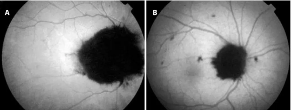
Figure 2. A) The initial fundus autoluorescent imaging of the RPE tear with two disc-sized hypoauto- luorescent areas. B) The initial spectral domain optical coherence
Evaluation of the choroid using spectral-domain optical coherence tomography (SD-OCT) was recently reported to be valuable because choroidal change has been associated with
Purpose : To evaluate the association between macular hole volume (MHV) and postoperative central macular thickness (CMT ) using spectral-domain optical coherence tomography
Purpose: To evaluate choroidal thickness (CT ) using spectral domain optical coherence tomography (SD-OCT ) imaging at baseline and 6 months after intravi- treal
A retrospective study was performed by reviewing medical records and spectral domain optical coherence tomography (SD-OCT ) findings of seven patients who were treated with a
The authors noted that “this is the first report of optical coherence tomography (OCT) imaging of macular atrophy in a child with presumed Zika virus infection-asso-
Optical coherence tomography of macular atrophy associated with microcephaly and presumed intrauterine Zika virus infection. Arq
