1
Original Article
Percutaneous Mitral Balloon Valvotomy. Long-term
Outcome and Assessment of Risk Factors for Death
and Major Events
Ivana Picone Borges, Edison Carvalho Sandoval Peixoto, Rodrigo Trajano Sandoval Peixoto,
Paulo Sergio de Oliveira, Mario Salles Netto, Pierre Labrunie, Marta Labrunie,
Ricardo Trajano Sandoval Peixoto, Ronaldo de Amorim Villela
Rio de Janeiro, RJ / Niterói, RJ - Brazil
Cinecor 4º Centenario, Rio de Janeiro e Universidade Federal Fuminense, Niterói, Rio de Janeiro
Mailing address: Edison Carvalho Sandoval Peixoto - Av. Epitácio Pessoa, 4986/301 - Cep 22471-003 - Rio de Janeiro, RJ - Brazil E-mail: [email protected]
Received for publication: 04/11/2004 Accepted for publication: 08/25/2004
Objective
To identify the factors that predict death and combined events, (death, new mitral balloon valvotomy, or mitral valve surgery) in long-term follow-up of patients undergoing percuta-neous mitral balloon valvotomy.
Methods
Follow-up was 49.0±31.0 (1 to 122) months. Techniques used were the single-balloon (84.4%), Inoue-balloon (13.8%), and double-balloon techniques (1.7%).
Results
Included in the study were 289 patients 38.0±12.6 years of age (range, 13 to 83). Before the procedure, 244 patients had echocardiographic score ≤ 8, and 45 patients had score >8. Females comprised 85%, and 84% patients were in sinus rhythm. During follow-up, survival of the total group was 95.5%, that of the group with ≤ 8 was 98.0%, finally that of the group with scores >8 was 82.2% (P<0.0001), whereas combined event-free survival was 83.4%, 86.1%, and 68.9%, respectively (P<0.0001). In the multivariate analysis, the factors that pre-dicted long-term death were a preprocedure echocardiographic score >8 and the presence of severe valvular mitral regurgitation during the procedure. The events that predicted combined events were a previous history of mitral valvular commissurotomy and atrial fibrillation and the presence of severe mitral valvular regurgitation during the procedure, and postprocedure mitral valvular area <1.5 m2 (failure).
Conclusion
Percutaneous mitral balloon valvotomy is an effective proce-dure, and over 2/3 of the patients were event-free at the end of follow-up. Survival in the group was high, even higher in the group with lower echocardiographic scores.
Key words
mitral valvular stenosis, mitral stenosis, mitral balloon valvo-tomy, rheumatic fever
Mitral balloon valvotomy was introduced in 1984 by Inoue et al 1. In 1986, McKay et al 2 and Palacios et al 3 put it into practice
in the United States. In Saudi Arabia, Al Zaibag et al 4, started
using the double-balloon with the transseptal technique. In Brazil, dilation with the retrograde technique 5,6 and the transseptal
te-chnique 7-9 was described in 1987.
Today, it is accepted that a similar valvular area can be obtained after percutaneous mitral-balloon valvotomy using any of these techniques 10-16.
Overall survival and event-free survival varied in the groups studied, due to the clinical and echocardiographic characteristics of the patients 17-24. Among the characteristics that favor a more
successful outcome are: young age, satisfactory valvular anatomy, echocardiographic score ≤ 8, presence of sinus rhythm, absence of mitral regurgitation before the procedure, and absence of surgical commissurotomy before the procedure.
The primary objective of this study was to establish the factors that predict death, and combined events, (death, new mitral-balloon valvotomy, and mitral valvular surgery), in the long-term follow-up of patients undergoing percutaneous mitral-balloon val-votomy, and the secondary is to compare the clinical and echo-cardiographic outcomes of a group with scores ≤ 8 and another group with scores > 8.
Methods
A prospective longitudinal study was performed in patients undergoing percutaneous balloon mitral valvotomy.
Patients excluded were those with unfinished procedures and those whose procedures were finished but the follow-up did not reach a month, either due to patients being lost to follow-up or because of complications or failure followed by major events, making 1-month follow-up impossible. Follow-up was discontinued in case of death, new mitral-balloon valvotomy, or mitral valve surgery. Patients were included again in the event of a new mitral-balloon valvuloplasty.
2
Percutaneous Mitral Balloon Valvotomy. Long-term Outcome and Assessment of Risk Factors for Death and Major Events
between 24 and 28 mm. If a new mitral regurgitation occurred or the level of previous mitral regurgitation worsened during the pro-cedure, regurgitation was quantified using Sellers et al’s classifi-cation25. The mean gradient before and after the procedure was
measured in 2 ways: 1st) by the 3-point method26 and 2nd) by
planimetry of the gradient area26. The mitral valvular area before
and after dilation was determined, assessing the cardiac output by thermal dilution and using Gorlin and Gorlin’s formula 27. The
follow-up was performed by telephone or letter. The following were assessed: functional class (FC) according to NYHA, mortality and the cause of death, medication used, performance of mitral valve surgery or of new balloon mitral valvotomy. After the ques-tionnaire was filled in, 1-dimensional and 2-dimensional Doppler echocardiogram was performed in 224 cases. Clinical evolvement of patients in the study was considered as of the first month after the procedure. Mitral valve area was assessed using planimetry or transmitral pressure half-time. Mitral valvular morphology was assessed using Wilkins’ score 28. The level of mitral regurgitation
was assessed by Doppler echocardiography according to the extension of the regurgitant flow in the left atrium, classified as mild, moderate, and severe 29. All patients underwent
echocar-diography before undergoing mitral valve valvuloplasty. At the beginning and end of the procedure, mitral valvular area was assessed by hemodynamic calculation 25,27 and the level of
regur-gitation by left ventriculography.
Patients were also divided into 2 subgroups A and B, using Wilkins’ score 28. Group A with scores ≤ 8 and group B with
scores > 8.
Success was defined as mitral valvular area ≥ 1.50 cm2,
ob-tained after the procedure by using hemodynamic assessment without severe mitral failure. Failure was defined as mitral valvular area < 1.5 cm2.
Severe mitral regurgitation grade 3 or 4+ was determined by using Sellers’ classification 25.
Comparison between continuous variables was performed using the Student t test, when the distribution was normal; otherwise, the Mann-Whitney test was used. Comparison between categorical va-riables was performed using the chi-square test, the chi-square test with Yates comparison, and Fisher’s exact test, according to the frequency of events, using EPI INFO’s software program 30 for the
calculation and as a databank. A multivariate model of Cox’s propor-tional risks was built to identify the independent factors predicting death and major events combined with death, new balloon mitral valvotomy, and mitral valvular surgery in a long-term follow-up, using SPSS 8 31 software program. Survival and event-free survival curves
were created using the Kaplan-Meier method 32, and compared,
using Log Rank, Breslow, and Tarone-Ware’s models. Multivariate analysis 33 on steps was used to identify the independent factors
predicting death and combined events in the long-term outcome. Relative risk of the variables related to time was assessed and the confidence Cox’s regression model 33. Variables that demonstrated
error probability < 7% (p<0.07) in the univariate analysis underwent multivariate analysis. Variables selected for multivariate analysis were age, success, presence of severe mitral regurgitation after procedure, effective balloon dilation area, the diameter of the dilation balloon of the mitral valve, cardiac rhythm, history of surgical mitral com-missurotomy, score ≤ 8 and > 8, and score ≤ 11 and < 11.
Results
Follow-up was completed of 289 patients. All of them had a completed procedure and at least 1 month of follow-up, 49.0±31.0 (1 to 122) months; 246 (85.1%) patients were females, with ages 38.0±12.6 (13 to 83) years old. Four patients were in NYHA FC I (1.4%), 72 patients (24.9%) were in FC II, 184 patients (63.7%) were in FC III, and 29 patients (10.0%) were in FC IV. Forty-five patients (15.6%) had atrial fibrillation, and 244 (84.4%) had sinus rhythm. Twenty-five patients (8.7%) had a history of previous surgical commissurotomy, and 9 patients (3.1%) had mitral balloon valvotomy. The echocardiographic score ranged from 4 to 14 (7.3±1.5), 244 patients (84.4%) had echocardio-graphic score ≤ 8, group A, and 45 (15.6%) > 8, group B. Echocardiographic mitral area, before mitral valvotomy, was 0.90±0.21 cm2; in group A it was 0.93±0.21 cm2, and in group
B it was 0.91±0.20 cm2 (p=0.5627). Of all the procedures,
244 (84.4%) were performed with the single-balloon, 40 (13.8%) with the Inoue, and 5 (1.7%) of 29.5±1.2 mm and a maximum dilation area 6.87±0.49 cm2. Mean pulmonary artery pressure,
mean mitral valvular gradient and mitral valvular area before and after balloon valvotomy were 38.0±14.3 and 26.7±10.1 mmHg (p<0.0001), 19.2±7.0 and 5.4±3.5 mmHg (p<0.0001), and 0.90±0.21 and 2.00±0.39 cm2 (p=0.0139), respectively. Of
all the procedures, 261 were successful (90.4%) and 19 (6.6%) were unsuccessful; mitral valvular area after mitral balloon valvoto-my was not assessed in 9 procedures (3.1%). Before mitral valvo-tomy, grade 1+ mitral regurgitation was present in 44 patients (15.2%), grade 2+ in 1 patient (0.3%), and the valve was compe-tent in 244 patients (84.4%). After mitral valvotomy, the valve was competent in 207 (71.6%) patients, and grade 1+ mitral regur-gitation occurred in 61 patients (21.1%), grade 2+ in 18 patients (6.2%), grade 3+ in 2 patients (0.7%), and grade 4+ in 1 patient (0.3%). Severe complications occurred in 5 patients of the 289 patients, 3 patients (1.0%) had severe mitral regurgitation, and 2 patients had cardiac tamponade, drained in the hemodynamic room. All patients remained with clinical evolvement for at least 1 month without a surgical procedure for mitral valve replacement.
In the final follow-up lasting 49.0±31.0 (1 to 122) months, NYHA FC was I in 141 (48.8%) patients, FC II in 82 patients (28.4%), FC III in 49 patients (17.0%), and FC IV in 4 patients (1.4%), with 13 (4.5%) deaths. Group A, with 244 patients, had, according to the NYHA, 124 patients in FC I, 69 in FC II, 43 in FC III, 3 in FC IV with 5 deaths, and group B, with 45 patients, had 17 in FC I, 13 in FC II, 6 FC III, 1 in FC IV with 8 deaths (p= 0.0001). Echocardiography was performed at the end of follow-up in 244 patients with mitral area 1.56±0.49 cm2; 97 (44.1%)
of the 244 patients had valvular area < 1.50 cm2; in group A it
was 1.58±0.50 cm2, and in group B it was 1.42±0.39 cm2
(p=0.1042). Preprocedure mean mitral valvular area assessed through echocardiography was 0.93±0.21cm2 and 0.91±0.20
(p=0.5627) in groups A and B, respectively (tab. I). Mean mitral valve area, assessed through the hemodynamic method (Gorlin), before balloon valvotomy was 0.90±0.22 and 0.94±0.21 (p=0.2075), respectively, in groups A and B (tab. I). Mitral valvular area (Gorlin), immediately after balloon mitral valvotomy was higher in group A 2.03±0.39 cm2 compared with that in group
3
echocardiography during follow-up, the mitral valve was competent in 60 patients (26.8%), and grade 1+ mitral regurgitation was present in 118 patients (52.7%), grade 2+ in 24 patients (10.7%), grade 3+ in 12 patients (5.4%), and grade 4+ in 10 patients (4.5%). Three patients with severe mitral regurgitation started follow-up, and all of them died, one during the surgical procedure. During follow-up, 19 new cases of severe mitral regurgitation occurred, 9 patients underwent surgery with one death, and 10 had clinical evolvement or waited for surgery. Fourteen patients (4.8%) underwent new mitral balloon valvotomy; 27 patients (9.3%) underwent mitral valve surgery, 16 due to mitral stenosis, 6 due to severe mitral regurgitation; and 5 due to double mitral lesion. During follow-up, 13 deaths (4.5%) occurred, 11 (3.8%) due to heart problems, 1 (0.7%) due to stroke, and 1 (0.7%) had no reported cause. Of the cardiac deaths, 6 occurred during mitral valve replacement, 4 due to acute pulmonary edema, and 1 due to heart failure. Late mortality was greater in group B, with 8 deaths (17.8%) in 45 patients, versus 5 deaths (3.5%) in 244 patients in group A (p=0.0001) (tab. I). In the total group, 48 (16.6%) combined events occurred. At the end of follow-up, however, number of combined events was lower in group A, 34 events (13.9%) compared with those in group B, 14 events (31.1%), p=0.0010 (tab. I). Survival of the total group at the end of the study was 95.5%. In group A, it was 98.0%, greater than in group B, where it was 82.2% (p=0.0001). Event-free survival of the total group was 83.4%, and in group A it was 86.1% greater than that in group B, 68.9% (p=0.0010).
Factors predicting long-term survival in the univariate analysis were echocardiographic score ≤ 8 (p=0.0001), echocardiographic score ≤ 11 (p<0.0001) age of the patient < 50 years old (p=0.0016), absence of severe mitral regurgitation per procedure (p<0.0001), sinus rhythm (p=0.0034), mitral balloon diameter ≥ 28 mm (p=0.0005), mitral dilation effective area > 6 cm2 (p=0.0005)
and mitral valve area (Gorlin) ≥ 1.5 cm2 (success) (p=0.0065).
Factors predicting combined event free survival in the univariate analysis were: sinus rhythm (p=0.0005); mitral dilation balloon dia-meter ≥ 28 mm (p=0.0005); effective area of mitral dilation ≥ 6 cm2 (p=0.0036) and mitral valve area ≥ 1.5 cm2 (p<0.0001),
echocardiographic score ≤ 8 (p=0.0017); echocardiographic score
≤ 11 (p<0.0001); absence of previous surgical mitral commissuro-tomy (p=0.0235), absence of severe mitral regurgitation per proce-dure (p<0.0001) and age of the patient < 50 years old (p=0.0013). In the multivariate analysis factors predicting long term survival
were: echocardiographic score ≤ 8 (p=0.0003) and absence of severe mitral regurgitation during mitral valvotomy (p=0.0001) and event free survival: absence of severe mitral regurgitation after balloon valvotomy (p=0.0038), absence of previous surgical mitral commissurotomy (p=0.0077), mitral valve area assessed through hemodynamic after the procedure ≥ 1.5 cm2 (p=0.0005)
and sinus rhythm (p=0.0220), (tab. II).
There was a significant difference in the survival Kaplan-Meier curves (log rank) for: echocardiographic score ≤ 8 and echocar-diographic score > 8 (p<0.0001), presence or absence of severe mitral regurgitation during procedure (p<0.0001) and event free survival for: presence or absence of previous commissurotomy (p=0.0191), presence or absence of severe mitral regurgitation per procedure (p<0.0001), sinus rhythm or atrial fibrillation (p= 0.0011), (fig. 1) and mitral valve area after the procedure using Gorlin’s method < 1.5 cm2 and ≥ 1.5 cm2 (p<0.0001), all of
them were significant variables in the multivariate analysis. Additionally the patients’ survival and event free survival curves were studied. They were divided into 3 score groups: echocardio-graphic score ≤ 8, from 9 to 11, and ≥ 12 (fig. 2 and 3), all of them with diverse outcomes and statistical significance.
Discussion
Mitral balloon valvotomy arose as an alternative to surgical treatment in patients with severe mitral stenosis. It is necessary to know the factors predicting death and major events in order to indicate this procedure as accurately as possible. Population is different in age, clinical and echocardiographic characteristics among the countries in Europe, North America, Asia, Africa, and South America.
Studies with the analysis of the outcome from different periods of follow-up are found in the literature, these studies range from 1 to 12 years of follow-up after the performance of mitral balloon valvotomy 17,18,21,24,34-42.
Mean age of our patients was 38.0±12.6 years old, it was in the middle compared to countries such as India43 (26 years old,
was the mean age in the study of Kaul et al43),Tunis16,17, and
Egypt44 (29 years old, was the mean age in the study of Zaki et
al44) with younger patients, and the mean ages of countries in
Europe20,37,45, United States24,46, and Japan38,47, with much older
patients. In the American studies of Palacios et al24, Cohen et
al48, and Pathan et al49 mean age of patients studied was 55, 59,
and 58 years old, respectively. Two Japanese studies38,47 presented
mean ages of patients 51 and 52 years old.
Women were predominantly, corresponding to 85.1% of the patients in this study, which is in accordance with the literatu-re16,17,24,36,38,40.
Previously to balloon mitral valvotomy, most patients were in NYHA FC III (63.7%), and IV (10%), 73.7% total, also in accor-dance with the literature 17,24,37,49. Currently, patients in NYHA FC
II and, exceptionally, those in FC I are accepted to undergo balloon mitral valvotomy17, 49, 50. In the end of follow-up, 77.2% of patients
were in FC I and II, 26.6% were not given any medication, and 18.4% of patients were in FC III, and IV. Farhat et al 16 observed
that 95% of patients were in FC I and II after 37±22 months of follow-up and. After a 7 year-follow-up of a selected population with favorable characteristics, 93.6% of the population was in
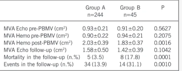
Table I - Valve area, mortality and events in the groups with
echocardiographic score ≤≤≤≤≤ 8 and > 8
Group A Group B P
n=244 n=45
MVA Echo pre-PBMV (cm2) 0.93±0.21 0.91±0.20 0.5627
MVA Hemo pre-PBMV (cm2) 0.90±0.22 0.94±0.21 0.2075
MVA Hemo post-PBMV (cm2) 2.03±0.39 1.83±0.37 0.0016
MVA Echo follow-up (cm2) 1.58±0.50 1.42±0.39 0.1042
Mortality in the follow-up (n.%) 5 (3.5) 8 (17.8) 0.0001 Events in the follow-up (n.%) 34 (13.9) 14 (31.1) 0.0010
4
Percutaneous Mitral Balloon Valvotomy. Long-term Outcome and Assessment of Risk Factors for Death and Major Events
FC I and FC II 17. Iung et al 20 found 56% of patients in FC I and FC
II, after a 10-year-follow-up. However, when they studied a po-pulation of patients with calcified valve22 they observed that only
36% of them were in FC I and II, after 8 year of follow up. Most patients (84%) in this study were in sinus rhythm when mitral valvotomy was indicated. Farhat et al 16 reported 71% of
patients in sinus rhythm before the procedure both studies had a young population. Studies with an older population24 usually have
a higher echocardiographic score and higher incidence of atrial fibrillation. Some authors stress that the presence of atrial fibrillation is a factor that would predict events in long-term follow-up 20,22,23,51
which is in accordance with our study (fig. 1), however, other authors do not agree with that premise 24,36,37,49,39,52,53.
In this study and in the literature, mitral balloon valvotomy immediately decreases left atrium pressure and pulmonary vessels, enabling relieve of symptoms immediately 16,18,23,24,48,54-57. There
was a decrease in pulmonary pressure in the presence of severe hypertension even in the presence of elevated systemic levels 57.
In the present study, 84.4% of patients presented echocardio-graphic score ≤ 8 before the procedure, in accordance with the other in the literature 24,54,58. The group of patients with
echocar-diographic score ≤ 8 presented more satisfactory immediate results (p=0.0016), just as those in the literature 24,36,54,58. When
func-tional class and long-term death were assessed, the group with lower score presented a more favorable outcome (p=0.0001). These patients were younger, with higher incidence of atrial fi-brillation, lower calcification; lower Functional Class, lower level of mitral regurgitation before the procedure, and lower incidence of previous surgical commissurotomy. Despite the less satisfactory results, in the group with echocardiographic score >8, when compared to the group with lower score, our study demonstrated that mitral balloon valvotomy is an alternative to mitral stenosis treatment, with good results, even though they may be inferior to
Cumulative Event Free Survival
1.1
1.0
.9
.8
.7
.6
.5
.4
.3
0 20 40 60 80 100 120 140
Sinus rhythm
Atrial fibrillation p=0.0011
Follow-up (months)
Fig. 1 - Event free survival curve (Kaplan-Meier). Sinus rhythm or atrial fibrillation (Log Rank, p=0.0011; Breslow, p=0.068 and Tarone-Ware, p=0.0023).
Cumulative Survival
1.2
1.0
.8
.6
.4
.2
0.0
-.2
0 20 40 60 80 100 120
Echo score ≤ 8
p<0.0001
Follow-up (months)
Fig. 2 - Survival curve (Kaplan-Meier). Echocardiographic score ≤ 8, from 9 to 11 and echocardiographic score ≥ 12 (Log Rank, Breslow and Tarone-Ware, 140 Echo score ≥9 and ≤ 11
Echo score ≥12
Cumulative Event Free Survival
1.2
1.0
.8
.6
.4
.2
0.0
-.2
0 20 40 60 80 100 120
Echo score ≤ 8
p<0.0001
Follow-up (months)
Fig. 3 - Event free survival curve (Kaplan-Meier). Echocardiographic score ≤ 8, from 9 to 11 (2) echocardiographic score ≥ 12 (Log Rank, Breslow and Tarone-Ware, p<0.0001).
140 Echo score ≥ 9 and ≤ 11
Echo score ≥12 Table II - Survival and event free survival - multivariate analysis
Variable Survival free of Significance Relative Risk Confidence interval (95%)
Inferior Superior
SCORE - score ≤ 8 Death 0.0003 0.1182 0.0372 0.3759
Severe mitral regurgitation - Absent Death 0.0001 0.0331 0.0059 0.1869
Previous commissurotomy
Absent Major Events 0.0077 0.3495 0.1613 0.7572
Mitral valve area (Gorlin) ≥ 1.5 cm2 Major Events 0.0005 0.2201 0.0943 0.5137
Absent severe mitral failure Major Events 0.0038 0.0783 0.0140 0.4385
Sinus Rhythm Major Events 0.0220 0.4564 0.2333 0.8931
Mitral valve area (Gorlin and Gorlin27) - mitral valve area after the procedure by Gorlin’s method (success) <1.5 cm2 and
≥ 1.5 cm2; SCORE - echocardiographic score
5
the other group, especially when the echocardiographic score is
≤ 11. Palacios et al 24 demonstrated statistically significant
difference in the success rate of the procedure for the group with score ≤ 8, related to mortality, need for mitral valve surgery and incidence of FC III and IV greater, in the long term, in the group with echocardiographic score >8 24. Hildick-Smith et al 36, had
successful results in 61% of cases, and a 6-year-event-free-survival in 56% of cases in a group of patients with unfavorable characte-ristics to procedure, including 59% of patients with echocardio-graphic score >8.
In this study, mitral valve was competent before the procedure in 84.4% of cases, and the rest were divided into grade 1+ or grade 2+ mitral regurgitation according to the classification of Sellers et al 25. After the procedure, 71.6% of patients remained
with competent mitral valve and 1% of patients followed-up for at least one month presented severe mitral regurgitation. This per-centage of mitral regurgitation does not reflect our total group, where the incidence of severe mitral regurgitation was 2.5%42.
Palacios et al 24 obtained 9.4% of severe mitral regurgitation after
the procedure higher in the group with echocardiographic score >8. In the study by Hernandez et al 37 the incidence of this
complication was 4%, in the study by Kaul et al 43 it was 3.3%,
in that by Iung et al 45 it was 4%, and in that by Farhat et al it was
4.6% 16.
Just as occurred in this study, the authors identified this compli-cation as a factor predicting long term events23,24,37,40,53,59. Zhang et
al 23 compared patients with or without mitral regurgitation before
the procedure, and observed that patients with pre-existing mitral regurgitation were older and presented more events in the long term outcome. Hernandez et al 37 found 4% of severe mitral
regur-gitation after the procedure unrelated to age or echocardiographic score and cases of mild or moderate mitral regurgitation after the procedure, with likelihood to regress or at least not worsen. Leaflets rupture led to severe mitral regurgitation and need for emergency surgery in all patients 43. There was a decrease in the level of
mitral regurgitation over time in 1.6% of patients, and probably the regurgitation mechanism was the consequence of the intensive stretching of mitral commissurotomy 37,43. Severe mitral regurgitation
may be predicted, through echocardiographic score specific to it60.
Mitral regurgitation before balloon valvuloplasty is a factor that pre-dicts lower long term event free survival23, in older patients usually
presenting mitral calcification and atrial fibrillation, compared to patients without concomitant mitral regurgitation. In our study, only 224 patients underwent echocardiogram in late follow-up and it was observed that 26.8% still presented competent mitral valve and there were 9.9% cases of severe mitral regurgitation. Three patients with severe mitral regurgitation in the post immediate period were maintained in this study for at least 1 month, and 19 new cases of severe mitral regurgitation were identified during our follow-up (7.79%). Kaul et al 43 found 3.3% cases of severe mitral
regurgitation in the post immediate period, 55% needed emergency valve replacement and, in the end of follow-up, they observed 8.4% of cases with severe mitral regurgitation, and 37.66% of cases requiring mitral valve surgery.
In the end of follow-up, through echocardiogram, mean mitral valve area was 1.56±0.49 cm2, 1.58±0.50 cm2 in the group
with echocardiographic score ≤ 8 and 1.42±0.39 cm2 in the
group with echocardiographic score >8 (p=0.1042). There was
a 0.44 cm2 loss of mitral valve area, on average in 49±31 months,
in the total group. Mitral valve area immediately after the procedu-re assessed by hemodynamic (Gorlin27) was on average 2.00±
0.39 cm2, with statistically significant difference between the 2
groups, higher in the group with echocardiographic score ≤ 8. Loss of mitral area may be a result of rheumatic disease develop-ment, and/or presence of blood flow turbulence in the deformed valves61. After mitral valvuloplasty, Hernandez et al 37 found a >0.3
cm2 mitral valve area loss in 12%, 22%, and 27% of patients in 3,
5, and 7 years respectively, on average, 0.13±0.21 cm2. Loss of
valve mitral area in the literature ranged from 0.16 to 0.4 cm2 for
diverse groups and in different outcome periods 17,38,49,62,63. Loss of
mitral valve area, in the study of Wang et al 46, was 0.06 cm2 per
year after the performance of balloon valvuloplasty. We could obser-ve in our study, in the end of the follow up period, 97 patients (44.1%) with mitral valve areas smaller than 1.50 cm2. This
criterion was used in the study to define restenosis, and it was the same adopted by other authors17,51. Mitral restenosis occurred in
44.1% of patients in a mean 49±31 follow-up period in this study, defined as mitral valve area < 1.50 cm2. In the literature we have
observed different restenosis frequency among the studies, ranging from 36 months to 7 years as well as different population and restenosis criteria. Restenosis definition may be done clinically or in terms of mitral valve area, by absolute loss or percentual loss, or loss of mitral valve gain after balloon valvuloplasty16-18,36,37,46,47,51;
the definition of restenosis varies among authors, usually it is referred as loss of 50% or over of the initial gain and/or mitral valve area < 1.5 cm2. Wang et al 46 observed gradual and progressive loss of
the mitral valve area, after balloon mitral valvuloplasty and absence of correlation between mitral valve area after the procedure and restenosis, suggesting that it is a part of an ongoing biological process rather than a mechanical or retraction process.
In the present study there were 13 cases (4.5%) of death in the end of the follow-up period, 11 (3.8%) had cardiac origin, and 46.15% of the cardiac deaths occurred in mitral valve surgery, performed in different hospitals. Farhat et al 16 found 7 cases
(1.6%) in the end of the 37±22 month-period, and another study of the same authors 17, with young patients with favorable
cha-racteristics, mortality was null in 7 years. Meneveau et al 21 found
a 6% mortality in 3 years, reaching 17% in 7 years and a half of follow-up. Palacios et al 24 found 13.03% of death in a follow-up
of 4.2±3.7 years, 10.07% had cardiac origin. Hildick-Smith et al 36 studied a population of patients with unfavorable characteristics
for the performance of balloon mitral valve valvuloplasty and observed a mortality of 3%, 12%, and 18% in 1, 3 and 6 years, respectively. Hernandez et al 37 found 5% of death in 39±23
months, 3.3% had cardiac origin. Hamasaki et al 38 found a
mortality of 2%, 7%, and 14% in 1, 5 and 10 years, respectively, of follow-up. Wang et al 46 found 11 deaths (3.58%), only 2
(18%) had cardiac origin. Iung et al 45 found 0.4% of deaths
during outcome. In this study when both echocardiographic score groups were compared, higher mortality was observed in the group with echocardiographic score >8, 17.8% against 3.5% in the group with echocardiographic score ≤ 8 (p=0.0001). Palacios et al 24 also found higher mortality in the group with higher
echo-cardiographic score. Thus, reported mortality during outcome is null in groups selected with favorable medium term 64,65, and even
6
Percutaneous Mitral Balloon Valvotomy. Long-term Outcome and Assessment of Risk Factors for Death and Major Events
References
1. Inoue K, Owki T, Kikamara T, Kitamura F, Miyamoto M. Clinical application of transvenous mitral comissurotomy by a new balloon catheter. J Thorac Cardiovasc Surg 1984; 87:394-402.
2. Mckay RG, Lock JE, Klane JF, Safian RD, Aroesty JM. Percutaneous mitral valvo-plasty in an adult patient with calcific rheumatic mitral stenosis. J Am Coll Cardiol 1986; 7:1410-5.
3. Palacios I, Lock JE, Klane JF, Block PC. Percutaneous transvenous balloon valvo-tomy in a patient with severe calcified mitral stenosis. J Am Coll Cardiol 1986; 7: 1416-9.
4. Al Zaibag M, Kasab JA, Ribeiro PA, Fagih MR. Percutaneous double balloon mitral valvotomy for rheumatic mitral valve stenosis. Lancet 1986; 1:757-61. 5. Mossmann RA, Blancher C, Koehler N et al. Valvoplastia mitral com cateter balão.
Experiência inicial com uma nova técnica. Arq Bras Cardiol 1987; 49:333-7. 6. Buchler JR, Assis Filho SF, Braga SLN, Souza JEMR. Percutaneous mitral
valvulo-plasty in rheumatic mitral stenosis by isolated transarterial approach. A new and feasible technique. Jpn Heart J 1987; 28:791-8.
7. Peixoto ECS. Valvoplastia mitral por via transeptal. Uma nova técnica de trata-mento da estenose mitral. Ars Cvrandi Cardiologia 1987; 9: 9-10.
8. Peixoto ECS, Oliveira PS, Salles Netto M et al. Valvoplastia mitral percutânea por balão. Resultados imediatos, complicações e evolução hospitalar. Arq Bras Cardiol 1995; 64:109-16.
9. Peixoto ECS, Oliveira PS, Salles Netto M et al. Valvoplastia mitral percutânea com a técnica do balão único. Resultados imediatos, complicações e evolução intra-hospitalar. Arq Bras Cardiol 1996; 66:267-73.
due to the unfavorable characteristics or when greater follow-up period were observed.
In the long-term follow-up, survival varied considerably, from 82% to 100% in 5 to 7 year-follow-up 17,18,21,34,36,37,38, probably
due to clinical and echocardiographic difference of the population studied. In a longer period between 10 to 12 years of follow-up, survival reported is 82% and 86%24,38. Long term results are less
satisfactory in Europe and North America,24,45,48,54, with older
pa-tients and more involved valve anatomy. Survival in this study, in the end of follow-up was 95.5% in the total group of patients, 98% in the echocardiographic score group ≤ 8, higher than that of the group with echocardiographic score >8, which was 82.8% (p<0.0001).
Event free survival in this study, in the end of follow-up was 83.4% greater in group A (86.1%) when compared to group B (68.9%), (p<0.0001). In the literature, we have found percentages ranging from 16% to 90% with follow-up ranging from 4 to 12 years17,18-24,37,45,47,48,66, due to the difference of the groups of patients.
Iung et al 45 found a 58% event free survival in 8 years of
follow-up, however those were patients undergoing balloon mitral valvu-loplasty because of reestenosis after surgical commissurotomy. Farhat et al 17 found a 90% event free survival in 7 years, in a
group of young patients, just as did Zaki et al 18, who found 91%
in 5 years. Sutaria et al 19 found event free survival of 36% of
elders in 5 years, in a 6-year- follow-up, Saeki et al 47 found 88%
of event free survival, and Iung et al.20, in a 10-year-follow-up
found 56.4% event free survival. Maneveau et al 21 compared
event free survival of the group with favorable mitral valve anatomy, with that of the group with unfavorable, and they found 70% and 16%, respectively, in 7 years of follow up. Iung et at 22, found
36% in 8 years, in patients with calcified valves. Zhang et al 23
found event free survival in a 6-year-period in a group with mitral regurgitation in 37% of patients and in 69% in the group without mitral regurgitation. Palacios et al 24, in a 12-year-follow-up, found
a 38% event free survival, in the group with echocardiographic score ≤ 8, and 22% in the group with increase score. Hernandez et al 37 found 69% event free survival in a 7-year-follow-up, in
patients with low score, whereas Cohen et al 48 found 51%, in a
6-year-follow-up, in patients with low echocardiographic score and increased age. Dean et al 66, in the Record of NHLBI, found
a 60% event free survival in a 4 year-follow-up.
Through multivariate analysis, we have observed in the present study that only echocardiographic score >8 and the presence of severe mitral regurgitation before mitral balloon valvuloplasty are independent factors predicting death in long term follow up In the
literature, Dean et al 66 found age >70 years old, FC IV before the
procedure and echocardiographic score >12 as factors predicting death using univariate analysis, in a 4-year-follow-up. Using mul-tivariate analysis, the independent factors predicting death found were echocardiographic score, FC IV before the procedure, high echocardiographic score, increased systolic pulmonary blood pres-sure, and end diastolic left ventricle pressure after the procedure. Palacios et al 54 found as independent factor predicting death in
the multivariate analysis, echocardiographic score, increased age of the patients and high functional class before the procedure.
In the multivariate analysis, independent factors predicting death in the long term follow up in this study were: history of surgical mitral commissurotomy, atrial fibrillation before the procedure, seve-re mitral seve-regurgitation during the proceduseve-re, and proceduseve-re failuseve-re (mitral valve area <1.50 cm2). In the literature independent factors
for events are: lower mitral valve area after the procedure20,37,40,45,59,67,
atrial fibrillation20,22,23,51, history of surgical mitral
commissuroto-my24,45,46,53,54, presence of severe mitral regurgitation after the
procedure20,24,37,40,46,53,59,67, increased functional class before the
pro-cedure20,22,24,48,54, increased echocardiographic score before the
pro-cedure23,24,36,37,40,48,54, increased age20,21,22,23,24,54, unfavorable mitral
valve anatomy20,21,22,23,38,59, increased mean pulmonary blood pressure
after the procedure21,24,40, increased transvalvular gradient after the
procedure20,21,22,46,51, increased left atrium pressure after the
proce-dure or enlarged left atrium36,46,59, male gender36, increased cardiac
thoracic index21,45, presence of co-morbidities36, and increased
fi-nal left ventricle pressure48.
The present study concluded that mitral balloon valvuloplasty is an efficient procedure. Survival and event free survival combined with death, new mitral balloon valvuloplasty and mitral surgery were high at the end of the follow-up period, and more than two third of the total group of patients were event free by the end of the study. In the patients with echocardiographic score ≤ 8 before the procedure compared with the score group > 8, survival and event free survival observed were significantly greater. Echocar-diographic score also separates 3 groups with different survival curves (≤ 8, between 9 and 11 and ≥ 12), (fig. 2 and 3). Echo-cardiographic score higher than 8 and the presence of severe mitral regurgitation were risk factors for death in the long term follow-up. The history of surgical mitral commissurotomy, presence of atrial fibrillation before the procedure, mitral valve regurgitation during the procedure, atrial fibrillation before the procedure, severe mitral regurgitation during the procedure and mitral valve area lower than 1.5 cm2 after the procedure, were risk factors for
7
10. Peixoto E, Oliveira P, Salles M et al. Inoue Balloon versus Monofoil Balloon in Mitral Valvuloplasty. Results and Complications. Am J Cardiol 1997; 80(supl.7A): 735. 11. Peixoto ECS, Oliveira PS, Salles Netto M et al. Balão único versus balão de Inoue
na valvoplastia mitral percutânea por balão. Resultados imediatos e complica-ções. Arq Bras Cardiol 1998; 71:59-64.
12. Peixoto ECS, Oliveira PS, Salles Netto M et al. Comparação dos resultados e com-plicações das técnicas do balão único e do balão de Inoue na valvoplastia mitral percutânea por balão. Rev Bras Cardiol Invas 1998; 6:6-12
13. Peixoto ECS, Oliveira PS, Salles Netto M et al. Inoue balloon versus single balloon in mitral valvuloplasty: Results and complications. Am J Cardiol 1998; 82 (supl. 7A): 1145.
14. Peixoto ECS, Peixoto RTS, Oliveira PS et al. Inoue balloon versus single balloon te-chnique in mitral valvuloplasty. Results, in-hospital evolution and cost. Am J Cardiol 2000; 86(supl. 8A): 67i.
15. Ribeiro PA, Fawzy ME, Arafat MA, Dunn B, Sriram R. Balloon Valvuloplasty Regis-try: Multicenter Experience with balloon mitral commissurotomy. Circulation 1992; 85: 448-61.
16. Farhat MB, Belbout F, Gamra H et al. Results of percutaneous double-balloon mitral commissurotomy in one medical center in Tunisia. Am J Cardiol 1995; 76: 1266-70. 17. Farhat MB, Ayari M, Maatouk F et al. Percutaneous balloon versus surgical closed and open mitral commissurotomy: seven-year follow-up results of a randomized trial. Circulation 1998; 97:245-50.
18. Zaki A, Salama M, El Masry M, Elhendy A. Five-year follow-up after percutaneous balloon mitral valvuloplasty in children and adolescents. Am J Cardiol 1999; 83:735-39.
19. Sutaria N, Elder AT, Shaw TR. Long term outcome of percutaneous mitral balloon valvotomy in patients aged 70 and over. Heart 2000; 83:374-5.
20. Iung B, Garbarz E, Michaud P et al. Late results of percutaneous mitral commissu-rotomy in a series of 1024 pacients. Analysis of late clinical deterioration: frequen-cy, anatomic findings, and predictive factors. Circulation 1999; 99:3272-8. 21. Meneveau N, Schiele F, Seronde MF et al. Predictors of event-free survival after
percutaneous mitral commissurotomy. Heart 1988; 80:359-64.
22. Iung B, Garbarz E, Doutrelant L et al. Late result of percutaneous mitral commis-surotomy for calcific mitral stenosis. Am J Cardiol 2000; 85:1308-14. 23. Zhang HP, Yen GS, Allen JW, Lau FY, Ruiz CE. Comparison of late results of
bal-loon valvotomy in mitral stenosis with versus without mitral regurgitation. Am J Cardiol 1998; 81:51-5.
24. Palacios IF, Tuzcu ME, Weyman AE, Newell JB, BlocK PC. Clinical follow-up of pa-tients undergoing percutaneous mitral balloon valvotomy. Circulation 1995; 91:671-6.
25. Sellers RD, Levy MJ, Amplatz K, Lillehei CW. Left retrogade cardioangiography in acquired cardiac disease. Technic, indication and interpretation in 700 cases. Am J Cardiol 1964; 14:437-47.
26. Yang SS, Bentivoglio L, Maranhão V, Goldberg H. From cardiac catheterization data to hemodynamic parameters. F A Davis company, 2nd edition, Philadelphia,
1978, p.1.
27. Gorlin R, Gorlin SG. Hydraulic formula for calculation of the area of the stenotic mitral valve, other cardiac values and central circulatory shunts. Am Heart J 1951; 41:1-29. 28. Wilkins GT, Weyman AE, Abascal VM, Block PC, Palacios IF. Percutaneous mitral valvotomy: An analysis of echocardiographic variables related to outcome and the mecanism of dilatation. Br Heart J 1988; 60:299-308.
29. Helmeke F, Nanda Nc, Hsiung MC et al. Color Doppler assessment of mitral regurgi-tation with orthogonal planes. Circulation 1987; 75:175-83.
30. Dean AG, Dean JA, Coulombier D et al. Epi-Info, version 6: A word processing, data-base and statistic program for public health on IBM-microcomputers. The division of surveillance and epidemiology. Epidemiology program office. Centers for disease control and prevention, Atlanta, 1995.
31. SPSS 8.0 for Windows, SPSS Inc, 1997.
32. Kaplan EL, Meier P. Non Parametric estimation from incomplete observations. J Am Stat Assos 1958; 53:457-81.
33. Cox DR. Regression models and life-tables. J R Stat Soc 1972; 34:197-220. 34. Cotrufo M, Renzulli A, Ismeno G et al. Percutaneous mitral commissurotomy
ver-sus open mitral commissurotomy: a comparative study. Eur J Cardiothorac Surg 1999; 15:646-651.
35. Mattos C, Braga SL, Esteves CA et al. Percutaneous mitral valvotomy in patients eighteen tears old and younger. Immediate and late results. Arq Bras Cardiol 1999; 73:373-81.
36. Hildick-smith DJ, Taylor GJ, Shapiro LM. Inoue balloon mitral valvuloplasty: long-term clinical and echocardiographic follow-up of a predominantly unfavourable po-pulation. Eur Heart J 2000; 21:1690-7.
37. Hernandez R, Banuelos C, Alfonso F et al. Long-term clinical and echocardiogra-phic follow-up after percutaneous mitral valvuloplasty with the Inoue balloon. Cir-culation 1999; 99:1580-6.
38. Hamasaki N, Nosaka H, Kimura T et al. Ten years clinical follow-up following suc-cessful percutaneous transvenous mitral commissurotomy: single-center experien-ce. Catheter Cardiovasc Interv 2000; 49:284-288.
39. Lau KW, Ding ZP, Quek S, Kwok V, Hung JS. Long-term (36-63 months) clinical and echocardiographic follow-up after Inoue balloon mitral commissurotomy. Cathet Cardivasc Diagn 1998; 43:33-8.
40. Gupta S, Vora A, Lokhandwalla Y et al. Percutaneous balloon mitral valvotomy in mitral restenosis. Eur Heart J 1996; 17:1560-4.
41. Borges IP, Peixoto ECS, Sena MA et al. Clinical and echocardiographic long-term follow-up in mitral balloon valvuloplasty. Echocardiographic score influence. J Am Col Cardiol 1998; 31(supl. C): 27C.
42. Peixoto E, Borges IP, Neves A et al. Clinical and echocardiographic long-term fol-low-up in mitral balloon valvuloplasty. Echocardiographic score influence. Am J Cardiol 1997; 80(supl. 7A):735.
43. Kaul UA, Singh S, Kalra GS et al. Mitral regurgitation following percutaneous transvenous mitral commissurotomy: a single-center experience. J Heart Valve Dis 2000; 9:262-6.
44. Zaki AM, Kassem HH, Bakhoum S et al. Comparison of early results of percuta-neous metallic mitral commissurotome with Inoue balloon technique in patients with high mitral echocardigraphic scores. Catheter Cardiovasc Interv 2002; 57:312-7.
45. Iung B, Garbarz E, Michaud A et al. Percutaneous mitral commissurotomy for res-tenosis after surgical commissurotomy: late efficacy and implications for patient selection. J Am Coll Cardiol 2000; 35:1295-302.
46. Wang A, Krasuski RA, Warner JJ et al. Serial echocardiographic evaluation of res-tenosis after successful percutaneous mitral commissurotomy. J Am Coll Cardiol 2002; 39:328-334.
47. Saeki F, Ishizaka Y, Tamura T. Long-term clinical and echocardiographic outcome in pacients with mitral stenosis treated with percutaneous transvenous mitral commissurotomy. Jpn Cir J 1999; 63:597-604.
48. Cohen DJ, Kuntz RE, Gordon SPF et al. Predictors of long-term outcome after per-cutaneous balloon mitral valvuloplasty. N Engl J Med 1992; 327:1329-35. 49. Chen CR, Cheng TO, Chen JY, Huang YG, Huang T, Zhang B. Long-term results of
percutaneous balloon mitral valvuloplasty for mitral stenosis: a follow-up study to 11 years in 202 patients. Cathet Cardiovasc Diagn 1998; 43:132-9. 50. Braunwald E. Valvular Heart Disease in BRAUNWALD E, ZYPES DP, LIBBY P, W B
Saunders Company, Philadelphia 2001, p.1643.
51. Langerveld J, Plokker HWT, Erns SM, Kelder JC, Jaarsma W. Predictors of clinical events or restenosis during follow-up after percutaneous mitral balloon valvoto-my. Eur Heart J 1999; 20:519-26.
52. Iung B, Garbarz E, Michaud P et al. Immediate and mid-term results of repeat per-cutaneous mitral commissurotomy for restenosis following earlier perper-cutaneous mitral commissurotomy. Eur Heart J 2000; 21:1683-4.
53. Tarka EA, Blitz LR, Herrmann HC. Hemodynamic effects and long-term outcome of percutaneous balloon valvuloplasty in patients with mitral stenosis and atrial fi-brillation. Clin Cardiol 2000; 23:673-7.
54. Palacios IF, Tuzcu ME, Weyman AE, Newell JB, Block PC. Clinical follow-up of patients undergoing percutaneous mitral balloon valvotomy. Circulation 1995; 91:671-676.
55. Fawzy ME, Kinsara AJ, Stefadouros M et al. Long term outcome of mitral balloon valvotomy in pregnant women. J Heart Valve Dis 2001; 10:153-157.
56. Sutaria N, Elder AT, Shaw TR. Mitral balloon valvotomy for the treatment of mitral stenosis in octogenarians. J Am Geriatr Soc 2000; 48:971-4.
57. Peixoto ECS, Peixoto RTS, Oliveira PS et al. Técnicas do balão único e do balão de Inoue na valvoplastia mitral por balão. Resultados, evolução intra-hospitalar e custo. Rev Bras Cardiol Invas 2002; 10:11-22.
58. Peixoto ECS, Peixoto RTS, Borges IP et al. Importância do estado anatômico da válvula mitral e não da comissurotomia cirúrgica prévia no resultado da valvo-plastia por balão. Rev Bras Cardiol Invas 2002; 10:23-9.
59. Osa A, Almenar L, Rincon de Arellano A et al. Long-term results of percutaneous mitral valvuloplasty. Rev Esp Cardiol 1998; 51:458-66.
60. Padial LR, Abascal VM, Moreno PR, Weyman AE, Levine RA, Palacios IF. Echocar-diography can predict the development of severe mitral regurgitation after percuta-neous mitral valvuloplasty by the Inoue technique. Am J Cardiol 1999; 83:1210-3. 61. Sutaria N, Elder AT, Shaw TR. Long term outcome of percutaneous mitral balloon
valvotomy in patients aged 70 and over. Heart 2000; 83:374-5.
62. Chen CR, Cheng TO, Chen JY, Zhou YL, Mei J, Ma TZ. Long-term results of percu-taneous mitral valvoplasty with Inoue balloon catheter. Am J Cardiol 1992; 70:1445-8.
63. Treviño AJ, Ibarra M, Garcia A et al. Immediate and long-term results of balloon mitral commissurotomy for rheumatic mitral stenosis: comparison between Inoue and double balloon techniques. Am Heart J 1996; 131:530-6.
64. Cardoso LC, Rati MAN, Pomerantzeff PMA et al Avaliação compativa entre valvo-plastia percutânea e comissurotomia a céu aberto na estenose mitral. Arq Bras Cardiol 1998; 70:415-21.
65. Cardoso LF, Grinberg M, Rati MA et al. Comparison between percutaneous bal-loon valvuloplasty and open commissurotomy for mitral stenosis. A prospective and randomized study. Cardiology 2002; 98:186-90.
66. Dean LS, Mickel M, Bonan R et al. Four-year follow-up of patients undergoing percutaneous balloon mitral commissurotomy: a report from the National Heart, Lung and Blood Institute balloon valuloplasty registry. J Am Coll Cardiol 1996; 28:1452-7.