Arq Neuropsiquiatr 2008;66(3-B):755-757
755 Clinical / Scientiic note
UnUSUal preSentation of Central
nervoUS SyStem metaStaSeS
Mechanisms of spread and radiological indings
Marcus André Acioly
1, Carlos Henrique Carvalho
1, João L. Pinheiro-Franco
1,
Jens Schittenhelm
2, Ulrike Ernemann
3, Michael Weller
4, Jürgen Honegger
1metáStaSeS inComUnS do SiStema nervoSo Central: meCaniSmoS de diSSeminação e aChadoS radiológiCoS
University Hospital, Tübingen, Germany: 1Department of Neurosurgery; 2Department of Neuropathology/Brain Research Institute; 3Department of
Neuroradiology; 4 Department of Neurology.
Received 22 January 2008, received in inal form 9 June 2008. Accepted 7 July 2008.
Dr. Marcus Andre Acioly – Department of Neurosurgery, University Hospital, Hoppe-Seyler-Strasse 3.D - 72076 Tübingen, Germany. E-mail: marcusacioly @yahoo.com.br
Brain metastases are a well-known complication of sys-temic cancer, occurring in 20–40% of the patients suffering from cancer1. In adults, the lungs represent the most
com-mon source of brain metastases comprising 36–64% of the cases1. The cerebral hemispheres, cerebellum, brainstem1,
pituitary (sellar region)2-4, cerebellopontine angle (CPA)/
internal auditory canal (IAC)2,5, and leptomeningeal
me-tastases (LM)1,2,6,7 are potential locations of dissemination.
We report an unusual case, in which solely the associ-ation of pituitary metastases (PM), bilateral CPA metasta-ses and LM was found in a patient affected of a large cell neuroendocrine carcinoma (LCNEC) of the lung.
CaSe
A 63-year-old woman presented with a 4-month history of emesis, anorexia, weight loss, and increasing thirst. After three months, the patient suddenly developed bilateral hearing loss, tinnitus and increased dizziness. Fluid intake and urine output
were consistent with diabetes insipidus. The neurological exam-ination revealed bilateral hearing loss, incomplete right periph-eral facial palsy and unstable gait. Other motor or sensory dei-cits were not noted. The tendon relexes were brisk.
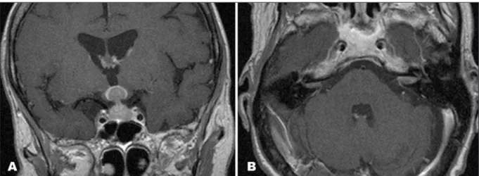
Magnetic resonance imaging (MRI) revealed an intrasellar and suprasellar space-occupying lesion isointense in T1-weight-ed precontrast study with strong enhancement in contrast im-ages iniltrating the pituitary gland, the pituitary stalk and the hypothalamus (Fig 1A and 1B), as well as bilateral CPA lesions. There was also loss of high-signal intensity of the pituitary pos-terior lobe.
A spinal tap showed a nonspeciic lymphocytic pleocyto-sis. The patient underwent a transsphenoidal biopsy revealing a high-grade pleomorphic epithelial tumour, like a large-cell car-cinoma. Besides, malignant cells were encountered on CSF cy-tology, conirming the diagnosis of LM (Fig 2A).
MRI also revealed the typical small nodular depot along the cauda, L4–L5 on the right side and L5–S1 on the left side, and
Arq Neuropsiquiatr 2008;66(3-B)
756
CNS metastases Acioly et al.
ventral of conus medullaris with strong enhancement in con-trast images consistent with the diagnosis of LM (Fig 2B). Tho-racic computed tomography (CT) scans showed a mass (measur-ing 1.4 × 1.1 cm) on the right side located within the retrotracheal space and a single increased lymph node (0.7 × 1.0 cm) near the right hilus. The bone scintigraphy revealed metastases exclu-sively to the skull base. A bronchoscopy was performed show-ing few nonspeciic inlammatory cells.
The immunohistochemical proile of the tumour cells re-vealed a large-cell neuroendocrine carcinoma (LCNEC) with ex-pression of cytokeratin 7 (CK7), thyroid transcription factor-1 (TTF-1) and synaptophysin with probable origin in the lung. The treatment was conducted with whole brain palliative radiother-apy and chemotherradiother-apy.
This publication was authorized by the ethical committee at our institution.
diSCUSSion
Morita and colleagues reported on a patient with PM who developed LM after a transsphenoidal procedure4. In
another study, Lee and colleagues2 described a case with
bilateral CPA lesions and a suprasellar mass, nonetheless without evidence of intrasellar or spinal involvement. Sev-eral combinations of PM and CPA metastases are encoun-tered in literature3,5 including postmortem studies3. We
de-scribed a unique case of metastases of LCNEC of the lung solely to the pituitary, LM and CPA at the time of diagno-sis. This is an unusual presentation of central nervous sys-tem metastases in a patient affected of LCNEC lung cancer. Metastases can reach the sella via several routes3,4: 1)
di-rect hematogenous spread to the pituitary parenchyma or diaphragma sellae; 2) spread from a hypothalamo-hypoph-yseal or stalk metastases through the portal vessels; 3)
di-rect extension from the skull base or juxtasellar metastases; and 4) meningeal spread through the suprasellar cistern. The most common route of spread for LM is hematog-enously to cerebrospinal luid (CSF) via small meningeal vessels2. Furthermore, direct extension, transport through
valveless venous plexus, escape from subependymal tu-mours or iatrogenic (postoperative) are also reported8.
Once in the CSF, tumour cells can also lodge in the sulci, the brain surface or on nerve roots growing into a thin diffuse coating of the meninges or focal nodules that are centered on the pia2,8, frequently involving regions in the
basilar cisterns and cauda equina, where the slow CSF low and gravity promote deposition of cells8. This direct
lep-tomeningeal involvement and/or dissemination through the CSF are the likely mechanism for metastatic CPA le-sions5. In the presented case, no single mechanism could
explain such an unusual presentation. Thus, we could as-sume that a combination of hematogenous route and CSF spreading is the potential mechanism of dissemination. Due to clinical presentation of pituitary involvement fol-lowed by cranial nerve impairment, it is possible that ma-lignant cells reached pituitary hematogenously and spread through CSF to CPA and LM.
Concerning the radiological indings, high-resolution cranial CT and MRI are sensitive for the diagnosis of PM9.
Rapid growth of a sellar tumour with invasion of the in-fundibular recess remits to the diagnosis of PM. MRI is the technique of choice for LM6. Cranial nerve enhancement
on cranial imaging and spinal intradural extramedullary (most frequently seen in cauda equina) enhancing nodules may be considered diagnostic of LM in cancer patients6.
Infundibular invasion, loss of high-signal intensity of the pituitary posterior lobe, along with cranial nerve and
Arq Neuropsiquiatr 2008;66(3-B)
757
CNS metastases Acioly et al.
nal enhancing nodules suggested metastatic disease in the presented case.
Of relevance is the concept of loculated intracranial LM well studied by Lee and colleagues2 in MRI, a pattern
that was also found in our patient imaging. Occasionally, collections of tumour cells can lodge in some portions of the intracranial arachnoidal space, including the ven-tricles, forming a rather distinct extra-axial lesion. When it occurs without diffuse pattern, it may cause a diagnostic dilemma and delay proper management2. The most
fre-quent locations are the suprasellar cistern, the lateral ven-tricles, the lateral recess of the fourth ventricle, the CPA cistern and the fourth ventricle, where there are abun-dant CSF collections. Initially the tumour cells iniltrate the leptomeninges as a single layer or as thicker multilay-ered aggregates2,8 that are not detectable by radiological
imaging. When a mass of tumour accumulates within the subarachnoid spaces cited above, a subarachnoidal extra-axial mass develops (Type A). Further tumour growth can lead to parenchymal iniltration. When the main tumour mass remains extra-axial and without edema, it is classi-ied as type B. Once the tumour extends beyond the pial surface with vasogenic edema, it is considered type C. The tumour can also grow along the cranial nerves, in a dumbbell fashion, mimicking schwannomas in type D. The loculated LM is observed in 80% of the patients and may present in an associated fashion2.
In conclusion, the association of PM, bilateral CPA/IAC metastases and LM is a rare clinical presentation of central nervous system metastases. The hypothesis of metastatic disease should be remembered in patients with sudden development of cranial nerve palsies, and CPA or sellar region lesions, alone or in combination. Besides, investiga-tion of CSF in such patients should be done routinely to evaluate the presence of concomitant LM.
Acknowledgments – We are grateful to Mr. Gerd Pister for his
assistance with igure production.
referenCeS
1. Sofietti R, Ruda R, Mutani R. Management of brain metastases. J Neu -rol 2002;249:1357-1369.
2. Lee YY, Tien RD, Bruner JM, De Pena CA, Van Tassel P. Loculated intra -cranial leptomeningeal metastases: CT and MR characteristics. AJNR Am J Neuroradiol 1989;10:1171-1179.
3. Matsuda R, Chiba E, Kawana I, et al. Central diabetes insipidus caused by pituitary metastasis of lung cancer. Intern Med 1995;34:913-918. 4. Morita A, Meyer FB, Laws ER Jr. Symptomatic pituitary metastases. J
Neurosurg 1998; 89:69-73.
5. Yuh WT, Mayr-Yuh NA, Koci TM, et al. Metastatic lesions involving the cerebellopontine angle. AJNR Am J Neuroradiol 1993;14:99-106. 6. Chamberlain MC. Neoplastic meningitis. J Clin Oncol 2005;23:3605-3613. 7. Straathof CS, de Bruin HG, Dippel DW, Vecht CJ. The diagnostic accu -racy of magnetic resonance imaging and cerebrospinal luid cytology in leptomeningeal metastasis. J Neurol 1999;246:810-814.
8. Kesari S, Batchelor TT. Leptomeningeal metastases. Neurol Clin 2003;21: 25-66.