Evaluation of final-year dental students
concerning therapeutic decision making
for proximal caries
Abstract: This cross-sectional study aimed to determine the radiograph-ic criteria used by i nal-year dental students when dei ning the need for restorative treatment for proximal caries, as well as investigating po-tentially associated factors in this therapeutic decision. A questionnaire with two schematic diagrams presenting i ve levels of proximal lesion penetration was administered to students attending the six private and three public dental schools in the state of Rio Grande do Sul, Southern Brazil. Absolute and relative frequencies were described and inferential statistics involving Chi-square and McNemar tests and simple logis-tic regression were carried out to assess variations in therapeulogis-tic deci-sions related to patient dentition (deciduous/permanent) and gender, age and dental school (public/private). Of the 346 dental students assessed, 28.6% (99/346) indicated restorative treatment for lesions restricted to the enamel in deciduous teeth and 38.2% (132/346) indicated the same for permanent teeth, revealing a statistically signii cant difference (p = 0.001). Student gender and age were not associated with the thera-peutic decision; however, a signii cant difference between dental schools was found when comparing restorative criteria in deciduous (p < 0.001) and permanent molars (p < 0.001). The odds of restorative decision in permanent teeth when the caries lesion was restricted to the enamel was 72% higher for students graduating from private schools compared to public schools (Odds Ratio: 1.72; 95% Coni dence Interval: 1.03-2.90). These data demonstrate a large variation between the therapeutic deci-sions regarding proximal caries reported by i nal-year dental students and suggest that deep rel ection is needed on the part of faculty in order to provide an evidence-based education.
Descriptors: Dental caries; Diagnosis; Radiography. Juliane Bervian(a)
Maximiano Ferreira Tovo(b) Carlos Alberto Feldens(c) Larissa Corrêa Brusco(d) Francinne Miranda da Rosa(e)
(a) MSc, Professor, School of Speech and Language Therapy, University of Passo Fundo, Passo Fundo, RS, Brazil.
(b) PhD; (c)MSc; (e)Graduate student
– Department of Pediatric Dentistry, School of Dentistry, Lutheran University of Brazil, Canoas, RS, Brazil.
(d) MSc, Private Practice.
Corresponding author:
Maximiano Ferreira Tovo
Rua Visconde do Herval, 850, Apto. 604, Menino de Deus
Porto Alegre - RS - Brazil CEP: 90130-150
E-mail: [email protected]
Introduction
For many years, therapeutic decision making in relation to proximal caries lesions was based on the radiographic aspects of the same. The presence of radiolucency, regardless of its extension, inexorably required a surgical restorative process.
Following the clinical advances and the new knowledge acquired on the events that lead to a demineralization process, the related literature has contributed to basing decisions on sustainable evi-dence.1-3 Even though several paradigmatic publica-tions exist regarding this subject, some studies reveal that, when using the radiographic criteria, decisions appear to rel ect the understanding of inevitable and rapid progress of caries lesions.1,2,4
Comprehension of this process by dental profes-sors and professionals has been the object of several studies. The foundation and evolution of the deci-sions made by i nal-year students has instigated new research.4-6
Thus, the objective of the present study was to evaluate the therapeutic decision making of i nal-year dental students from universities in Rio Grande do Sul, when approaching radiographic images of proximal caries lesions in the deciduous and perma-nent dentitions. Associations between the decision made and the dental school, gender and age of the student were also investigated.
Material and Methods
This cross-sectional study was approved by the Ethics Committee for Research on Humans of the Lutheran University of Brazil (protocol n. 025/2002). The dental schools and students autho-rized the realization of this study by signing a term of free informed consent.
Study population
The study population was composed of i nal-year dental students enrolled in six private and three public schools in the state of Rio Grande do Sul. Final-year students were dei ned as those who were studying in the i nal semester or year of undergradu-ate school who, following the proposed curriculum, had concluded the disciplines Pediatric Dentistry I and II and Radiology.
Calculations regarding the sample size indicated that interviewing 309 i nal-year students was re-quired, using the following parameters: 95% con-i dence level and power of 80% to detect a 60% difference in the restorative treatment decision cri-teria between students from public (unexposed) and private (exposed) institutions (exposed and unex-posed ratio of 5:2, the frequency of the outcome un-exposed being 20%, which was detected in a pilot study). However, sample selection was abandoned after considering the logistical possibility of collect-ing data from the entire i nal-year population.
Questionnaire
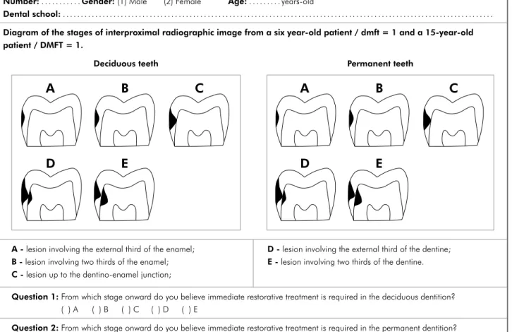
A questionnaire to determine age, gender and dental school attended by the i nal-year students was applied (Figure 1). Next, the students were present-ed to two schematic diagrams presenting i ve differ-ent levels of radiographic penetration in proximal caries lesions, related to two patients: a 6-year-old presenting dmft = 1; and a 15-year-old presenting DMFT = 1. The students were required to indicate at what level they would opt for restorative treat-ment in deciduous and permanent dentitions.5,7,8
Data analysis
Initially, the absolute and relative frequencies of the restorative decisions made by the students were described. The outcome was dichotomized, so the i ve levels of radiographic penetration of the lesions in both dentitions were grouped into two categories: lesions penetrating up to the dentino-enamel junc-tion (DEJ) and lesions penetrating from the external third of the dentine onwards.
for confounding factors9 in this study. Analyses were performed using the Epi-Info (version 3.3.2, CDC, Atlanta, GA, USA) and SPSS (version 13.0, SPSS Inc., Chicago, IL, USA) software.
Results
Of a total of 355 i nal-year students, the replies of 346 (response rate of 97.5%) were analyzed, in-cluding 38.2% (n = 132) males and 61.8% (n = 214) females. Age varied between 20 and 43 years, with a mean (standard deviation) of 24.5 (3.8) years and median (Q25-Q75) value of 23 (22-25) years. The number of i nal-year students per dental school var-ied between 26 and 66.
The replies of the 346 students regarding the stage at which restorative treatment should occur in the deciduous dentition were distributed as
fol-lows: external 1/3 of enamel: n = 7 (2.0%); 2/3 of enamel: n = 14 (4%); DEJ: n = 78 (22.5%); exter-nal 1/3 of dentine: n = 172 (49.7%); 2/3 of dentine: n = 75 (21.7%). In the permanent dentition the deci-sions were as follows: external 1/3 of enamel: n = 9 (2.6%); 2/3 of enamel: n = 23 (6.6%); DEJ: n = 100 (28.9%); external 1/3 of dentine: n = 140 (40.5%); 2/3 of dentine: n = 74 (21.4%).
Considering the dichotomized outcome, obser-vation showed that 181 i nal-year students (52.3%) indicated restorative treatment in both dentitions only when the lesion reached the dentine, while 66 (19.1%) suggested restorative treatment in both den-titions for lesions extending up to the DEJ (Table 1). The remaining students consisted of discordant pairs, since they indicated distinct cutoff points for each of the dentitions: 33 (9.5%) students indicated Questionnaire concerning therapeutic decision
Number: . . . .... Gender: (1) Male (2) Female Age: . . . years-old
Dental school: . . . .. . . .. . . .. . . .. . . .. . . .. . . .. . . .. .. . . .. . . .. . . . .. . . . .. . . .. . . . .. . . . .. . .
Diagram of the stages of interproximal radiographic image from a six year-old patient / dmft = 1 and a 15-year-old patient / DMFT = 1.
Deciduous teeth Permanent teeth
A
B
D
E
C
A
B
D
E
C
A - lesion involving the external third of the enamel;
B - lesion involving two thirds of the enamel;
C - lesion up to the dentino-enamel junction;
D - lesion involving the external third of the dentine;
E - lesion involving two thirds of the dentine.
Question 1: From which stage onward do you believe immediate restorative treatment is required in the deciduous dentition? ( ) A ( ) B ( ) C ( ) D ( ) E
Question 2: From which stage onward do you believe immediate restorative treatment is required in the permanent dentition? ( ) A ( ) B ( ) C ( ) D ( ) E
that enamel should be restored in deciduous denti-tion lesions, but that in the permanent dentidenti-tion the lesion would have to reach the dentine to character-ize immediately required restorative treatment. The inverse option was indicated by 66 students and the difference between these groups was statistically signii cant (McNemar test: p = 0.001).
Overall, the results showed that restorative treat-ment in lesions extending up to the DEJ was pro-posed by 28.6% (99/346) of the i nal-year students for the deciduous dentition and by 38.2% (132/346) for the permanent dentition.
Table 2 shows that opting for treatment pre-sented no difference between genders, in both the deciduous (p = 0.429) and permanent dentitions (p = 0.884); nor was there any difference for the
student age groups concerning the decision for re-storative treatment in the deciduous (p = 0.602) and permanent dentitions (p = 0.314).
Observation revealed a large variation in the cri-teria used to determine the need for restorative treat-ment between dental schools, both for the deciduous (p < 0.001) and permanent dentitions (p < 0.001). In lesions extending up to the DEJ in the deciduous den-tition, there was one dental school in which no stu-dent (0%) opted for restorative treatment, while in another school, 55.6% of the students indicated this option. In the permanent dentition, these frequencies varied between dental schools from 3% to 51.5%.
Logistic regression analysis demonstrated that in the deciduous dentition, no statistically signii -cant difference occurred in the restorative decision
Table 1 - Absolute and relative frequencies concerning the decision for restorative treatment in the deciduous and permanent dentitions by final-year dental students.
Permanent teeth
Total
p*
Up to DEJ Dentine
n (%) n (%) n (%)
Deciduous teeth
Up to DEJ 66 (19.1) 33 (9.5) 99 (28.6)
0.001
Dentine 66 (19.1) 181 (52.3) 247 (71.4)
Total 132 (38.2) 214 (61.8)
*McNemar test.
Table 2 - Absolute and relative frequencies concerning the decision for restorative treatment on the proximal surfaces of the deciduous and permanent dentitions by final-year dental students, according to gender.
Gender
Restorative criteria in deciduous teeth
p*
Restorative criteria in permanent teeth
p*
Up to DEJ In dentine Up to DEJ In dentine
n (%) n (%) n (%) n (%)
Male 41 (31.1) 91 (68.9)
0.429 51 (38.6) 81 (61.4) 0.884
Female 58 (27.1) 156 (72.9) 81 (37.9) 133 (62.1)
*Chi-square test.
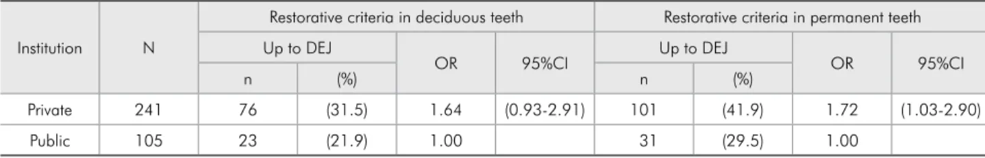
Table 3 - Absolute and relative frequencies, odds ratios (OR) and 95% confidence intervals (95% CI) regarding the decision for restorative treatment applied to the deciduous and permanent dentitions, according to the type of institution.
Institution N
Restorative criteria in deciduous teeth Restorative criteria in permanent teeth
Up to DEJ
OR 95%CI Up to DEJ OR 95%CI
n (%) n (%)
Private 241 76 (31.5) 1.64 (0.93-2.91) 101 (41.9) 1.72 (1.03-2.90)
criteria between public and private i nal-year den-tal students (Table 3). However, the probability of opting for restorative treatment in lesions extending up to the DEJ in the permanent dentition was 72% greater in students graduating from private dental schools in relation to public institutions (OR: 1.72; 95% CI: 1.03-2.90).
Discussion
The application of a clinical simulation as an in-strument to capture diagnostic and therapeutic de-cisions is frequently used in studies of this nature. Despite the subjectivity of the method, the stan-dardization of a model and its replication in relation to the methodological design allows comparison between results obtained in different investiga-tions.4,5,10,11 In certain studies, the use of a question-naire seeks to base the i ndings on a gold standard. However, in the present study, the intention was not to compare the replies with a previously established standard, but rather to evaluate the therapeutic de-cision making of a i nal-year student population in the state of Rio Grande do Sul. Difi culties in deter-mining coherent information in relation to diagno-sis and therapeutic decision making have been previ-ously reported.11,12 In the questionnaire applied here, the clinical proi le of the patient was outlined, in-cluding information regarding patient age, dmft and DMFT, thus providing the student with information that would aid in the decision to indicate restorative treatment. The information of the radiographic exam was complemented by those indicators.13
The use of radiographic images for diagnosing proximal lesions in studies that analyzed the valid-ity of the method is recognized as highly specii c. Although sensitivity is compromised due to the amount of demineralized material required to pro-duce a radiographic record, false-positive diagnoses appear not to compromise the performance of the method since the observation of a radiolucent image shows a strong correlation with demineralized tis-sue. The validity of the method, however, does not consolidate its use as an exclusive element of analy-sis in the therapeutic decision making process.3,14
Classii cation of a caries lesion, according to its penetration level, differs among the reports found in
the related literature. In the present study, the pen-etration levels used followed a progressing lesion sequence: enamel, subdivided into external and in-ternal, involvement of the dentino-enamel junction, and then the dentine, subdivided into external and internal involvement, for both the deciduous7 and permanent teeth.2,5,7,8,15
The indication for restorative treatment invari-ably entails the elimination of the dental structure affected. Although many of the students’ responses only indicated the treatment of dentine lesions, ther-apeutic treatment for the enamel was indicated by a signii cant proportion of i nal-year students. Recent literature tends to consider the indication of imme-diate restorative treatment as adequate for dentine lesions.5,10,11 During the 1980s, the criteria were dif-ferent from those established in the 1990s. This fact was demonstrated by a group of researchers who compared the results of a questionnaire applied to Norwegian dentists on two different occasions, in 1983 and 1995. On the second occasion the dentists were shown to be more cautious regarding immedi-ate restorative treatment in permanent teeth. In 1995, 81% of the professionals opted to indicate restorative treatment when the radiographic images showed that the lesion had already reached the dentine.8
The indication of immediate restorative treat-ment in dentine is mainly justii ed by the fact that the procedure should be limited to lesions that pres-ent clinical cavities and proven demineralization on the tooth surface.2 Restorative treatment by itself does not prevent or eliminate disease. However, this is apparently not the understanding of part of the population studied.
etio-logical factors involved, while seeking to reverse the caries process. In the majority of the teaching insti-tutions evaluated, it would appear that this is not the understanding of the events surrounding this disease and its manifestations.
The indication of distinct restorative treatments for the deciduous and permanent dentitions was found in the present results, with fewer indications for immediate restorative treatment in deciduous teeth than in permanent teeth. Thus, the students proved to be more conservative in their approach to the deciduous dentition compared to the permanent dentition. Two possibilities could be inferred from this: either the students based their answers on as-pects which are characteristic of the deciduous den-tition (morphology) and the biological cycle of the same, or the students were unaware of the dynamic process of caries disease and, fearing its progres-sion in the permanent teeth, indicated restorative treatment believing that this would stop it. In their study, Nuttall, Pitts15 (1990) showed that dentists also showed different attitudes in relation to the de-ciduous and permanent dentitions when questioned regarding diagnosis and the decision to indicate re-storative treatment, mainly when considering lesions radiographically coni ned to the enamel. Concerning the indication of restorative treatment, a large varia-tion between the dental schools regarding the crite-ria that dei ne such treatment was observed, a fact also noted in research performed previously.4 The students’ replies indicated a restorative approach for lesions located outside the dentine in both decidu-ous and permanent teeth. Notably, the probability of opting for restorative treatment in permanent dentition when the lesion was still restricted to the enamel was 72% greater among students attending
private dental schools in relation to public institu-tions. Based on an initial analysis, one could infer from that that different philosophical differences in teaching exist between the private and public insti-tutions. However, data that compare therapeutic ap-proaches between private and public dental schools are inexistent in the related literature, and the re-sults of this study do not contribute toward explain-ing the reasons for such differences.
Given the occurrence of large disparities in the results concerning therapeutic decision making, as revealed by the i nal-year students, we suggest ongo-ing analysis of the available clinical approaches to the caries disease process, so that academic training and education might include committed procedures regarding this theme.
Conclusions
The results of the present study permit the con-clusion that a signii cant proportion of the i nal-year dental students indicated the restoration of proxi-mal surfaces when the radiographic images of the lesions were still restricted to the enamel in both the deciduous and permanent dentitions; a lack of uniformity between the teaching institutions exists regarding the radiographic criteria that dei ne the need for restorative treatment in such lesions; the option to proceed with a restorative treatment of the permanent teeth for lesions that are radiographically restricted to the enamel was greater among students graduating from private dental schools than those from public schools.
These data suggest that deep rel ection is needed on the part of faculty regarding treatment approach-es to the cariapproach-es disease procapproach-ess in order to provide an evidence-based education.
References
1. Araújo RM, Araújo MAM, Vannucci MG. Comprovação clínica de cáries interproximais diagnosticadas radiograficamente. Rev Odontol UNESP. 1998;27(2):553-65.
2. Bille J, Thylstrup A. Radiographic diagnosis and clinical tissue changes in relation to treatment of approximal carious lesions. Caries Res. 1982;16(1):1-6.
3. Feldens CA, Tovo MF, Kramer PF, Feldens EG, Ferreira SH, Finkler M. An in vitro study of the correlation between clinical
and radiographic examinations of proximal carious lesions in primary molars. J Clin Pediatr Dent. 2003;27(2):143-8. 4. Maupomé G, Sheiham A. Radiographic criteria employed
5. Maupomé G, Sheiham A. Decisions on diagnosis and man-agement of approximal caries by final-year dental students. Dentomaxillofac Radiol. 1997;26(2):107-11.
6. Tveit AB, Espelid L, Skodje F. Restorative treatment decisions on approximal caries in Norway. Int Dent J. 1999;49(3):165-72.
7. Pitts NB, Rimmer PA. An in vivo comparison of radiographic and directly assessed clinical caries status of posterior ap-proximal surfaces in primary and permanent teeth. Caries Res. 1992(2);26:146-52.
8. Thylstrup A, Fejerskov O. Diagnóstico radiológico no tratamen-to da cárie dentária. In: Thylstrup A, Fejerskov O. Cariologia Clínica. 2ª ed. São Paulo: Santos; 2001. p. 367-82.
9. Rothman KJ, Greenland S. Modern Epidemiology. Philadel-phia: Lippincott-Williams & Wilkins; 1998.
10. Lith A, Pettersson LG, Gröndahl HG. Radiographic study of approximal restorative treatment in children and adolescents
in two Swedish communities differing in caries prevalence. Community Dent Oral Epidemiol. 1995;23(4):211-6. 11. Mejàre I, Sundberg H, Espelid I, Tveit B. Caries assessment
and restorative treatment thresholds reported by Swedish den-tists. Acta Odontol Scand. 1999;57(3):149-54.
12. Gabre P, Birring E, Gahnberg L. A 20-year study of dentists’ and dental hygienists’ assessment of dental caries lesions in bite-wing radiographs. Swed Dent J. 2006;30(1):35-42. 13. Pitts NB. Regression of approximal carious lesion diagnosed
from serial standardized bitewing radiographs. Caries Res. 1986;20(1):85-90.
14. Araújo FB, Araújo DR, Santos CK, Souza MA. Diagnosis of approximal caries in primary teeth: Radiographic versus
clinical examination using tooth separation. Am J Dent. 1996;9(2):54-6.