Cláudia Carolina de Almeida Mendes
Dissertation presented to obtain the Ph.D degree in Developmental Biology
Instituto de Tecnologia Química e Biológica António Xavier | Universidade Nova de Lisboa
Oeiras,
May 2015
Cláudia Carolina de Almeida Mendes
Dissertation presented to obtain the Ph.D degree in Developmental Biology
Instituto de Tecnologia Química e Biológica António Xavier | Universidade Nova de Lisboa
Oeiras,
May 2015
Nutritional plasticity and
evolutionary divergence in the
Drosophila
ovary
Drosophilaovary
PhD thesis, Instituto Gulbenkian de Ciência, Universidade Nova de Lisboa, 2015
In English, with summary in Portuguese
This thesis has been scanned for plagiarism on April 29th 2015 and there was no conflict with published works.
Cover image by the author © 2015 Drawings of adult ovaries of Drosophila.
This thesis is dedicated to
My parents, António Mendes and Luciana Cruz,
Declaração/Declaration
Declaro que esta dissertação é o resultado do meu próprio trabalho desenvolvido entre Abril de 2011 e Janeiro de 2015 no laboratório da Dra. Christen Mirth, Instituto Gulbenkian de Ciência em Oeiras, Portugal, com co-orientação do Dr. Élio Sucena do Instituto Gulbenkian de Ciência, Oeiras, Portugal. Este doutoramento foi realizado no âmbito do Programa Gulbenkian de Doutoramento (edição 2010-2011). Parte do capítulo 1 foi publicado no Frontiers in Physiology como “Mechanisms regulating nutrition-dependent developmental plasticity through organ-specific effects in insects”, T. Koyama, C.C. Mendes and C.K. Mirth (2013). O capítulo 2 e 3 estão integrados num manuscrito submetido para publicação com autoria de C.C. Mendes e C.K. Mirth. O capítulo 4 está integrado num manuscrito em preparação com autoria C.C. Mendes, E. Sucena e C.K. Mirth.
I declare that this dissertation is a result of my own research carried out between April 2011 and January 2015 in the laboratory of Dr. Christen Mirth, Instituto Gulbenkian de Ciência in Oeiras, Portugal, with the co-supervision of Dr. Élio Sucena of the Instituto Gulbenkian de Ciência, Oeiras, Portugal. Part of chapter 1 has been published in Frontiers in Physiology entitled “Mechanisms regulating nutrition-dependent developmental plasticity through organ-specific effects in insects”, T. Koyama, C.C. Mendes and C.K. Mirth (2013). Chapter 2 and 3 are part of a manuscript submitted for publication, authored by C.C. Mendes and C.K. Mirth. Chapter 4 is part of a manuscript in preparation, authored by C.C. Mendes, E. Sucena and C.K. Mirth.
Apoio Financeiro/Financial Support
Esta dissertação teve o apoio financeiro da Fundação para a Ciência e a Tecnologia, bolsa de doutoramento #SFRH/BD/51624/2011 e da Fundação Calouste Gulbenkian.
This dissertation had the financial support from Fundação para a Ciência e a Tecnologia, doctoral fellowship #SFRH/BD/51624/2011 and Fundação Calouste Gulbenkian.
Acknowledgments
At the end of this journey, I look back at the last four and half years and I am thankful for many people that crossed my path and shaped the way I do science, and most importantly, how they empower the discovery of my inner self.
First of all, to my mom and dad for their unconditional support and love. This would not have been possible without you. You always encouraged my pursuit of knowledge and creativity. You gave me freedom and strength to choose my own path and allowed me to learn from my own mistakes. Words are not enough to express my deepest gratitude to you. To my brother for all the games we played together, the movies and TV shows we watched together, the laughs and adventures we shared and the discussions about physics, maths, airplanes, biology and astronomy we had throughout the years. Thank you for caring and defending your little sis. To my beloved Ricardo, for always standing by me in the hardest moments of this thesis. Thank you for you love and for the peace you bring to my heart.
To my supervisor, Christen Mirth, thank you for taking me into your group and allowing me to grow as a young scientist. I admire your enthusiasm towards science and life in general. One of the best advices I ever received came from you - respect should be earned. It helped me go through some tough obstacles during my PhD and I am very grateful for your advice. To my co-supervisor, Élio Sucena, thank you for introducing me the, not so new anymore, ‘evo-devo’ field and encouraging me to be critical to every research, especially to my own work. I truly respect your critical and hypothesis-driven mind and your constant ´fight´ for a better ‘PhD life’ at IGC. Everything I know about fly pushing I learned with Beatriz Garcia-Fernández when I was a master student in Florence Janody’s Lab at IGC. I embraced her scientific rigor throughout my PhD thesis and it definitely paid off.
To present and past members of the Mirth Lab: Sara Lennox for her kind friendship. I truly miss our cultural trips in Lisbon and our ‘movie dates’. Marisa Oliveira and Takashi Koyama for scientific discussions. Maria João Carvalho for all the laughs and discussions about almost everything: science, movies, dance, love, future. The ‘four little ones’, Nuno Soares, Marisa Rodrigues, Andreia Oliveira and André Alves, thank you for the endless discussions in the fly room.
matter: Maria Adelina Jerónimo, Marta Marialva, Elvira LaFuente, Leila Shirai, Roberto Keller, Inês Conceição, Filipa Marta, Barbara Vreede, Vítor Faria, Alexandre Leitão, Diogo Manoel, Kohtaro Tanaka, Rui Castanhinha, Nelson Martins, Luiz Gonzalez, Gastón Guilgur, Pedro Prudêncio, Paulo Navarro Costa, Triin Laos, Patrícia Silva and Raquel Santos. To the amazing class 2010 PIBS for their friendship and for starting this journey with me: Özlem Aybüke Işık, Leonor Duarte Margalha, Madalena Carneiro, Jorge Sousa, Marc Gouw, Jordi Salmona, Ewa Chrostek and Krzysztof Kus. To the fly community at IGC for making the fly room a fun place to work on: Ana Rita Marques, Sascha Werner, Swadhin Jana and Catarina Brás-Pereira.
Throughout my PhD I have attended several conferences and courses and I have met many wonderful people that enhanced my enthusiasm towards science. Thanks to Alistair McGregor and Casper Breuker for organizing the Eco-Evo-Devo Postgraduate Summer School at Oxford Brookes University. I attended the first edition when I was completely frustrated with my PhD project. After attending this course, I gained more confidence to overcome the obstacles in front of me. I must mention Karin Olsson and Onuralp Soylemez for keeping in touch. I would also like to thank Madan Babu, Aapo Kahilainen and Ossi Nokelainen, who I met during the European Meeting of PhD Students in Evolutionary Biology in Portugal, for their kind friendship and thoughtful advices about life, love, science and dance. To João Alpedrinha for giving me the opportunity to attend my very first conference dinner at the 2013 meeting of the European Society of Evolutionary Biology in Portugal.
To my friends from college, who I kept in touch since, for continuously showing me ‘the bright side of life’: Sara Falcão, Diana Antunes, Andreia Penado, Ana Sofia Carvalho, Diana Rodrigues, Ana Marcelino and Pedro Almada. To my ‘dance crew’, the Crocodile Crew, for all our crazy and genuine moments in the dance classes, competitions and shows: Alexandra Guerreiro, Ana Almeida, Cátia Sá, Moniki and Rita Torrão. Thank you for keeping alive my love for dance.
CONTENTS
Table of Contents i
List of Figures iii
List of Tables vi
Summary vii
Sumário xi
1 General Introduction 1
1.1 The mystery of biodiversity . . . 2 1.2 Developmental plasticity and evolutionary diversification . . 3 1.3 Developmental sensitivity to environmental cues . . . 5 1.4 Hormonal mechanisms of developmental plasticity . . . 8
1.4.1 Nutrition and the insulin/insulin-like growth factor signalling (IIS) pathway . . . 9 1.4.2 Nutrition and the ecdysone signalling pathway . . . 12 1.4.3 Organ-specific sensitivities to nutrition . . . 14 1.5 Aims and thesis scope . . . 17
2 Nutritional plasticity in ovariole number in Drosophila
melanogaster 21
time, female weight, early female fecundity and
ovariole number . . . 26
2.2.4 Immunocytochemistry . . . 26
2.2.5 Image Acquisition and Analysis . . . 27
2.2.6 Statistical Analysis . . . 27
2.3 Results . . . 28
2.3.1 Two phases of sensitivity to nutrition regulate the plastic response of ovariole number . . . 28
2.3.2 Ovary development during L3 larval stages . . . 31
2.3.3 TF formation and ovary growth respond differently to pre- and post-critical weight nutrition . . . 33
2.4 Discussion . . . 35
2.5 Conclusions . . . 39
3 Hormonal signalling regulates plasticity in ovariole number in Drosophila melanogaster 43 3.1 Introduction . . . 45
3.2 Material and Methods . . . 47
3.2.1 Fly Stocks . . . 47
3.2.2 Larval staging, dietary manipulations and ecdysone feeding experiments . . . 47
3.2.3 Measurements of life-history traits: developmental time, female weight and adult ovariole number . . . 49
3.2.4 Immunocytochemistry, imaging and analysis . . . 49
3.2.5 Statistical Analysis . . . 50
3.3 Results . . . 50
3.3.1 Ovariole number is regulated by IIS and ecdysone signalling . . . 50
3.3.2 Role of IIS during ovary development . . . 53
3.3.3 Role of ecdysone signalling during ovary development 55 3.3.4 The interplay between IIS and ecdysone signalling pathways . . . 59
3.4 Discussion . . . 64
3.5 Conclusions . . . 66
4 Evolutionary divergence in ovariole number in Drosophila mojavensis subspecies 69 4.1 Introduction . . . 71
4.2 Material and Methods . . . 74
4.2.2 Larval staging . . . 74 4.2.3 Immunocytochemistry, imaging and analysis . . . 76 4.2.4 Dietary manipulations . . . 76 4.2.5 Experimental crosses to generate F1 and F2 hybrids 76 4.2.6 Measurements of life history traits: developmental
time, adult body size, ovariole number and female fecundity . . . 77 4.2.7 Statistical Analysis . . . 77 4.3 Results . . . 78
4.3.1 Ovariole number diversity in species of the
Drosophila mulleri subgroup . . . 78
4.3.2 Divergence of life history traits in D. moj. sonorensis and D. moj. wrigleyi . . . 80
4.3.3 The dynamics of TF formation and ovary growth during L3 larval stages . . . 83 4.3.4 The effects of larval nutrition in developmental time,
female body size, and ovariole number . . . 84 4.3.5 Phenotypic analysis of F1 and F2 hybrids . . . 85 4.4 Discussion . . . 89
4.4.1 Plastic responses and evolved variation in ovariole number . . . 89 4.4.2 The relationship between ovariole number and
female body size . . . 93 4.4.3 Generating a hypothesis on egg laying behaviour . . 94 4.5 Conclusions . . . 95
5 General Discussion 97
5.1 Ovariole number shows distinct phases of nutritional sensitivity . . . 98 5.2 A hypothesis for variation in the onset of TFC differentiation100 5.3 Plasticity and evolution in ovariole number and body size . 103
LIST OF FIGURES
1.1 The relantionship between phenotype and the environment. 7 1.2 The IIS pathway in D. melanogaster. . . 11 1.3 The activation and derepression functions of ecdysone
signalling. . . 13 1.4 Organs differ in their sensitivity to nutrition. . . 15
2.1 Changes in nutrition during the first phase of sensitivity have greater effects in ovariole number than in the second phase of sensitivity. . . 29 2.2 Ovariole number is positively correlated with early female
fecundity. . . 31 2.3 Ovary development during L3 larval stages under optimal
nutritional conditions. . . 32 2.4 Distinct stage-specific developmental processes during
ovary development are regulated by nutrition. . . 34 2.5 TF formation and ovary growth respond differently to
pre-and post-critical weight nutrition. . . 36 2.6 Critical weight separates two phases of sensitivity to
nutrition in ovariole number. . . 37 2.1 Control test for En antibody. . . 40 2.2 Larval nutrition affects development time and female weight. 41
3.1 Experimental design of nutritional manipulations. . . 48 3.2 traffic jam-GAL4 is expressed in ovarian somatic cells
3.4 Manipulating IIS or ecdysone signalling in the larval ovary reduces adult ovariole number and female weight. . . 54 3.5 Role of IIS during ovary development. . . 56 3.6 Role of ecdysone signalling during ovary development. . . . 58 3.7 Feeding wild-type larvae with 20E-supplemented
sucrose-only food increases TF number and ovary volume. . . 60 3.8 The interplay between IIS and ecdysone signalling pathways. 62
4.1 Distribution of the fourD. mojavensis subspecies. . . 73
4.2 Ovariole number in females of D. mojavensis wrigleyi is
reduced relative to otherD. mojavensis subspecies. . . 79 4.3 Daily egg production in D. moj. sonorensis and D. moj.
wrigleyi. . . 81
4.4 Adult body size and duration of L3 development inD. moj. sonorensis and D. moj. wrigleyi. . . 82
4.5 The dynamics of TF formation and ovary growth inD. moj. sonorensis and D. moj. wrigleyi. . . 84 4.6 The effects of larval nutrition in developmental time, female
body size and ovariole number in D. moj. sonorensis and D. moj. wrigleyi. . . 86 4.7 Ovariole number and female body size in F1 and F2 hybrids. 88 4.8 Changes in distinct developmental processes underlie
nutritional-induced and subspecies-specific variation in ovariole number. . . 90
5.1 A hypothetical threshold of ecdysone sensitivity controlled by IIS. . . 101 5.2 Alternative mechanisms might underlie differences in the
onset of TFC differentiation between the twoD. mojavensis
subspecies. . . 103 5.3 The relationship between IIS activity and nutritional
LIST OF TABLES
3.1 Pairwise comparisons of the rate of ovary growth in larvae fed on standard food. . . 63 3.2 Pairwise comparisons of the rate of TF formation in larvae
fed on standard food. . . 63 3.3 Pairwise comparisons of the rate of TF formation in larvae
fed on sucrose alone. . . 63
4.1 Species stocks used in this study. . . 75 4.2 Generalized linear model (family=Poisson) for
SUMMARY
The environment can modify developmental trajectories and generate a range of distinct phenotypes without altering an organism’s genome, a widespread phenomenon called developmental plasticity. The past decades have seen a resurgent interest in understanding how developmental plasticity contributes to evolutionary processes, as it can produce phenotypic variation among individuals and facilitate diversification among populations that inhabit distinct ecological niches. To better understand the importance of plastic responses for evolutionary change, we need to explore how the environment alters development to produce phenotypic variation and then compare this to how genetic variation influences these same developmental processes.
My thesis work explored the developmental mechanisms underlying both plasticity and subspecies-specific variation in ovariole number, a major determinant of female reproductive capacity, in Drosophila. Ovariole
number is determined during third instar (L3) larval stages and begins with the differentiation of terminal filament cells (TFCs) that gradually intercalate into stacks called terminal filaments (TFs). The number of TFs at pupariation directly determines the number of ovarioles. The developmental processes underlying TF formation are know to vary both with environmental conditions, like nutrition, and between species.
I first addressed how nutrition influences ovariole number in D. melanogaster. By manipulating nutrition at specific stages during L3
growth arrests and the onset of TFC differentiation is strongly delayed, resulting in a severe reduction in ovariole number. On the other hand, the effects on ovariole number in larvae that are malnourished during the second phase are more modest; ovary growth and the formation of new TFs continue, although at a reduced rate relative to well-fed larvae. Secondly, I determined the role of two hormonal pathways, the insulin/insulin-like growth factor signalling (IIS) and ecdysone signalling pathways, in regulating the nutritional sensitivity of the ovary (Chapter 3). My results indicate that both pathways regulate the nutritional-sensitive onset of TFC differentiation, with ecdysone signalling playing a pivotal role in this process. Conversely, IIS, and to a lesser extent, ecdysone signalling coordinate the rate of TF formation and of ovary growth with nutritional conditions.
Lastly, I investigated the developmental changes that give rise to differences in ovariole number between two subspecies of D. mojavensis, the D. moj. sonorensis and D. moj. wrigleyi (Chapter 4). As these
subspecies inhabit geographically isolated areas and breed in distinct host cacti, they provide a unique opportunity to investigate the early events associated with morphological diversification. Based on my detailed characterizations of ovary development, I found that differences in the rate of ovary growth can explain much of the variation in ovariole number betweenD. moj. sonorensis andD. moj. wrigleyi. From these findings, I propose that evolutionary changes in the activity of IIS could underlie the differences in ovary growth, and consequently ovariole number, between these subspecies (Chapter 5).
In summary, my results underscore the importance of hormonal pathways in coordinating stage-specific developmental processes with environmental conditions, and specifically suggest that changes in the activity of hormonal pathways can account for plastic responses, and potentially also for evolutionary diversification.
SUMÁRIO
O ambiente pode alterar o desenvolvimento de um organismo e criar uma variedade de fenótipos sem alterar o seu genoma. Este fenómeno, extremamente comum na natureza, é denominado plasticidade. Nas últimas décadas, o interesse em compreender como a plasticidade pode contribuir para os processos evolutivos tem vindo a crescer. É um interesse preenchido ao facto de a plasticidade ser capaz de produzir variação fenotípica entre indivíduos e facilitar a diversificação entre populações que habitam diferentes nichos ecológicos. Para melhor compreender a importância da plasticidade na evolução, é necessário explorar de que forma o ambiente altera o desenvolvimento para produzir diversos fenótipos, e identificar se alterações semelhantes no desenvolvimento são responsáveis pela variação fenotípica entre espécies.
Esta tese teve como objetivo interpretar os mecanismos de desenvolvimento, que estão na base de, quer de respostas plásticas no número de ovaríolos de Drosophila, quer de diferenças neste número
resultante de variação genética entre subespécies deDrosophila. O número
alturas do terceiro estágio larval, demonstrei que o número de ovaríolos exibe dois períodos sensíveis à nutrição (Capítulo 2). Estes são separados pela transição de desenvolvimento conhecida como peso crítico. Quando as larvas são submetidas a um défice alimentar durante o primeiro período sensível, o crescimento do ovário é reprimido e o início da diferenciação dos TFCs é extremamente atrasado, resultando daí, uma severa redução no número de ovaríolos. Por outro lado, os efeitos no número de ovaríolos em, larvas que são mal nutridas, durante o segundo período sensível à nutrição são mais moderados; o crescimento do ovário e a formação de novos TFs continua. No entanto, a sua taxa de progressão é reduzida relativamente a larvas bem nutridas. De seguida, explorei a função de duas vias de sinalização hormonal, a via da insulina e a via da ecdisona, na regulação da resposta nutricional do ovário (Capítulo 3). Estes resultados indicam que ambas as vias regulam o início da diferenciação dos TFCs, tendo a via da ecdisona um papel fulcral neste processo. Contrariamente, ambas as vias, a da insulina, e, em menor grau a da ecdisona, regulam as taxas de formação de TFs e do crescimento do ovário em resposta às condições nutricionais.
Finalmente, explorei possíveis alterações no desenvolvimento que pudessem explicar as diferenças observadas no número de ovaríolos entre duas subespecies de D. mojavensis, a D. moj. sonorensis e a D. moj. wrigleyi (Capítulo 4). Estas subespecies habitam áreas geograficamente
isoladas e desenvolvem-se em cactus distintos, providenciando uma oportunidade única para investigar os primeiros eventos associados com a diversificação morfológica. Baseado nas caracterizações detalhadas do desenvolvimento ovárico que efetuei, demonstrei que as diferenças na taxa de crescimento do ovário podem explicar, em grande parte, as diferenças no número de ovaríolos entre a D. moj. sonorensis e a D. moj. wrigleyi.
Tendo em conta esta observação, propus que mudanças evolutivas na atividade da via da insulina poderão estar na base das diferenças da taxa de crescimento do ovário, e, consequentemente, no número de ovaríolos entre as duas subespecies (Capítulo 5).
1
GENERAL INTRODUCTION
“Fasten your seatbelts. It’s going to be a bumpy night.”
1.1
The mystery of biodiversity
When we look carefully at the natural world, we cannot help but notice the wonder of living things. From the astonishing beauty and diversity of species that inhabit even some of the most inhospitable places on Earth to the spectacular and intricate machinery of the cells that can only be appreciated at the molecular level. But, how did such diversity and complexity come to be? The theory of evolution developed by Charles Darwin and Alfred Russel Wallace drastically changed our perception of how life forms diversify. After the widely scientific acceptance of the theory of evolution during the 1930s and 1940s – when empirical and theoretical work recognized genes as the unit of evolutionary change by means of natural selection –, “most evolutionary geneticists would agree that the major problems of the field have been solved” (Charlesworth, 1996). Yet, an emerging paradigm of how living things diversify has recently put forward by researchers from different disciplines, including genetics, developmental biology, physiology and ecology. This comprehensive view argues that changes that occur during an organism’s development as a result of the delicate interplay between genes and the external environment should be recognized as causes of evolutionary change (West-Eberhard, 2003; Stearns, 1989; Moczek, 2012; Laland et al., 2014). From this broader vision of evolution, a particularly exciting concept has resurged that enhances our understanding of the origins of phenotypic variation; that of developmental plasticity. This widespread phenomena refers to the ability of an organism to change its developmental trajectories in response to environmental variation and generate a range of phenotypes without altering its genome (West-Eberhard, 2003; Stearns, 1989). Such environmentally-induced changes were once seen as nuisance and oddities that complicated evolutionary and developmental studies, but within the last decade, a renewed interest in how developmental plasticity might contribute to evolutionary diversification has grown tremendously (West-Eberhard, 2003; Stearns, 1989; Wund, 2012; Laland et al., 2014; Beldade et al., 2011).
Chapter 1
between species. In this introductory chapter, I therefore introduce the concept of developmental plasticity and discuss what is known about its contribution to evolutionary processes. I then focus on the importance of environmental sensitivity during development (with emphasis on insects), and how changes in the timing and amount of hormone production account for many, if not all, environmentally-induced phenotypes. Finally, I conclude the chapter with a discussion regarding some unsolved issues in our understanding of developmental plasticity and how this thesis will address those gaps.
1.2
Developmental plasticity and evolutionary
diversification
A plethora of environmental cues can mould the developmental programs of an organism and lead to the production of distinct phenotypes (Beldade et al., 2011). The resulting phenotypes can range from gradual changes, such as temperature-induced differences in body and wing size in the fruit fly (Partridge et al., 1994), to dramatically distinct polyphenic morphs, such as the gregarious and solitarious forms of several locusts species (Rogers et al., 2014). Such morphological responses are induced during specific developmental stages and are typically irreversible, while behavioural and physiological traits tend to be flexible and can be rapidly reversed (reviewed in (Whitman and Agrawal, 2009)). Importantly, not all environmentally-induced phenotypes are adaptive, and some can even be maladaptive (Price et al., 2003; Whitman and Agrawal, 2009). Nevertheless, because it provides a range of phenotypic responses to changes in the environment, plasticity can facilitate phenotypic divergence among individuals and ultimately guide evolutionary change (reviewed in (Pfennig et al., 2010)).
outline a potential scenario by which plasticity promotes phenotypic diversification. As selection acts on phenotypes rather than genotypes, buffering mechanisms during development, usually referred as canalization or robustness, may prevent selection from acting on newly arising mutations of small effect and facilitate their accumulation within a range of environmental conditions (Flatt, 2005). Such genetic variation is often described as cryptic genetic variation, in which genetic variants have little or no effect on phenotypic outcome (Gibson and Dworkin, 2004; Paaby and Rockman, 2014; Schlichting, 2008). In response to novel environmental stimuli (e.g. after migration to a new ecological niche), buffering mechanisms could be disrupted allowing previously cryptic genetic variation to become expressed as a broad range of novel phenotypic variants: some will be maladaptive, while other may allow a population to persist in the new environment. Such unmasking of heritable variation enables natural selection to operate; that is, gene combinations and regulatory networks that stabilize and integrate the induced, favoured phenotype are gradually spread and fixed across the population through a process called genetic accommodation 1 (Moczek, 2007; Pfennig et al., 2010; West-Eberhard, 2003).
Under some circumstances, the environmental stimuli may no longer be required for the expression of the induced, favoured phenotype, resulting in the loss of environmental sensitivity by means of genetic assimilation (Pigliucci et al., 2006; Waddington, 1959). This further suggests that developmental plasticity itself can evolve. In fact, the ability of living things to alter their development in response to environmental cues is probably the ancestral state, with selection then acting to maintain or buffer environmental effects (Nijhout, 2003b).
Taken together, the environment can unravel hidden and novel phenotypes that become expressed constitutively in a population, thus promoting diversity among populations inhabiting alternative niches and ultimately guiding evolutionary change. Importantly, the phenotypic divergence between individuals “begins not with genetic change, but
1
Chapter 1
with environmentally induced change to the phenotype” ((Whitman and Agrawal, 2009), pp.39). Current empirical and theoretical studies support a role of developmental plasticity in evolution (Price et al., 2003; Standen et al., 2014; Susoy et al., 2015), however, there are still many open questions, particularly, on the nature and release of cryptic genetic variation (Gibson and Dworkin, 2004; Paaby and Rockman, 2014; Schlichting, 2008) and on the molecular and developmental mechanisms that allow an induced phenotype to become constitutively expressed (Moczek, 2007; Suzuki and Nijhout, 2006; Nijhout, 2008; West-Eberhard, 2003).
The study of developmental plasticity is not just a scientific dispute on how life forms evolved (Laland et al., 2014). A better comprehension of how the environment moulds phenotypes will have important implications in our understanding on how organisms can cope with global climate change (Beldade et al., 2011; Chevin et al., 2010); how invasive species can rapidly colonize new environments and threaten the diversity of local species (Davidson et al., 2011; Moczek, 2007); and how complex human diseases, such as cancer and diabetes, arise (Feinberg, 2007). Exciting and insightful understanding can therefore be drawn from the study of developmental plasticity and shed a new light on current issues in modern science.
1.3
Developmental sensitivity to environmental
cues
Developmental processes are modified by numerous
array of environments (Figure 1.1) (Schlichting and Pigliucci, 1998). In the fruit flyDrosophila melanogaster, for instance, different organs respond differently to the same environmental signal (Shingleton et al., 2009). Larvae reared in poor nutritional conditions show dramatic reductions in the size of their wings, palps and legs (Shingleton et al., 2005, 2009), whereas other organs, such as the male genitalia and the central nervous system (CNS), vary little with nutritional changes (Shingleton et al., 2005; Tang et al., 2011).
On the other hand, a given trait may show different plastic responses depending on the environmental cue (Shingleton et al., 2009). The size of the Drosophila ovary is determined by the number of ovarioles, which
are the functional and discrete units where oogenesis takes place (King et al., 1968). While nutrition and ovariole number exhibit a linear and positive reaction norm (i.e. rich nutritional environments enable the formation of an optimal number of ovarioles) (Bergland et al., 2008), rearing larvae either at higher or at lower temperatures than the optimal (25ºC) reduces ovariole number, resulting in a relationship with a bell shaped form (Klepsatel et al., 2013a).
The degree of plasticity of an individual trait also greatly depends on developmental windows of environmental sensitivity during which environmental cues can alter the course of developmental trajectories (Pigliucci, 2001). When such developmental windows, also known as critical periods, are surpassed, changes in environmental conditions induce more modest alterations in the related trait. Critical periods may exist simply because developmental processes are often continuous and irreversible (Nijhout, 1999). This is particularly relevant in holometabolous insects, such as lepidopterans (e.g. butterflies and moths) and dipterans (e.g. fruit flies), where adult body size and many other adult traits are determined during larval stages.
One of the best examples of the importance of critical periods in determining phenotypic outcomes is the butterflyBicyclus anynana. This
Chapter 1
Figure 1.1: The relantionship between phenotype and the environment.
small eyespots. Temperature shift experiments at specific developmental points revealed that the critical period in which temperature can induce changes in wing pattern occurs late in larval development (Kooi and Brakefield, 1999). Further examples have been described in other polyphenic butterflies (Nijhout, 2003b). These studies are beginning to allow us to piece together how critical periods of environmental sensitivity are regulated.
1.4
Hormonal
mechanisms
of
developmental
plasticity
Chapter 1
whole body growth. Therefore, like adult body size, the size of many adult organs is determined by the amount of growth that imaginal discs achieve during development.
Although significant progress has been made in understanding the hormonal mechanisms underlying nutritional plasticity of organ size in non-model insects (Beldade et al., 2011), recent advances in D. melanogaster have opened up unique opportunities to generate insight
into the hormonal mechanisms through which nutrition changes organ size and produces novel and diverse morphologies. In D. melanogaster, like many holometabolous insects, three developmental hormones – the insulin-like peptides, juvenile hormone (JH), and the steroid moulting hormone ecdysone – translate signals from the nutritional environment to regulate body and organ growth (Mirth and Shingleton, 2012; Nijhout, 2003a). Although JH is a key regulator of growth in the tobacco hornworm Manduca sexta (Nijhout and Williams, 1974) and the dung
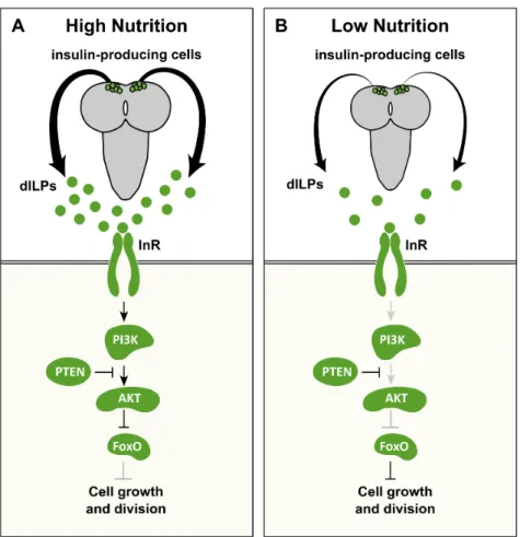
beetlesOnthophagus taurus (Emlen and Nijhout, 1999), its role in growth D. melanogaster was, until recently, controversial (Flatt, 2005; Mirth et al., 2014; Riddiford and Ashburner, 1991; Riddiford et al., 2010). In the following pages, I will therefore focus on what is known about the role of the D. melanogaster insulin-like peptides (dILPs), the insulin/insulin-like growth factor signalling (IIS) pathway, and the ecdysone signalling pathway in regulating nutritional plasticity in body and organ size.
1.4.1 Nutrition and the insulin/insulin-like growth factor signalling (IIS) pathway
InD. melanogaster, and many other animals, nutrition modifies body and
of the eight dILPs – dILP2, dILP3, and dILP5 – are exclusively expressed in the insulin-producing cells (Ikeya et al., 2002; Rulifson et al., 2002). The expression of these dILPs is nutrient dependent; starvation represses both their synthesis and secretion (Brogiolo et al., 2001; Ikeya et al., 2002). Further, ablation of the insulin-producing cells reduces adult body size in a similar fashion to starvation (Rulifson et al., 2002). These findings indicate that most of the nutrition-dependent growth is presumably regulated by the dILP production in the insulin-producing cells. The additional dILPs are expressed in several different tissues, including the imaginal discs, the mid gut, and the ventral nerve cord, and are thought to have systemic effects on growth (Brogiolo et al., 2001; Colombani et al., 2012; Garelli et al., 2012).
After being released into the insect bloodstream, dILPs act on target tissues by binding to the insulin receptor (InR) (Brogiolo et al., 2001). Once InR is activated, a highly conserved phospho-kinase signal transduction cascade, the IIS, is induced ultimately regulating cell growth and division. This is mainly achieved by activating positive growth regulators, such as the protein kinase Akt, and suppressing negative growth regulators, such as the transcription factor Forkhead Box class (FOXO) and the Tuberous Sclerosis Complex 1 and 2 (TSC1/2) (reviewed in (Taniguchi et al., 2006).
The suppression of TSC1/2 allows an additional nutrient-sensitive pathway, the target of rapamycin (TOR) signalling pathway, to remain active. The TOR pathway responds directly to intracellular amino acid concentrations via the TOR complex and regulates a number of cellular processes to promote growth (Gao et al., 2002; Sarbassov et al., 2005). In addition, the TOR complex itself regulates the IIS pathway by activating Akt (Sarbassov et al., 2005), which illustrates the extensive crosstalk between the two nutrition-sensitive pathways. Suppressing any component in the IIS pathway slows growth and results in smaller adults in a similar manner as starvation (Britton et al., 2002; Brogiolo et al., 2001). Combined, these findings illustrate that the circulating levels of dILPs and the IIS pathway coordinate growth rate with nutritional inputs.
Chapter 1
period – another crucial determinant of body and organ size in insects. The IIS pathway controls the growth period primarily by regulating the timing of the pulses of the steroid hormone ecdysone at specific stages in development (Koyama et al., 2014). How does ecdysone, in turn, regulate the duration of the growth period and ultimately body and organ size?
1.4.2 Nutrition and the ecdysone signalling pathway
Ecdysone is synthetized and secreted by the prothoracic glands in a series of discrete peaks throughout larval and pupal development. This periodic release of ecdysone together with a temporal- and tissue-specific expression of the ecdysone receptor complex, a heterodimer between Ecdysone Receptor (EcR) and Ultraspiracle (Usp), orchestrate many aspects of larval development: from larval molts and metamorphosis to growth and differentiation of target tissues (reviewed in (Yamanaka et al., 2013).
Chapter 1
Figure 1.3: The activation and derepression functions of ecdysone signalling.
Ecdysone binds to its receptor, a heterodimer between EcR and Usp, to induce two types of functions.(A) Activation function: ecdysone binds to EcR-Usp and directly activates gene transcription. (B) Derepression function: ecdysone binds to EcR-Usp and relieves the repressive action of the EcR-Usp, allowing gene transcription. Knocking down either EcR or Usp partially activates gene transcription, while overexpressing a dominant negative form of EcR with a mutated ligand-binding domain (EcRDN
) prevents gene transcription.
Ecdysone exerts its effects by binding to the EcR/Usp heterodimer complex. This complex represses the transcription of a subset of ecdysone target genes in the absence of ecdysone (Figure 1.3B) (Brown et al., 2006; Cherbas, 2003; Schubiger and Truman, 2000; Schubiger et al., 2005). Once ecdysone binds to EcR/Usp, it induces target gene transcription either by direct activation via EcR/Usp (Figure 1.3A) or by relieving the repressive action of the EcR/Usp (Figure 1.3B). Several genetic tools inD. melanogaster allow us to explore the specific roles of ecdysone signalling
Usp, using RNAi, eliminates the repressive function of EcR/Usp, thereby partially inducing ecdysone function (Figure 1.3A) (Brown et al., 2006; Cherbas, 2003; Mirth et al., 2009; Schubiger et al., 2005). Conversely, overexpressing a dominant negative form of EcR with a mutated ligand-binding domain abolishes both the derepression and activation functions of ecdysone (Figure 1.3B)(Brown et al., 2006; Cherbas, 2003; Hu et al., 2003). In Chapter 3, I took advantage of these two well-described genetic tools to investigate the role of ecdysone signalling in regulating nutritional plasticity in organ size.
1.4.3 Organ-specific sensitivities to nutrition
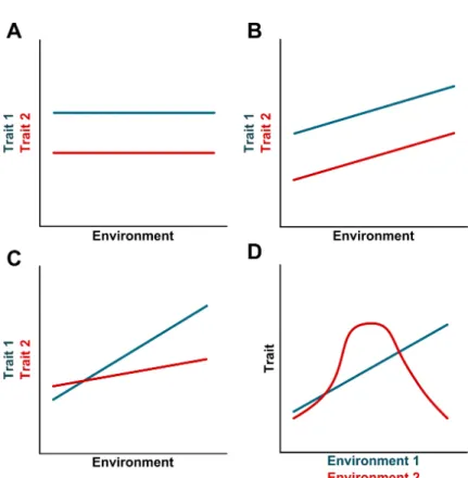
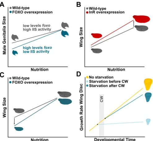
If the levels of circulating dILPs reflect the nutritional status of an insect, how do different organs respond differentially to nutritional variation? As discussed above, the size of the male genitalia and the CNS is relatively invariant across nutritional conditions (Cheng et al., 2011; Shingleton et al., 2005; Tang et al., 2011). This low sensitivity to nutrition is achieved through different mechanisms. In the case of the CNS, InR-independent activation of the IIS pathway allows the CNS to maintain its growth rate even when circulating dILPs are low (Cheng et al., 2011). Alternatively, the male genitalia reduces its plasticity in response to nutrition by expressing low levels offoxo mRNA (Tang et al., 2011). When circulating dILPs and the activity of the IIS pathway are reduced, FOXO remains in the nucleus and supresses growth (Jünger et al., 2003). As the male genitalia expresses low levels of foxo, it is able to maintain its size even when larvae are malnourished (Figure 1.4A) (Shingleton et al., 2005, 2009; Tang et al., 2011). Overexpressing FOXO in the male genitalia increases its sensitivity to nutrition and results in smaller genitalia (Figure 1.4A) (Tang et al., 2011). Despite the differences in mechanisms between the CNS and the male genitalia, ultimately these organs are protected from the effects of poor nutrition by retaining high levels of activity of the IIS pathway irrespective of nutritional conditions.
Chapter 1
Figure 1.4: Organs differ in their sensitivity to nutrition. (A) The male genital disc maintains its size even when larvae are poorly fed. This reduction in nutritional sensitivity is achieved by reducing the levels of foxo mRNA and retaining high IIS activity in low nutritional environments (grey line). Overexpressing FOXO in the male genitalia increases its sensitivity to nutrition (blue line). (B, C) Nutrition affects the size of the wings in proportion with body size (grey line). (B) An increase ofInRexpression results in an increase in nutritional sensitivity by enhancing wing size in large flies (red line). (C) An increase of foxo expression enhances the nutritional sensitivity of the wing by supressing wing size in small individuals (blue line). (D) The sensitivity to nutrition of the wing discs varies with developmental time. Before critical weight, starvation severely reduces the growth of the wing disc. On the other hand, discs grow considerably even in post-critical weight larvae that are malnourished.CW:critical weight. Adapted from (Shingleton and Frankino, 2013; Shingleton and Tang, 2012; Shingleton et al., 2008)
an exaggerated response to nutrition (Shingleton and Frankino, 2013; Shingleton and Tang, 2012). Overexpressing either FOXO or InR specifically in the wing disc increases its sensitivity to nutrition making it hyperallometric (i.e. disproportionally larger) in relation to body size (Figure 1.4B, C). However, this hyperallometry is achieved through different ways: increasing InR expression resulted in an exaggerated
increase in the wing size of larger individuals, but has little or no effects in the wing size in smaller individuals (Figure 1.4B) (Shingleton and Tang, 2012). On the other hand, an increase in foxo expression led to
a disproportionally small wing size in smaller individuals, but almost no effect in larger individuals (Figure 1.4C) (Shingleton and Tang, 2012). Thus it appears that organs can display exaggerated responses to nutrition by modulating the IIS pathway at several levels of its action (Shingleton and Frankino, 2013).
Organs can also change their sensitivity to nutrition with developmental time. For instance, starving pre-critical weight larvae compromises wing disc growth and differentiation, but after critical weight starvation has a more modest effect on the development of the wings discs; that is, discs grow considerably and continue to differentiate even when post-critical weight larvae are poorly fed (Figure 1.4D) (Mirth et al., 2009; Shingleton et al., 2008). This switch in sensitivity to nutrition at critical weight seems to be mediated by changes in the IIS pathway. Supressing the IIS pathway just after critical weigh abolishes body growth, but the wing discs continue to grow presumably until their size is appropriate for the much reduced body size (Figure 1.4D)(Shingleton et al., 2005, 2008). These findings have led some authors to hypothesize that an intrinsic growth rate that does not require nutritional inputs may enable further growth of the developing organs when nutrition, and accordingly the IIS, is severely reduced (Nijhout et al., 2014; Shingleton et al., 2008).
Chapter 1
(Lanet et al., 2013). These findings suggest that ecdysone signalling acts in target tissues and allows their development to proceed even in the absence of nutritional inputs.
1.5
Aims and thesis scope
Even though developmental plasticity has gradually become a fundamental aspect in our evolutionary thinking, there are still many issues to be solved. To fully comprehend the role of plasticity in evolution, we first need to understand how developmental processes are regulated by environmental signals to generate a diverse range of phenotypes. Developmental plasticity should be seen as a fundamental source of phenotypic variation, a key condition for natural selection to operate. In this thesis, I therefore implemented a simple, yet powerful, developmental approach to investigate how the environment shapes the developmental trajectory of a developing organ to generate distinct morphologies. Specifically, the aim of this thesis was to investigate i) how developmental processes are modified by the environment, particularly nutrition, within a species and ii) compare whether similar developmental changes account for differences between species.
For this purpose, I used ovariole number, an important determinant of ovary size and female fecundity, in Drosophila. Ovariole number is determined during larval stages and is highly plastic in response to several environmental conditions, including nutrition and temperature. Moreover, ovariole number shows remarkable diversity among Drosophila species. Thus, ovariole number is an excellent model to address the aims of this thesis.
Despite the potential relevance of developmental windows of environmental sensitivity on the outcome of plasticity, few studies in insects have taken this factor in consideration when addressing the effects of nutrition in body and organ growth. This issue will be addressed in Chapter 2. Here I examined the existence of critical periods of sensitivity to nutrition during ovary development in D. melanogaster, with special
sensitivity. This will be the first step in elucidating which development processes during ovary development are modified by nutrition to generate differences in ovariole number.
The role of hormonal pathways in regulating nutritional plasticity in ovariole number will be explored in Chapter 3. Much of the current research has underscored hormonal signals as key regulators of developmental plasticity. However, how hormones regulate the nutritional sensitivity of a developing organ with developmental time has not been fully addressed. In this chapter, I asked whether IIS and/or ecdysone signalling pathways act at critical weight to regulate nutritional plasticity in ovariole number. I carefully characterized the contribution of each signalling pathway in the regulation of three developmental processes that I previously described to account for the nutritional-induced differences in ovariole number (Chapter 2). This powerful approach sheds an interesting light on the hormonal regulation of nutritional plasticity in ovariole number.
Chapter 1
Finally, in Chapter 5, I will discuss the main contributions of my thesis work and present future avenues of research that would further demonstrate that the environment not only acts as a selective agent, but it also contributes to the creation of novel and diverse life forms.
Acknowledgements
2
NUTRITIONAL PLASTICITY IN
OVARIOLE NUMBER IN
DROSOPHILA MELANOGASTER
“A world of endless possibilities and infinite outcomes. Countless choices define our fate: each choice, each moment, a moment in the ripple of time. Enough ripple, and you change the tide. . . For the future is never truly set.”
Abstract
The extent to which an organism can adjust its developmental trajectory in response to environmental conditions, known as developmental plasticity, often depends on critical periods of environmental sensitivity. Here, I identify two phases of sensitivity to nutrition that regulate plasticity in ovariole number, an important determinant of fecundity and ovary size, in Drosophila. These two phases are separated by the developmental transition at critical weight. In the first highly-sensitive phase, optimal nutrition is required to promote ovary growth and to induce the onset of terminal filament cell (TFC) differentiation, which serves as a starting point for ovariole development. The second phase begins after TFC differentiation is initiated at critical weight. In this phase, the formation of terminal filaments (TFs) through intercalation of TFCs and ovary growth continue, albeit at reduced rates, in larvae that are poorly fed. These results shed a new light on how organs change their sensitivity to environmental variation at critical weight.
Publication
Chapter 2 and 3 are part of a manuscript submitted for publication, authored by C.C. Mendes and C.K. Mirth.
Authors’ contributions
Chapter 2
2.1
Introduction
Developmental plasticity, the ability of an organism to adjust its developmental trajectory in response to environmental variation, is a seemingly universal property of all multicellular organisms. Often, the extent of developmental plasticity depends not only on the traits and environmental conditions considered (Mirth and Shingleton, 2012), but also on the existence of phases of environmental sensitivity, commonly referred as critical periods, during which developmental processes can respond plastically (Koyama et al., 2013; Nijhout, 2003a). In the most extreme cases, an environmental cue within a critical period triggers a developmental switch between alternative developmental trajectories, giving rise to distinct phenotypes, such as dramatic seasonal differences in the pigmentation of butterfly wing patterns and different body sizes and shapes in honeybee castes (Brakefield et al., 1996; Smith et al., 2008). Understanding how developing organs change their sensitivity to environmental conditions, and how this influences their plastic response, is an important step towards a comprehensive knowledge of how the environment generates new phenotypic variants.
Critical weight also regulates the sensitivity of developing organs to nutrition over developmental time. Larvae starved before reaching critical weight delay the patterning of their presumptive adult tissues, the imaginal discs (Mirth et al., 2009). Conversely, starvation after critical weight allows continued patterning and growth of the imaginal discs (Mirth et al., 2005, 2009; Shingleton et al., 2008). This additional role of critical weight has been overlooked in current research, and importantly, whether critical weight determines periods of nutritional sensitivity has not yet been fully investigated. In this chapter, I attempt to elucidate how developing organs change their sensitivity to nutrition over developmental time, with special emphasis on the potential role of critical weight in mediating nutritional sensitivity.
To address this issue, I used ovariole number in D. melanogaster as a
model. Ovarioles are egg-producing structures in the insect ovary that directly affect female reproductive capacity (Boulétreau-Merle et al., 1982; R’ kha et al., 1997; Klepsatel et al., 2013b,a). Although little is known about the genetic cascades involved in ovariole development (Cheng et al., 2011; Forbes et al., 1996; Godt and Laski, 1995; Patel et al., 1989; Sahut-Barnola et al., 1995; Sarikaya and Extavour, 2015), the cellular events mediating this process are better characterized. Ovariole development occurs during the third instar (L3) larval and early pupal stages (Kerkis, 1931; King, 1970; King et al., 1968) through the intercalation of terminal filament cells (TFCs) into stacks of seven to ten flattened cells, called terminal filaments (TFs) (Godt and Laski, 1995; Sahut-Barnola et al., 1995, 1996). Each TF defines the position of one ovariole and thus, the number of TFs at pupariation is equivalent to the number of ovarioles in the adult (Godt and Laski, 1995; Hodin and Riddiford, 1998; Sahut-Barnola et al., 1995; Sarikaya et al., 2012).
Chapter 2
and Riddiford, 1998). However, it was unclear whether ovary development exhibits critical periods of nutritional sensitivity, and importantly, how the developmental processes are modified by nutrition at different periods of sensitivity. I therefore examined whether changes in nutrition at specific stages during L3 larvae influence the plastic response of ovariole number. I further investigated how distinct stage-specific developmental processes during ovary development respond to changes in nutrition and account for nutritional-induced differences in ovariole number.
2.2
Materials and Methods
2.2.1 Fly stock
To assess the effects of larval nutrition on ovariole number, I used an outbred population (wild type) of Drosophila melanogaster established in the laboratory of Dr. Élio Sucena in 2007, originating from 160 fertilized females collected in Azeitão, Portugal (Martins et al., 2013). The population was kept in laboratory cages with high census (> 1500 individuals) and maintained at constant temperature (25°C) on standard fly food (4.5% molasses, 7.2% sugar, 7% cornmeal, 2% yeast extract, 1% agar and 2.5% Nipagin solution).
2.2.2 Larval staging and dietary manipulations
Adults were allowed to lay eggs for two to six hours on fresh food plates (60×15 mm Petri dish). Egg density was controlled to prevent
eclosion. To obtain L3 ovaries, larvae of the appropriate age were dissected and processed for immunocytochemistry (Figure 2.1C, 2.3A, 2.5A). All experiments were performed at 25°C.
2.2.3 Measurements of life-history traits: developmental
time, female weight, early female fecundity and ovariole number
To determine the average time to pupariation, newly ecdysed L3 larvae were transferred to vials (20-30 larvae per vial) containing standard food. The number of larvae pupariating (immobile larvae with evaginated spiracles) was counted in 2 h intervals until all larvae pupariated. I used pharate weight as a proxy of adult body size (Mirth et al., 2005). Pharate adults were collected from food vials and food residuals were carefully cleaned off from the pupal cases using distilled water and a paintbrush. I distinguish females from males by the presence or absence of male-specific sex combs through the pupal case. Female pharate adults were individually weighed on a Sartorius SE2 ultramicrobalance.
To determine early fecundity, newly eclosed females were individually maintained in vials on standard food with one male of the same food/time point. Individuals were transferred to fresh vials every day during the first three days after eclosion. All eggs were counted daily. To count adult ovariole number, newly eclosed flies were maintained in vials (ten females and five males per vial) on standard food until the time of dissection (4-6 days after eclosion) (Figure 2.1A). Ovaries were dissected in cold phosphate buffered saline containing 1% Triton X-100 (PBT) and ovarioles were teased apart and counted under a dissecting microscope.
2.2.4 Immunocytochemistry
Chapter 2
with PBT and blocked in 2% normal donkey serum in PBT for 30 minutes. Primary antibody incubation in mouse anti-Engrailed (Developmental Studies Hybridoma Bank 4D9, 1:40) diluted into 2% normal donkey serum in PBT was conducted overnight at 4°C. After washing three times for 20 minutes in PBT, larvae were incubated in the dark with goat anti-mouse Alexa 568 (Invitrogen, 1:200) and TRICT-Phalloidin (Sigma, 1:200) diluted into 2% normal donkey serum in PBT overnight at 4°C. Larvae were rinsed with PBT and ovaries were mounted on a poly-L-lysine-coated coverslip using Fluoromount-G (SouthernBiotech).
2.2.5 Image Acquisition and Analysis
Samples were imaged using a Zeiss LSM 510 Meta confocal microscope using a 40x 1.3NA oil objective lens. During confocal image acquisition, the detection parameters were adjusted to avoid under- or overexposed pixels, and images were acquired through the full thickness of the ovary at 1 µm. Images were processed and analysed using ImageJ (NIH) and Adobe Photoshop (Adobe Systems). For each time point/genotype/food treatment, terminal filament cells (TFC) were identified by Engrailed expression. Forming terminal filaments (TFs) were identified by the presence of TFC in stacks with the characteristic flattened cell morphology, and total number of forming TFs were counted. For ovary volume, the ImageJ Volumest plugin was used (Merzin, 2008).
2.2.6 Statistical Analysis
function ‘sm.ancova’ under ‘sm’ library. All data analyses and statistics were conducted using R v3.1.2 (R Development Core Team, 2014). Plots were made using GraphPad Prism v6 (GraphPad Software). p-values are
indicated in the text and figures.
2.3
Results
2.3.1 Two phases of sensitivity to nutrition regulate the plastic response of ovariole number
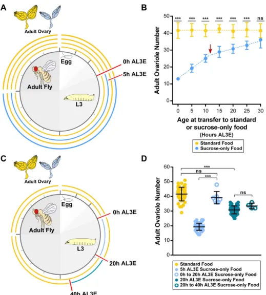
To determine critical periods of sensitivity to nutrition in ovariole number, I fed L3 larvae either on standard food or on sucrose-only food at timed intervals starting between 0 h to 30 h AL3E until the end of the feeding period (Figure 2.1A). Larvae fed on sucrose-only food are starved of protein, lipids and other micronutrients present in yeast, yet show higher rates of survival than when starved completely. Overall, larvae transferred to sucrose-only food between 0 and 25 h AL3E showed a significant reduction in ovariole number when compared to the controls transferred to standard food (Figure 2.1B). In contrast, transferring larvae to sucrose-only food at 30 h AL3E did not cause a significant reduction in ovariole number (Figure 2.1B).
Chapter 2
Nevertheless, the effects of the sucrose-only food in ovariole number could also be a direct consequence of different lengths of exposure to the sucrose-only food. To test this hypothesis, I performed a preliminary experiment where L3 larvae were fed on sucrose-only food for 20 h starting either at 0 h AL3E or at 20 h AL3E and then returned to standard food until the end of the feeding period (Figure 2.1C). As described above, ovariole number is severely reduced when larvae were transferred to sucrose-only food at 5 h AL3E (Figure 2.1B, D). Surprisingly, ovariole number in larvae fed on sucrose alone for a short period between 0 h to 20 h AL3E was similar to standard food control (Figure 2.1D). In contrast, when larvae were fed on sucrose-only food from 20 h to 40 h AL3E, ovariole number was significantly reduced (Figure 2.1D). This reduction in ovariole number was similar when compared to larvae transferred to sucrose alone at 20 h AL3E until the end of development (Figure 2.1B, D). These observations corroborate a previous study where re-feeding pre-critical weight larvae after a brief period of starvation delays pupariation for longer than the length of the starvation period, but does not affect final body size. After critical weight, when the duration of the larval growth period is fixed, short periods of starvation have no effect on the timing of pupariation and thus, larvae are unable to reach their optimal body size even after re-feeding (Beadle et al., 1938).
Chapter 2
Figure 2.2: Ovariole number is positively correlated with early female fecundity. Number of eggs laid was counted in the first three days after eclosion (diamond: 1st day after eclosion; square: 2nd day after eclosion; circle: 3rd day after eclosion) from females fed on standard food as larvae (yellow symbols) and females fed on sucrose-only food as larvae at timed intervals starting between 5 h to 25 h AL3E (symbols with different shades of blue) until the end of the feeding period. Plotted values represent means and error bars show 95% confidence intervals of means. L3: third instar larvae; AL3E: after L3 ecdysis.
2.3.2 Ovary development during L3 larval stages
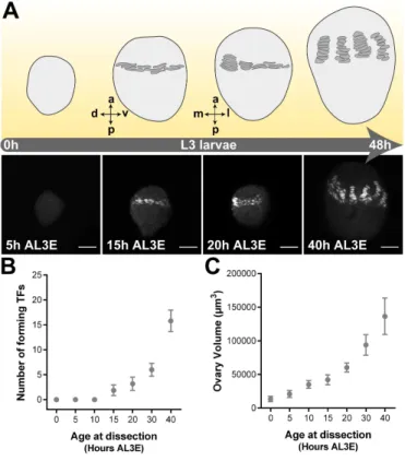
To further understand how nutrition regulates ovariole number, I first analysed ovary development in L3 larvae raised in standard food. When TFCs differentiate from the surrounding ovarian somatic cells, they upregulate expression of the transcription factor Engrailed (En) (Patel et al., 1989). Thus, I used En as a marker for TFC differentiation and TF formation.
Figure 2.3: Ovary development during L3 larval stages under optimal nutritional conditions.(A) Schematic drawings representing ovary development in L3 larvae reared in standard food. Terminal filaments (TFs) are represented as dark grey symbols. Axis are presented as A-P, anterior-posterior; D-V, dorsal-ventral; M-L, medial-lateral. Pictures show developing ovary during L3 larval stages under standard food. Engrailed (grey) marks terminal filament cells (TFCs). Scale bar: 20µm. (B) Number of forming terminal filaments (TFs). (C) Ovary volume. Plotted values represent means and error bars show 95% confidence intervals of means. L3: third instar larvae; AL3E: after L3 ecdysis.
Chapter 2
2.3.3 TF formation and ovary growth respond differently
to pre- and post-critical weight nutrition
From my description of ovary development during L3 larval stages, I hypothesized that larval nutrition regulates one or all of the three developmental processes: i) the onset of differentiation of the first TFCs, representing the first step in ovariole development, ii) the rate at which new TFs emerged through intercalation of TFCs (referred as the rate of TF formation), and iii) the rate of ovary growth.
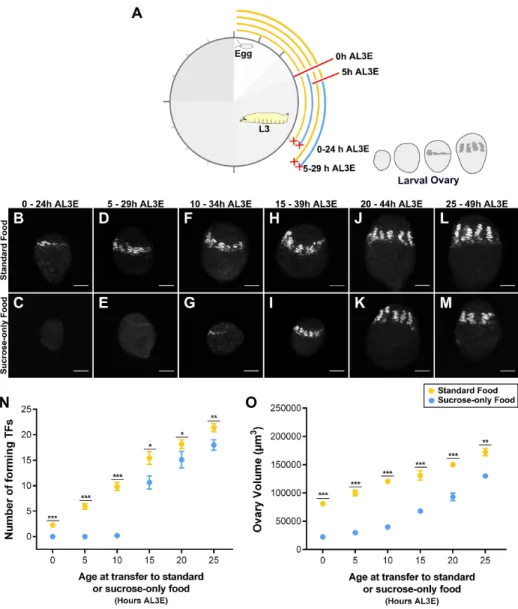
To test which of these processes respond to changes in nutrition, I fed larvae on sucrose-only food for 24 h, starting at 5 h intervals between 0 h to 25 h AL3E, and quantified the number of TFs and ovary volume for each condition at the end of this one-day starvation period (Figure 2.4A). When larvae were fed on sucrose-only food before reaching critical weight (before 10 h AL3E), I failed to observe any En-positive cells in the ovaries, indicating that the onset of TFC differentiation is delayed (Figure 2.4B-E, N). Wing discs and central nervous systems of larvae staged before 10 h AL3E did show En expression, indicating that this antigen was detectable in other tissues (Supplementary Figure S2.1). In addition, the ovary volume was severely reduced in larvae fed on sucrose-only food before 10 h AL3E (Figure 2.4O). In contrast, when larvae were transferred to sucrose-only food around the time of the critical weight transition (at 10 h AL3E), the majority of ovaries had few TFCs (Figure 2.4F, G) and in some ovaries TFCs were organized into short TFs (Figure 2.4N). Ovary volume was still greatly reduced in these larvae (Figure 2.4O). Finally, ovaries from larvae transferred to sucrose-only food after reaching critical weight (after 15 h AL3E), all had forming TFs (Figure 2.4H-M). Nevertheless, both TF number and ovary volume were moderately reduced when compared with larvae fed on standard food (Figure 2.4N, O).
Figure 2.4: Distinct stage-specific developmental processes during ovary development are regulated by nutrition.((A) Experimental design to examine how developmental processes respond to changes in nutrition during L3 larval stages. Only the first two time points are shown (0 h and 5 h AL3E). Dissection times are marked with red crosses. (B-M) Shown is terminal filaments (TFs) marked with Engrailed (grey) in ovaries from larvae fed on (B, D, F, H, J, L) standard food or (C, E, G, I, K, M) sucrose-only food for 24 h starting between 0 h to 25 h AL3E. Scale bar: 20µm. (N) Number of forming terminal filaments (TFs) and (O) ovary volume of ovaries from larvae fed on standard food (yellow circles) or sucrose-only food (blue circles). Plotted values represent means and error bars show 95% confidence intervals of means. In some cases, error bars are too small to be shown. Welch Two sample t-test using Holm’s
Chapter 2
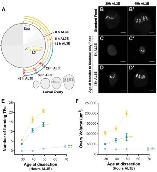
Figure S2.2A). Therefore, I presumed that TF formation eventually occurs in these ovaries, even if its onset is delayed. To test this hypothesis, I fed pre-critical weight larvae on sucrose-only food and dissected the larval ovaries at three different time points (Figure 2.5A). Indeed, TFCs and few short TFs were observed at 49 h AL3E (Figure 2.5C-C´, E) and new TFs were still forming at 69 h AL3E, albeit at a significantly reduced rate (Figure 2.5E). A slower rate of TF formation relative to standard food controls was also found in ovaries from post-critical weight larvae fed on sucrose-only food (Figure 2.5D-D’, E). These post-critical weight larvae pupariate at the same time as the standard food controls, but are smaller in body size (Supplementary Figure S2.2B). Furthermore, while ovaries from post-critical weight larvae fed on sucrose-only food showed a slight increase in ovary volume over L3 larval stages, feeding on sucrose-only food strongly arrested ovary growth in pre-critical weight larvae (Figure 2.5F).
2.4
Discussion
An important step towards a better understanding how the environment modifies the developmental trajectory of an organism to produce distinct phenotypes is to determine critical periods of environmental sensitivity. The present work identified such critical periods in the developing fly ovary. With my detailed characterization of the effects of nutrition on ovary development, I identified two phases of sensitivity to nutrition during L3 larval stage that regulate the plastic response of ovariole number. This switch in sensitivity coincides with the timing of critical weight (Koyama et al., 2014; Mirth et al., 2005, 2009; Shingleton et al., 2005). I further found that distinct developmental processes during ovary development respond differentially to changes in nutrition in each phase of sensitivity (Figure 2.6).
Figure 2.5: TF formation and ovary growth respond differently to pre- and post-critical weight nutrition.(A) Experimental design to examine the dynamics of TF formation and ovary growth in larvae transferred to standard food (yellow line) and in larvae transferred to sucrose-only food either at 5 h AL3E (light blue line) or at 15 h AL3E (dark blue line). Dissection times are marked with red crosses. (B-D’) Shown is terminal filaments (TFs) marked with En (grey). (B-B’) Ovaries from larvae reared on standard food. (C-D’) Ovaries from larvae transferred to sucrose-only food from: (C-C’) 5 h or (D-D”) 5 h AL3E. Larvae dissected at (B, C, D) 29 h or (B´, C’, D’) 49 h AL3E. Scale bar: 20µm. (E) Number of forming terminal filaments (TFs) and (F) ovary volume of ovaries from larvae fed on standard food (yellow circles or transferred to sucrose-only food either at 5 h AL3E (light blue circles) or at 15 h AL3E (dark blue points). Plotted values represent means and error bars show 95% confidence intervals of means. In some cases, error bars are too small to be seen. ANCOVAs using Holm’s
Chapter 2
Figure 2.6: Critical weight separates two phases of sensitivity to nutrition in ovariole number.In the first, highly sensitive phase, poor nutrition arrests ovary growth and strongly suppresses the onset of terminal filament (TF) formation. Once TF formation is initiated around critical weight, TF formation and ovary growth proceed, although at a slower rate, in response to poor nutrition. These two phases of sensitivity influence the plastic response of ovariole number; changes in nutrition during the first phase of sensitivity have greater effects in ovariole number than in the second phase of sensitivity.
may only occur when the timing of critical weight is also affected. Thus, the attainment of critical weight and the onset of TF formation appear to be intimately connected and both are critical steps in regulating the plastic response of ovariole number.
Interestingly, I found that changes in nutrition during the second phase of sensitivity did not supress the formation of new TFs. In fact, new TFs continue to form in malnourished larvae. This suggests that while the initial trigger for TFC differentiation is highly sensitive to nutrition, subsequent TFCs continue to differentiate from the pool of TFC precursors irrespective of the nutritional conditions. Several other tissues also exhibit a similar switch in sensitivity to nutrition that allows progression of cell differentiation when larvae are poorly fed (Lanet et al., 2013; Mirth et al., 2009). For instance, after critical weight, neuroblasts in the optic lobe are able to generate the full repertoire of neuronal types independently of nutritional variation (Lanet et al., 2013). The attainment of critical weight may serve as a signal that ensures that sufficient endogenous nutritional reserves exist to sustain neuronal diversity (Lanet and Maurange, 2014). A similar mechanism may be employed to promote TFC differentiation under conditions of poor nutrition.
Even though TF formation proceeded under poor nutritional conditions, the rate at which new TFs were formed was significantly slower than standard food controls, resulting in a reduced number of TFs. Such reduction in the rate of TF formation alludes to changes in the production of new TFC precursors. However, our current knowledge on when and how TFC precursors are produced have thus far been limited (Lengil et al., 2015; Sahut-Barnola et al., 1996). Future work on identifying additional TFC markers may help us understand whether changes in nutrition during L3 larval stages affect the production of TFC precursors and how this may influence the rate of TF formation.
Chapter 2
modifies the growth trajectories of the wing imaginal discs (Garcia-Bellido and Merriam, 1971; Martin, 1982; Bryant and Levinson, 1985). After attainment of critical weight, wing discs have an intrinsic growth rate that promotes considerable growth under poor nutritional conditions. This intrinsic growth is not present before critical weight, and thus, wing discs arrest growth when pre-critical weight larvae are poorly fed (Shingleton et al., 2008). In light of these observations, I propose that developing ovaries may also have an intrinsic growth rate after critical weight that allows progression of growth in poorly-fed larvae.
2.5
Conclusions
The results described in this chapter revealed that critical weight plays a fundamental role in reprograming the developing ovary’s response to nutrition. Furthermore, this work contributes to a better understanding of the developmental processes that regulate ovariole number, and provides the developmental tools that will be used throughout this thesis.
Acknowledgements
Supplementary Figures
Chapter 2