Effect of the Transient Pharmacological
Inhibition of Mapk3/1 Pathway on Ovulation
in Mice
Dayananda Siddappa1, Élaine Beaulieu2, Nicolas Gévry2, Philippe P. Roux3,
Vilceu Bordignon1, Raj Duggavathi1 *
1Department of Animal Science, McGill University, Ste-Anne-de-Bellevue, QC H9X 3V9, Canada, 2Département de Biologie, Université de Sherbrooke, Sherbrooke, QC J1K 2R1, Canada,3Institute for Research in Immunology and Cancer, Faculty of Medicine, Université de Montréal, Montreal, QC H3C 3J7, Canada
Abstract
Mitogen-activated protein kinase 3/1 (Mapk3/1) pathway is critical for LH signal transduction during ovulation. However, the mechanisms remain incompletely understood. We hypothe-sized that Mapk pathway regulates ovulation through transcriptional regulation of ovulatory genes. To test this hypothesis we used immature mice superovulated with equine and human chorionic gonadotropins (eCG and hCG) and PD0325901, to inhibit hCG-induced Mapk3/1 activity. Mice received either the inhibitor PD0325901 (25μg/g, i.p.) or vehicle at 2h before hCG stimulation. Administration of the inhibitor abolished Mapk3/1 phosphoryla-tion in granulosa cells. While vehicle-treated mice ovulated normally, there were no ovula-tions in inhibitor-treated mice. First, we analyzed gene expression in granulosa cells at 0h, 1h and 4h post-hCG. There was expected hCG-driven increase in mRNA abundance of many ovulation-related genes includingPtgs2in vehicle-treated granulosa cells, but not (P<0.05) in inhibitor-treated group. There was also reduced mRNA and protein abundance of the transcription factor, early growth response 1 (Egr1) in inhibitor-treated granulosa cells. We then used GRMO2 cell-line to test if Egr1 is recruited to promoter ofPtgs2 fol-lowed by chromatin immunoprecipitation with either Egr1 or control antibody. Enrichment of the promoter regions in immunoprecipitants of Egr1 antibody indicated that Egr1 binds to thePtgs2promoter. We then knocked down Egr1 expression in mouse primary granulosa cells using siRNA technology. Treatment withEgr1-siRNA inhibited Egr1 transcript accumu-lation, which was associated with reduced expression ofPtgs2when compared to control-siRNA treated granulosa cells. These data demonstrate that transient inhibition of LH-stimu-lated MAPK3/1 activity abrogates ovulation in mice. We conclude that Mapk3/1 regulates ovulation, at least in part, through Egr1 and its target gene,Ptgs2in granulosa cells of ovu-lating follicles in mice.
OPEN ACCESS
Citation:Siddappa D, Beaulieu É, Gévry N, Roux PP, Bordignon V, Duggavathi R (2015) Effect of the Transient Pharmacological Inhibition of Mapk3/1 Pathway on Ovulation in Mice. PLoS ONE 10(3): e0119387. doi:10.1371/journal.pone.0119387
Academic Editor:Jean-Marc A Lobaccaro, Clermont Université, FRANCE
Received:February 28, 2014
Accepted:January 30, 2015
Published:March 24, 2015
Copyright:© 2015 Siddappa et al. This is an open access article distributed under the terms of the
Creative Commons Attribution License, which permits unrestricted use, distribution, and reproduction in any medium, provided the original author and source are credited.
Funding:DS was funded by McGill Graduate Student Fellowship, Department of Animal Science Graduate Excellence Fellowship and RQR. This study was also partly funded by Réseau Québécois en Reproduction. The funders had no role in study design, data collection and analysis, decision to publish, or preparation of the manuscript.
Introduction
Ovulation is a multi-gene, multi-step process involving complex signaling pathways, which fa-cilitates synchronization of oocyte maturation and cumulus expansion with that of follicular rupture. It is unequivocal that preovulatory luteinizing hormone (LH) surge initiates these pro-cesses through remarkable changes in gene expression program of granulosa cells within ovu-lating follicles. Some of the important signaling pathways through which LH brings about ovulatory events are cAMP/Protein Kinase A (PKA) pathway, Mitogen-activated protein ki-nase 3/1 (Mapk3/1; ERK1/2) pathway and phosphatidylinositide 3-kiki-nases (PI3K) pathway [1– 4]. A recent study using granulosa-specificMapk3/1knockout (KO) mice [5] providedin vivo
evidence for the importance of Mapk3/1 signaling in LH signaling during ovulation.
Granulosa cells fromMapk3/1KO mice showed altered expression of hundreds of LH regu-lated genes [5], but which transcription factors act as mediators of their signals have not been completely identified [6]. Many transcription factors including nuclear receptor 5a2 (Nr5a2) [7] (CAAT/enhancer binding protein beta (Cebpb) [6], early growth response-1 (Egr1) [8] and Progesterone receptor (Pgr) [9] are critical LH signaling during ovulation. It was reported that 19% of the LH-driven genes were regulated in granulosa cells of bothMapk3/1andCebpa/b
conditional KO mice at 4h hCG [6]. This indicates that the rest 81% Mapk3/1-dependent genes are regulated by transcription factors other than Cebpa/b, which are yet to be identified.
While conditional KO model is a powerful tool to study physiological processes in vivo, it is not devoid of limitations. For example expression of the Cre-recombinase may not be active in all cells of interest, therefore, leading to incomplete gene deletion. On the other hand, pharma-cologic method of inhibition of a protein’s activity is economical, less time consuming and rela-tively simple compared to genetic manipulation. Moreover, using pharmacologic method one can inhibit protein activity activity transiently at a precise physiological stage. Limitations of pharmacologic methods include potential“off-target”effects. PD0325901 is specific inhibitor of Mapk-kinase (Map2k; MEK), which abrogates Mapk3/1 activity without cytotoxicity when administered as a single dose of 25μg/g bodyweight in mice [10,11]. More importantly,
PD0325901 does not have off-target effects shown by other Map2k inhibitors, U0126 and PD98059 [12]. Therefore, PD0325901 treatment is an excellent alterative method to inhibit Mapk3/1 activity at precise time-points during follicular development.
The aim of our study was to identify novel transcription factors that play an important role downstream of Mapk3/1 signaling in the process of ovulation. We hypothesized that Mapk3/1 pathway regulates ovulation through transcriptional regulation of ovulatory genes. To test this hypothesis we employed anin vivopharmacologic method of inhibition of Mapk3/1 activity with out disrupting theMapk3/1gene expression. Here we report our study exploring the effect of PD0325901 on ovulation in superovulated immature mice.
Materials and Methods
Animals and treatments
Husbandry. Inbred C57BL/6NCrl mice (Charles River) were housed in standard plastic rodent cages and maintained on a 12-h light/dark cycle withad libitumfeed (Teklad-Rodent ir-radiated Diet, Harlan) and water. The animal use protocol was approved by the Animal Care and Use Committee, McGill University.
Life Sciences; 5 IU i.p.) to induce ovulation and luteinization. In this protocol, the ovulation oc-curs at 12–14h post-hCG [7,13,14].
Inhibition of Mapk3/1 activity. A potent selective Map2k (MEK) inhibitor PD0325901 (Selleckchem) was initially dissolved in DMSO (Fisher Scientific) to prepare a stock solution of 100μg/μl concentration. A dosing solution of 2.5μg/μl in 5% DMSO in saline was prepared
just before treatment. For inhibition of Mapk3/1 activity, mice were administered with a single dose of PD0325901 (25μg/g body weight, i.p.) 2h before hCG treatment. PD0325901 will be
referred to as Map2k-inibitor from here on for readers’simplicity. Mice treated with 5% DMSO in saline served as vehicle controls. We determined Mapk3/1 activity by determining the abundance of phosphorylated isoform of Mapk3/1 relative to its total isoform. The dose of Map2k-inhibitor was determined based on a preliminary experiment with doses of 10 and 25μg/g 2 h before hCG treatment, of which the later dose resulted in consistent inhibition of
Mapk3/1 activity.
Ovulation rate and histology
To study the effect of inhibition of Mapk3/1 activity on ovulation rate, we counted cumulus oo-cyte complexes (COCs) from both oviducts of Map2k-inhibitor and vehicle treated mice at 18h post-hCG. Ovaries collected at 18h post-hCG from these mice were fixed in 10% neutral buff-ered formalin at 4°C for 2 days. Paraffin embedded ovaries were cut (4μthickness) and sections
were stained by hematoxylin and eosin for histological observation under Leica DM200 micro-scope attached with Leica EC3 camera.
Granulosa cell collection and real-time PCR
Granulosa cells were collected by follicle puncture [15] at 0h, 1h and 4h post-hCG
(N = 3 mice/group). Briefly, granulosa cells were collected by follicular puncture using 27G needle. The cell suspension was passed through a cell strainer (BD Falcon, Mississauga, ON, Canada; 40μm) to filter out cumulus–oocyte complexes. Pure populations of mural granulosa
cells from both ovaries of each mouse were pooled together. Total RNA was purified from granulosa cells using Picopure RNA isolation kit (Arcturus Biosciences) followed by cDNA synthesis from 500 ng of RNA using iScript kit (Bio-Rad). We analyzed mRNA expression by real-time PCR as previously described [14] using the primers shown inTable 1. Expression data for each gene of interest was normalized to mean expression levels of four reference genes (B2m,Sdha,L19 and Gapdh).
Protein extraction and immunoblot analyses
GRMO2 cell culture and cAMP treatment
Mouse granulosa cell line, GRMO2 cells were cultured in DMEM/F12 (Wisent) supplemented with 5μg/ml insulin, 10μg/ml transferrin, 30pM selenite solution (Wisent), 2% fetal bovine
serum (Wisent), penicillin-streptomycin (Wisent) and 3μg/ml BSA (Sigma) at 37°C and 5%
CO2 in a humidified incubator [16]. GRMO2 cells were treated with 1mM 8Br-cAMP (Sigma) or vehicle for 4 hours to simulate preovulatory LH treatment. Cells were then harvested after 4h (3–5 wells per treatment) for either gene expression analyses by qPCR detailed above or chromatin immmunoprecipitation described below.
Chromatin immunoprecipitation
Chromatin immunoprecipitation (ChIP) assays were performed as described previously [17]. Briefly, cells were cross-linked for 10 min at room temperature with 1% formaldehyde in PBS. Cells were then washed in PBS, resuspended in 200μl of ChIP lysis buffer [1% SDS, 10 mm
EDTA, 50 mm Tris-HCl (pH 8.0), and protease inhibitors], and sonicated. The chromatin so-lution was diluted 10-fold in ChIP diso-lution buffer [0.01% SDS, 1.1% Triton X-100, 1.2 mm EDTA, 16.7 mm Tris (pH 8.1), 16.7 mm NaCl, and protease inhibitors]; 5% of the lysate was used for purification of input DNA. Each sample was precleared by incubating with 2μg of
salmon sperm DNA/protein A-agarose 50% gel slurry (Roche Diagnostics) for 2 h at 4°C. Two to 4μg of the Egr1 antibody or rabbit IgG was added and immunoprecipitated at 4°C
over-night. The immunoprecipitant was collected using salmon sperm DNA/protein A-agarose and washed sequentially with the following buffers: low-salt wash buffer [0.1% SDS, 1% Triton X-100, 2 mm EDTA, 20 mm Tri-HCl (pH 8.1), and 150 mm NaCl], high-salt wash buffer [0.1% SDS, 1% Triton X-100, 2 mm EDTA, 20 mm Tris-HCl (pH 8.1), and LiCl wash buffer [0.25 m LiCl, 1% Nonidet P-40, 1% sodium deoxycholate, 1 mm EDTA, and 10 mm Tris-HCl (pH 8.1)], and Tris-EDTA buffer [10 mm Tris-HCl (pH 8.0) and 1 mm EDTA]. DNA-protein cross-links were reversed by incubation at 65°C overnight followed by proteinase K treatment. DNA was purified with Qiaquik PCR purification column (QIAGEN).
Table 1. Primer sequence for real time PCR.
Gene Forward Reverse
Adamts1 CATAACAATGCTGCTATGTGCG TGTCCGGCTGCAACTTCAG
Areg AGGGGACTACGACTACTCAG GAAACTTGGCAGTGCATGGA
Egr1 TCGGCTCCTTTCCTCACTCA CTCATAGGGTTGTTCGCTCGG
Fshr GTGCTCACCAAGCTTCGAGCTAT AAGGCCTCAGGGTTGATGTACAG
Has2 TGTGAGAGGTTTCTATGTGTCCT ACCGTACAGTCCAAATGAGAAGT
Mapk1 CAGGTGTTCGACGTAGGGC TCTGGTGCTCAAAAGGACTGA
Mapk3 TCCGCCATGAGAATGTTATAGGC GGTGTTGATAAGCAGATTGG
Nr5a2 TCATGCTGCCCAAAGTGGAGA TGGTTTTGGACAGTTCGCTT
Pappa CACAGGCAGAGCATCAGGAAG TGCTTGCCATGAGGTAACCAG
Pgr GGTGGAGGTCGTACAAGCAT CTCATGGGTCACCTGGAGTT
Ptgs2 TGAGCAACTATTCCAAACCAGC GCACGTAGTCTTCGATCACTATC
Ptgs2-ChIP CGCAACTCACTGAAGCAGAG ATGGGGAGAACCTTGCTTTT
Ptx3 CCTGCGATCCTGCTTTGTG GGTGGGATGAAGTCCATTGTC
Scarb1 TTTGGAGTGGTAGTAAAAAGGGC TGACATCAGGGACTCAGAGTAG
Star CCGGGTGGATGGGTCAA CACCTCTCCCTGCTGGATGTA
Tnfaip6 GGGATTCAAGAACGGGATCTTT TCAAATTCACATACGGCCTTGG
Primary granulosa cell culture and siRNA-induced Egr1 gene
knockdown
Granulosa cells from immature mice were collected, as described above, at 40h after eCG ad-ministration under aseptic conditions. Granulosa cell suspension was centrifuged at 1000g for 5 min at 37°C and pellet was re-suspended in electroporation medium (DMEM/F12 glutamax, Gibco 10565–018). Following cell counting using a hemocytometer, granulosa cells were dilut-ed to a final concentration of 0.2 X10⁶cells in 10μl of DMEM/F12 media. Homogenous
granu-losa cell suspension was split into three parts. Each part was then mixed with either medium or control siRNA (at 20nM conc, Dharmacon, D-001210–05–05) or Egr1 siRNA smart pool (20nM, Dharmacon, M-040286–01). Electroporation was done using the Neon Transfection System (MPK 1096–772) and MBI microporator (Digital Bio) with the settings of 1000 volts, 30 millisecond and 3 pulses. Electroporated granulosa cells were plated in 24-well tissue culture plate (Sarstedt; 0.2 X10⁶cell/well) containing pre-warmed medium and incubated for 6 h at 37°C and 5% CO2. At the end of 6h incubation period, granulosa cells were treated with either forskolin (Fo; 10μM, Sigma, P3917) and phorbol-12-myristate (PMA; 20μM, Sigma, P1585) or
medium for 4 h. Combined treatment with Fo and PMA (Fo+PMA) was used to mimic LH or hCG treatmentin vivo. Following this treatment period, granulosa cells were harvested for ei-ther transcript or protein analysis.
Statistical analysis
For data involving time and treatment, we used SigmaPlot 12.3 software two-way ANOVA fol-lowed by Holm-Sidak analysis for multiple comparisons. All data are represented as
mean ± SEM and p<0.05 was considered statistically significant.
Results
Map2k-inhibitor abolishes LH induced Mapk3/1 activity in granulosa
cells
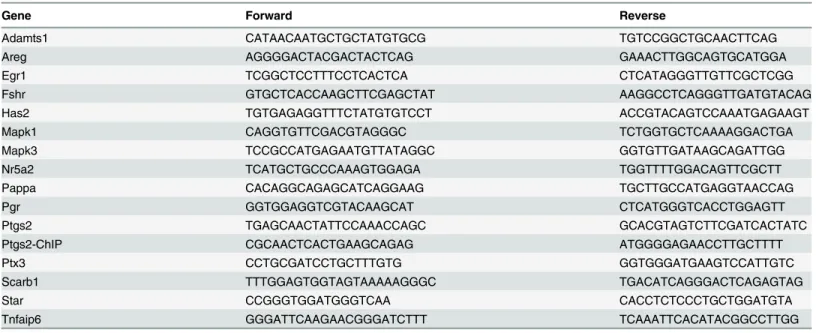
We first determined whether Map2k-inhibitor could be used as pharmacological inhibitor of granulosa cell Mapk3/1 activity during hCG-induced preovulatory follicle maturation in mice. Treatment with the inhbitor at 2h before hCG stimulation dramatically reduced the relative abundance of phospho-Mapk3/1 in granulosa cells of ovulating follicles compared to vehicle treatment (Fig. 1).
Inhibition of Mapk3/1 activity in preovulatory follicles abrogates ovulation
Pharmacological inhibition of Mapk3/1 activity does not inhibit global
transcription in granulosa cells, nor does it cause toxicity in mice
To investigate the mechanisms underpinning anovulation in the absence of Mapk3/1 activity, we undertook molecular phenotyping of granulosa cells collected at 0, 1 and 4h post-hCG in control and Map2k-inhibitor treated mice. Even though Map2k-inhibitor completely abol-ished Mapk3/1 activity in granulosa cells, it did not alter the relative mRNA abundance of
Mapk3/1in granulosa cells (Fig. 3A). Expression pattern ofFshrandNr5a2from 0h to 4h post-hCG was similar in granulosa cells of both control and Map2k-inhibitor treated mice (Fig. 3B). Interestingly, relative mRNA abundance ofScarb1andPappaincreased from 0h to 4h post-hCG in granulosa cells of both groups of mice (Fig. 3C). Further administration of ve-hicle or Map2k-inhibitor to mice did not produce any general signs of toxicity. These mice were active and appeared healthy upon physical examination. Paraffin sections of the liver and kidney of vehicle or Map2k-inhibitor treated mice were similar histologically and did not show any obvious signs of toxicity at 18h post-hCG (data not shown). Histologically ovary from Vehicle or Map2k-inhibitor treated mice did not showed any signs of toxicity, except ab-sence of ovulation.
Expression of hCG-induced ovulatory genes is decreased in granulosa
cells lacking Mapk3/1 activity
Expression patterns of the genes implicated in the process of ovulation in vehicle and Map2k-inhibitor treated mice are shown inFig. 4. One of the most important genes induced in granu-losa cells by preovulatory LH surge is the prostaglandin synthase 2 (Ptgs2), which is critical for both follicular rupture and cumulus expansion [18–20]. At 4h post-hCG a 545-fold in-crease inPtgs2mRNA was observed in granulosa cells of vehicle treated mice, but such a dra-matic increase was absent (P<0.001) in granulosa cells of Map2k-inhibitor treated mice. The
two other genes that play an important role in follicular rupture are progesterone receptor (Pgr) and A disintegrin and metalloproteinase with thrombospondin motifs 1 (Adamts1). In granulosa cells of vehicle treated micePgrmRNA was induced by 876-fold at 4h post-hCG compared to 0h hCG. Such a remarkable induction was completely absent in Map2k-inhibitor treated mice (P<0.001). Likewise, in Map2k-inhibitor treated mice, lack of Mapk3/1 activity
abolished (P<0.001) a 9-fold increase inAdamts1mRNA observed in granulosa cells of
Fig 1. Transient inhibition of hCG induced Mapk 3/1 activity by Map2k inhibitor treatment during ovarian superstimulation protocol.Granulosa cells collected by follicular puncture from mice treated with PD0325901 showed absence of phosphorylation of Mapk3/1 at Thr302/Tyr204 compared to vehicle treated mice (n = 3 mice/group/time point).
vehicle treated mice. Apart from Ptgs2, genes that mediate cumulus expansion include Hya-luronan synthase 2 (Has2), TNFα-induced protein 6 (Tnfaip6) and pentraxin 3 (Ptx3). In
ve-hicle treated mice increase inHas2mRNA abundance was 8-fold at 4h post-hCG. Map2k-inhibitor treatment abrogated such an increase in the mRNA expression ofHas2at 4h hCG (P<0.001). Similarly, 822-fold increase inPtx3and 476-fold increase inTnfaip6mRNA
ex-pressions seen in granulosa cells of vehicle treated mice was absent (P<0.001) in
Map2k-in-hibitor treated mice. Epidermal growth Factor (EGF)-like growth factors namelyAreg
(amphiregulin),Ereg(Epiregulin) andBtc(Betacellulin) have been implicated in oocyte mei-otic maturation [3], of which we examined the expression pattern ofAreg. Expression ofAreg
mRNA was highest at 1h hCG in vehicle treated granulosa cells, while its induction was re-duced (P<0.05) by 50% in Map2k-inhibitor treated granulosa cells.Cebpbhas been implicated
in regulation of the terminal differentiation of granulosa cells during ovulation[6]. In vehicle treated mice granulosa cellsCebpbmRNA expression was highest at 1h hCG and it was down-regulated (P<0.001) in Map2k-inhibitor treated mice.
Fig 2. Inhibition of hCG induced cumulus expansion, oocyte maturation and follicular rupture due to Map2k inhibitor treatment.Treatment of immature mice with single dose of 25μg/g of PD0325901 resulted in anovulation compared to vehicle treated mice (A) during superovulation (n = 5 mice/ group). This anovulation as evidenced by trapped GV stage oocytes with compact cumulus cells (C&D) inside the preovulatory follicles at 18h hCG (n = 5 mice/ group). Vehicle treated mice showed well-developed corpus luteum (B) evidencing follicular rupture, ovulation and luteinization. CL—corpus luteum;
GV—germinal vesicle.
Inhibition of Mapk3/1 activity reduces the expression of transcription
factor Egr1
Of the major transcription factors involved in ovulation mentioned in the introduction, Nr5a2 was not affected by Mapk3/1 inhibition (Fig. 3);Pgrwas not expressed at 1h (Fig. 4), and Cebpb appears to be dispensable for early events of preovulatory LH signaling [6]. Therefore, we asked the question as to which transcription factor could mediate Mapk3/1 regulated tran-scription of LH-induced genes like Ptgs2. As it has been well established that LH/hCG induces
Fig 3. Map2k inhibitor treatment does not shutdown the global transcription in granulosa cells.Treatment of immature mice with single dose of 25μg/g of PD0325901 did not alter relative mRNA abundance of Mapk1 and Mapk3 (A). Expression of known hCG regulated genes likeFshrandNr5a2(B) as well as hCG induced genes likeScarb1andPappa(C) were also not altered in PD0325901 treated mice (n = 3mice/group/time point).
Fig 4. Map2k inhibitor treatment reduces the expression of hCG induced ovulatory genes.Treatment of immature mice with single dose of 25μg/g of PD0325901 inhibited the expression of hCG-induced genes involved in follicular rupture likePtgs2,Pgr,Adamts1; genes involved in cumulus expansion like Has2,Ptx3,Tnfaip6; gene involved in oocyte meiotic maturation like Areg and luteinization likeCebpb(n = 3mice/group/time point). Mapk3/1 regulated LH induced genes likePtgs2,Ptx3 and Tnfaip6were not regulated by Cebpb.
Egr1 expression in granulosa cells during ovulation [21,22], we hypothesized that this Mapk3/1 activity would be indispensible for the expression of the transcription factor, early growth re-sponse-factor 1 (Egr1). Indeed, inhibition of Mapk3/1 activity abolished (P<0.001)
LH-in-duced mRNA expression ofEgr1at 1h post-hCG as compared to vehicle treatment (Fig. 5A). Confirming these mRNA data, relative abundance of Egr1 protein was remarkably absent in granulosa cells lacking Mapk3/1 activity at 1h and 4h post-hCG as compared to control granu-losa cells (Fig. 5B).
Egr1 binds to promoter regions of
Ptgs2
In light of the data describe above, the obvious question was whether Egr1 acts as a down-stream effector of Mapk3/1 for induction of the ovulatory genes such asPtgs2. To address this question, we performed ChIP analysis using Egr-1specific antibody. Because ChIP assays re-quire large number of cells, we used a mouse granulosa cell line, GRMO2 cells. First, we
Fig 5. Transient inhibition of Mapk3/1 reduces transcription and translation of transcription factor Egr1.A) Treatment of immature mice with single dose of 25μg/g of PD0325901 inhibited the expression of hCG-induced expression ofEgr11h and 4h hCG. B) Abundance of Egr1 protein in granulosa cells of the inhibitor and vehicle treated mice. Actb was used as loading control.
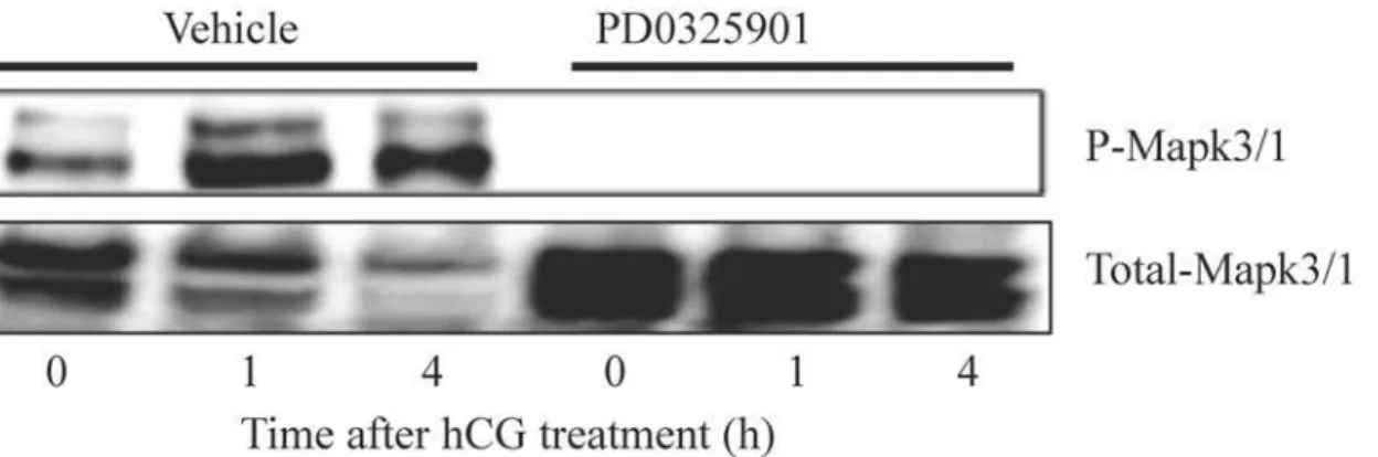
characterized gene expression pattern of GRMO2 cells in response to cAMP. GRMO2 cells stimulated with cAMP and granulosa cells from mice treated with hCG showed similar relative expression pattern genes likeStar,Ptgs2andEgr1(Fig. 6A). To determine whether Egr1 binds to the promoters of ovulatory genes, we performed ChIP and qPCR using cAMP stimulated GRMO2 cells. Immunoprecipitation with Egr1-specific antibody showed that cAMP treatment of GRMO2 cells for 4 h increased the enrichment of the promoter ofPtgs2(region-159 to -33 relative to the transcription start site) compared to GRMO2 cells that were not treated with cAMP (Fig. 6B). However, there was no significant increase in amplification of the regions in GRMO2 cells with or with out cAMP treatment when immunprecipitation was done using rab-bit IgG. These data clearly indicated that Egr1 is recruited to the promoter region ofPtgs2in re-sponse to cAMP treatment that is analogous to LH stimulation.
Knockdown of Egr1 reduces
Ptgs2
transcript abundance
To further demonstrate the importance of Egr1 forPtgs2expression, we used siRNA to knock-down Egr1 expression in mouse primary granulosa cells. Treatment with forskolin and PMA (Fo+PMA) dramatically induced the expression of ovulatory genes includingEgr1andPtgs2
when compared to medium-treated granulosa cells (data not shown). Immunoblot assays re-vealed that inhibition of Mapk3/1 activity using PD0325901 abolished Ptgs2 induction by treatment with Fo+PMA (Fig. 7A). Treatment of primary granulosa cells withEgr1-siRNA re-duced Egr1 mRNA and protein abundance compared to levels of control siRNA treated cells (Fig. 7B). Relative mRNA levels of Ptgs2 were 55% lowerEgr1-siRNA treated granulosa cells as compared to control siRNA treated cells (Fig. 7B).
Discussion
In the present study we show that transient pharmacological inhibition of Mapk3/1 activity using a single dose of the Map2k inhibitor, PD0325901 during preovulatory follicle maturation resulted in anovulation. This phenotype is similar to the one observed in granulosa-specific
Mapk3/1KO mice [5]. Our results confirm that this anovulatory phenotype is due to downre-gulation of numerous genes involved in ovulation. The novel aspect this study is that we have identified the Egr1 as the downstream transcription factor that mediates transcription of genes involved in follicular rupture namely,Ptgs2andAdamts1.
Ovulation is a process of release of oocyte through rupture of preovulatory follicles[13], regulated by the preovulatory LH-surge. This complex process involves a remarkable gene ex-pression program initiated by LH through multiple signaling pathways and numerous tran-scription factors. Mapk3/1 are highly conserved Ser/Thr kinases, which regulate cellar proliferation and differentiation [23] by modulating transcription, translation and post trans-lational modifications of their targets [24]. A recent study [5] demonstrated, using granulosa-specificMapk3/1-KO mice, that Mapk3/1 are critical for LH signaling in granulosa cells during ovulation. That study showed Cebpb as a downstream transcription factor responsible for sev-eral Mapk3/1 target genes.
Fig 6. A) Comparison of expression profile of ovulatory genes in granulosa cells from hCG treated mice and cAMP treated GRMO2 cells. Expression profiles of ovulatory genes likeStar,Ptgs2andEgr1were similar at 0h, 1h and 4h after hCG or cAMP treatment in granulosa cells or cultured GRMO2 cells respectively. B) Binding of Egr1 transcription factor to Ptgs2 promoter region (-159 to -33 relative to the transcription start site). Chromatin immunoprecipitation and qPCR using 4h cAMP treated GRMO2 cells showed the enrichment of promoter region containing Egr1 binding site (identified by bioinformatic analysis) in
immunoprecipitants of Egr1 antibody indicating that Egr1 binds to Ptgs2 promoter. Rabbit IgG was used as a control antibody.
Fig 7. Inhibition of Mapk3/1 activity and knockdown of Egr1 results in reduced induction of Ptgs2 in primary granulosa cells in vitro.A) Treatment with PD0325901 decreased Fo+PMA induced increases in the abundance of Ptgs2 protein relative to vehicle treated primary granulosa cells. B) Pre-treatment with Egr1-siRNA inhibited Fo+PMA induced expression of Egr1 when compared to control siRNA treatment in primary granulosa cells in vitro. This was associated with reduced abundance of Pgts2 transcript in Egr1-siRNA treated cells. Fo—forskolin; PMA—phorbol-12-myristate.
indicating the Pappa may be important for ovulation. Given that both Scarb1 and Pappa, were normally induced in Map2k-inhibitor treated mice, anovulation in these mice does not appear to be due to global abrogation of LH signaling. Indeed, it was previously reported that
PD0325901 treatment at 25 ug/g dose, similar to the dose used in this study, did not have any toxic effects [10,11]. Therefore, it is possible to conclude that inhibition of Mapk3/1 activity in granulosa cells resulted in anovulation through inhibition of a specific set of gene involved in the ovulatory processes.
Genes that are induced by hCG and implicated in ovulatory process were remarkably down-regulated in granulosa cells of Map2k-inhibitor treated mice. Similar downregulation of multi-ple genes including,Ptgs2,PgrandHas2was reported in granulosa-specificMapk3/1KO mice [5]. Ptgs2 appears to play a pleiotropic role during ovulation as ovaries ofPtgs2null mice pre-sented absence of CLs, compromised cumulus expansion but normal follicle development with oocyte maturation [18]. On the other hand, Adamts1 and Pgr have been implicated mainly in follicular rupture as mice lackingAdamts1[28] orPgr[9] showed ovulatory defects with nor-mal cumulus expansion and luteinization. Even pharmacological inhibition of Pgr using RU486 significantly decreased the number of ovulations [29]. Thus in light of theses reports, our data of lack on LH-induced expression ofPtgs2,Adamts1andPgras a result of inhibition of Mapk3/1 activity indicate that Mapk3/1 regulate follicular rupture through transcriptional regulation of the aforementioned genes in granulosa cells.
In the present study Map2k-inhibitor treatment downregulated hCG-induced expression of
Has2,Tnfaip6andPtx3suggesting that Mapk3/1 play a critical role in cumulus expansion through transcriptional regulation of the genes involved in the process. Likewise, granulosa-spe-cificMapk3/1KO mice also showed down regulated cumulus expansion after hCG stimulation. This conclusion is well supported by observations that cumulus expansion is severely compro-mised in mice null forTnfaip6[30], Ptx3 [31] andHas2[32]. Further, inhibition of Mapk3/1 downregulated hCG-induced expression ofAregandBtc, which are important for cumulus ex-pansion and oocyte meiotic maturation. Indeed, pharmacologic inhibition of Mapk3/1 using U0126 reduced the expression of LH-inducedAregandEregin luteinized human granulosa cells [1]. Also, U0126 treatment inhibited oocyte maturation in cultured large antral follicles [33]. Overall, our mRNA expression data confirm the importance of Mapk3/1 activity in the transcriptional regulation of ovulation-related genes in granulosa cells of ovulating follicles. Therefore, it is important to determine the transcription factors that act as downstream effec-tors of Mapk3/1 signaling in order to decipher the molecular mechanisms of the regulation of ovulation by this pathway.
Granulosa-specificMapk3/1KO mice showed decreased expression of Cebpb, which was proposed to be one of the downstream effectors of Mapk3/1 signaling in regulating ovulation [5]. It was further demonstrated that granulosa-specific deletion ofCebpa/bgenes resulted in complete anovulation [6], confirming the mediatory role of these transcription factors in Mapk3/1 signaling. However, this study also revealed that Cebpa/b accounted for 19% of Mapk3/1-regulated genes during early hours of preovulatory differentiation. Most importantly, LH-induced genes likeAreg,Ereg,Ptgs2,Tnfaip6andPtex3were unaffected in the absence of Cebpa/b in granulosa cells [6]. We also observed downregulation ofCebpbexpression, which could not explain the dramatic perturbation of the ovulatory gene expression in Map2k-inhibi-tor treated mice. Therefore, our results along with the genetic studies above clearly demonstrate that Mapk3/1 signaling is complex and may involve several other transcription factors as downstream effectors to regulate gene expression program during ovulation.
not corpus luteum [8,34], indicating that it may play a significant role in ovulation. Multiple signaling pathways including Mapk, protein kinase A and C signaling pathways are known to induceEgr1expression [35]. Most importantly, LH/hCG can transiently induceEgr1 exclusive-ly in granulosa cells preovulatory follicles mediating proliferation and/or differentiation during follicular growth, ovulation and luteinization [21,22]. In line with our hypothesis, inhibition of Mapk3/1 activity resulted in remarkable downregulation of hCG-induced Egr1 mRNA and protein. These results clearly indicate that Mapk3/1 activity is indispensable for LH-induced Egr1 expression in granulosa cells.
Several lines of evidence show that Mapk pathway is critical forEgr1expression in multiple cell types. Inhibition of Mapk3/1 pathway by Map2k inhibitor PD98059, but not Mapk14 or PI3K pathway, decreased hypoxia induced Egr1 expression in mouse and human alveolar epi-thelial cells [36]. In bovine luteal cells PGF2αtreatment inducedEGR1through Mapk3/1
path-way and PD098059 challenge inhibited thisEGR1induction [37]. In rat granulosa cells hCG-induced Egr1 expression was co-regulated by Mapk3/1, cAMP response element binding pro-tein (Creb) and Egr1 itself [21]. However, Creb activity was not altered in granulosa cells null forMapk3/1[5]. Similarly, inhibition of Mapk3/1 activity in the present study showed com-plete downregulation of Egr1 despite unalteredCrebexpression (data not shown). Taken to-gether, these data suggest that Mapk3/1 signaling may be more critical than the classical cAMP/Creb pathway for LH-induced Egr1 expression in granulosa cells. Further it has been shown that Egr1 expression is regulated by the transcription factor, serum response factor (Srf) in multiple cell types including granulosa cells [28], gonadotropes [38] and endothelial cells [39]. Since Srf is a known target of Mapk3/1 [40], it is possible to speculate from our data that hCG-induced Egr1 expression in granulosa cells is requires Mapk3/1 kinase activity and the transcription factor Srf during ovulation.
To further link Mapk3/1 dependent Egr1 expression to genes important for follicle rupture, we hypothesized that Egr1 regulates LH-induced transcription of Ptgs2. To test this hypothesis we first used cAMP primed GRMO2 cells. GRMO2 cells have been previously used [16] to study LH-stimulated events in granulosa cells. Systematic analyses revealed that they faithfully recapitulate LH-induced gene expression program in response to cAMP challenge. ChIP analy-ses using Egr1 antibody clearly demonstrated that Egr1 binds to the promoter ofPtgs2. Inhibi-tion of Mapk3/1 activity in primary mouse granulosa cells in vitro resulted in reduced
expression of Ptgs2 confirming that Mak3/1 activity is indispensable for Ptgs2 induction dur-ing ovulation. Finally, inhibition of Egr1 induction usdur-ing siRNA technology resulted in reduced expression of Ptgs2 in primary granulosa cells, further confirming that Egr1 regulates Ptgs2 ex-pression in granulosa cells. Our results are further supported by a previous study, which showed that overexpression of EGR1 in bovine granulosa cells increased expression ofPTGS2
expression [41]. Even thoughEgr1KO mice are known to be infertile along with multiple ovar-ian defects [8], Egr1 target genes have not been explored. With our study demonstrating regu-lation of Ptgs2 expression by Egr1, it would be very interesting to explore these genes in granulosa cellsEgr1KO mice.
here demonstrate that single dose of PD0325901 has an overwhelming inhibitory effect on ovu-lation in superovulated mice. These dramatic results warrant further detailed studies to test the effect of such inhibitors on ovarian function.
Acknowledgments
We thank Profs. Sarah Kimmins, Jaswinder Singh and Luis Agellon for allowing us to use their laboratory facilities for this study. We also thank Ms. Eliza Rossi Komninou, Ms. Melissa Pan-sera, Ms. Yasmin Schuermann, Ms. Lisa Dupuis and Ms. Maaike Hibbein-Kirby for excellent technical assistance.
Author Contributions
Conceived and designed the experiments: DS NG VB RD. Performed the experiments: DS EB. Analyzed the data: DS NG PPR VB RD. Contributed reagents/materials/analysis tools: NG PPR VB RD. Wrote the paper: DS RD.
References
1. Ben-Ami I, Armon L, Freimann S, Strassburger D, Ron-El R, Amsterdam A. (2009) EGF-like growth fac-tors as LH mediafac-tors in the human corpus luteum. Hum Reprod 24: 176–184. doi:10.1093/humrep/
den359PMID:18835871
Fig 8. Transcriptional regulation of ovulatory genes by Mapk3/1 through Egr1 transcription factor. Preovulatory LH surge induced Mapk3/1 activity regulates transcription of ovulatory genes such asPtgs2 trough transcription factor Egr1. Inhibition of this LH-induced Mapk3/1 activity by Map2k inhibitor PD0325901 results in anovulation.
2. Ashkenazi H, Cao X, Motola S, Popliker M, Conti M, Tsafriri A. (2005) Epidermal growth factor family members: endogenous mediators of the ovulatory response. Endocrinology 146: 77–84. PMID:
15459120
3. Park JY, Su YQ, Ariga M, Law E, Jin SL, Conti M. (2004) EGF-like growth factors as mediators of LH ac-tion in the ovulatory follicle. Science 303: 682–684. PMID:14726596
4. Fan HY, Liu Z, Cahill N, Richards JS (2008) Targeted disruption of Pten in ovarian granulosa cells en-hances ovulation and extends the life span of luteal cells. Mol Endocrinol 22: 2128–2140. doi:10.1210/
me.2008-0095PMID:18606860
5. Fan HY, Liu Z, Shimada M, Sterneck E, Johnson PF, Hedrick SM, et al. (2009) MAPK3/1 (ERK1/2) in ovarian granulosa cells are essential for female fertility. Science 324: 938–941. doi:10.1126/science.
1171396PMID:19443782
6. Fan HY, Liu Z, Johnson PF, Richards JS (2011) CCAAT/Enhancer-Binding Proteins (C/EBP)-{alpha} and-{beta} Are Essential for Ovulation, Luteinization, and the Expression of Key Target Genes. Mol Endocrinol 25: 253–268. doi:10.1210/me.2010-0318PMID:21177758
7. Duggavathi R, Volle DH, Mataki C, Antal MC, Messaddeq N, Auwerx J, et al. (2008) Liver receptor ho-molog 1 is essential for ovulation. Genes Dev 22: 1871–1876. doi:10.1101/gad.472008PMID:
18628394
8. Topilko P, Schneider-Maunoury S, Levi G, Trembleau A, Gourdji D, Driancourt MA, et al. (1998) Multi-ple pituitary and ovarian defects in Krox-24 (NGFI-A, Egr-1)-targeted mice. Mol Endocrinol 12: 107–
122. PMID:9440815
9. Lydon JP, DeMayo FJ, Funk CR, Mani SK, Hughes AR, Montgomery CA Jr, et al. (1995) Mice lacking progesterone receptor exhibit pleiotropic reproductive abnormalities. Genes Dev 9: 2266–2278. PMID:
7557380
10. Barrett SD, Bridges AJ, Dudley DT, Saltiel AR, Fergus JH, Flamme CM, et al. (2008) The discovery of the benzhydroxamate MEK inhibitors CI-1040 and PD 0325901. Bioorg Med Chem Lett 18: 6501–
6504. doi:10.1016/j.bmcl.2008.10.054PMID:18952427
11. Solit DB, Garraway LA, Pratilas CA, Sawai A, Getz G, Basso A, et al. (2006) BRAF mutation predicts sensitivity to MEK inhibition. Nature 439: 358–362. PMID:16273091
12. Wauson EM, Guerra ML, Barylko B, Albanesi JP, Cobb MH (2013) Off-target effects of MEK inhibitors. Biochemistry 52: 5164–5166. doi:10.1021/bi4007644PMID:23848362
13. Richards JS, Russell DL, Ochsner S, Espey LL (2002) OVULATION: New Dimensions and New Regu-lators of the Inflammatory-Like Response. Annual Review of Physiology 64: 69–92. PMID:11826264
14. Dupuis L, Schuermann Y, Cohen T, Siddappa D, Kalaiselvanraja A, Pansera M, et al. (2014) Role of leptin receptors in granulosa cells during ovulation. Reproduction 147: 221–229. doi:
10.1530/REP-13-0356PMID:24256641
15. Deroo BJ, Rodriguez KF, Couse JF, Hamilton KJ, Collins JB, Grissom SF, et al. (2009) Estrogen recep-tor beta is required for optimal cAMP production in mouse granulosa cells. Molecular endocrinology 23: 955–965. doi:10.1210/me.2008-0213PMID:19324971
16. Weck J, Mayo KE (2006) Switching of NR5A proteins associated with the inhibin alpha-subunit gene promoter after activation of the gene in granulosa cells. Mol Endocrinol 20: 1090–1103. PMID:
16423880
17. Svotelis A, Bianco S, Madore J, Huppe G, Nordell-Markovits A, Mes-Masson AM, et al. (2011) H3K27 demethylation by JMJD3 at a poised enhancer of anti-apoptotic gene BCL2 determines ERalpha ligand dependency. EMBO J 30: 3947–3961. doi:10.1038/emboj.2011.284PMID:21841772
18. Dinchuk JE, Car BD, Focht RJ, Johnston JJ, Jaffee BD, Covington MB, et al. (1995) Renal abnormali-ties and an altered inflammatory response in mice lacking cyclooxygenase II. Nature 378: 406–409.
PMID:7477380
19. Lim H, Paria BC, Das SK, Dinchuk JE, Langenbach R, Trzaskos JM, et al. (1997) Multiple female repro-ductive failures in cyclooxygenase 2-deficient mice. Cell 91: 197–208. PMID:9346237
20. Micu MC, Micu R, Ostensen M (2011) Luteinized unruptured follicle syndrome increased by inactive disease and selective cyclooxygenase 2 inhibitors in women with inflammatory arthropathies. Arthritis Care Res (Hoboken) 63: 1334–1338. doi:10.1002/acr.20510PMID:21618455
21. Russell DL, Doyle KM, Gonzales-Robayna I, Pipaon C, Richards JS (2003) Egr-1 induction in rat granu-losa cells by follicle-stimulating hormone and luteinizing hormone: combinatorial regulation by tran-scription factors cyclic adenosine 3',5'-monophosphate regulatory element binding protein, serum response factor, sp1, and early growth response factor-1. Mol Endocrinol 17: 520–533. PMID:
22. Espey LL, Ujioka T, Russell DL, Skelsey M, Vladu B, Robker RL, et al. (2000) Induction of early growth response protein-1 gene expression in the rat ovary in response to an ovulatory dose of human chorion-ic gonadotropin. Endocrinology 141: 2385–2391. PMID:10875238
23. Ramos JW (2008) The regulation of extracellular signal-regulated kinase (ERK) in mammalian cells. Int J Biochem Cell Biol 40: 2707–2719. doi:10.1016/j.biocel.2008.04.009PMID:18562239
24. Sugden PH, Clerk A (1997) Regulation of the ERK subgroup of MAP kinase cascades through G pro-tein-coupled receptors. Cell Signal 9: 337–351. PMID:9376213
25. Jimenez LM, Binelli M, Bertolin K, Pelletier RM, Murphy BD (2010) Scavenger receptor-B1 and luteal function in mice. J Lipid Res 51: 2362–2371. doi:10.1194/jlr.M006973PMID:20404351
26. Cherian-Shaw M, Puttabyatappa M, Greason E, Rodriguez A, VandeVoort CA, Chaffin CL. (2009) Ex-pression of scavenger receptor-BI and low-density lipoprotein receptor and differential use of lipopro-teins to support early steroidogenesis in luteinizing macaque granulosa cells. Endocrinology 150: 957–
965. doi:10.1210/en.2008-0619PMID:18832102
27. Nyegaard M, Overgaard MT, Su YQ, Hamilton AE, Kwintkiewicz J, Hsieh M, et al. (2010) Lack of func-tional pregnancy-associated plasma protein-A (PAPPA) compromises mouse ovarian steroidogenesis and female fertility. Biol Reprod 82: 1129–1138. doi:10.1095/biolreprod.109.079517PMID:20130263
28. Mittaz L, Russell DL, Wilson T, Brasted M, Tkalcevic J, Salamonsen LA, et al. (2004) Adamts-1 is es-sential for the development and function of the urogenital system. Biol Reprod 70: 1096–1105. PMID:
14668204
29. Iwamasa J, Shibata S, Tanaka N, Matsuura K, Okamura H (1992) The relationship between ovarian progesterone and proteolytic enzyme activity during ovulation in the gonadotropin-treated immature rat. Biol Reprod 46: 309–313. PMID:1536908
30. Fulop C, Szanto S, Mukhopadhyay D, Bardos T, Kamath RV, Rugg MS, et al. (2003) Impaired cumulus mucification and female sterility in tumor necrosis factor-induced protein-6 deficient mice. Development 130: 2253–2261. PMID:12668637
31. Varani S, Elvin JA, Yan C, DeMayo J, DeMayo FJ, Horton HF, et al. (2002) Knockout of pentraxin 3, a downstream target of growth differentiation factor-9, causes female subfertility. Mol Endocrinol 16: 1154–1167. PMID:12040004
32. Su YQ, Sugiura K, Eppig JJ (2009) Mouse oocyte control of granulosa cell development and function: paracrine regulation of cumulus cell metabolism. Semin Reprod Med 27: 32–42. doi:
10.1055/s-0028-1108008PMID:19197803
33. Su YQ, Denegre JM, Wigglesworth K, Pendola FL, O'Brien MJ, Eppig JJ. (2003) Oocyte-dependent ac-tivation of mitogen-activated protein kinase (ERK1/2) in cumulus cells is required for the maturation of the mouse oocyte-cumulus cell complex. Dev Biol 263: 126–138. PMID:14568551
34. Lee GH, Proenca R, Montez JM, Carroll KM, Darvishzadeh JG, Eppig JJ. (1996) Abnormal splicing of the leptin receptor in diabetic mice. Nature 379: 632–635. PMID:8628397
35. Pagel JI, Deindl E (2011) Early growth response 1—a transcription factor in the crossfire of signal
trans-duction cascades. Indian J Biochem Biophys 48: 226–235. PMID:22053691
36. Jones N, Agani FH (2003) Hyperoxia induces Egr-1 expression through activation of extracellular sig-nal-regulated kinase 1/2 pathway. J Cell Physiol 196: 326–333. PMID:12811826
37. Hou X, Arvisais EW, Jiang C, Chen DB, Roy SK, Pate JL, et al. (2008) Prostaglandin F2alpha stimu-lates the expression and secretion of transforming growth factor B1 via induction of the early growth re-sponse 1 gene (EGR1) in the bovine corpus luteum. Mol Endocrinol 22: 403–414. PMID:17916653
38. Duan WR, Ito M, Park Y, Maizels ET, Hunzicker-Dunn M, Jameson JL. (2002) GnRH regulates early growth response protein 1 transcription through multiple promoter elements. Mol Endocrinol 16: 221–
233. PMID:11818496
39. Wu SQ, Minami T, Donovan DJ, Aird WC (2002) The proximal serum response element in the Egr-1 promoter mediates response to thrombin in primary human endothelial cells. Blood 100: 4454–4461.
PMID:12393577
40. Yoon S, Seger R (2006) The extracellular signal-regulated kinase: multiple substrates regulate diverse cellular functions. Growth Factors 24: 21–44. PMID:16393692