Inhibition of DNA topoisomerase I activity and induction of apoptosis by
thiazacridine derivatives
Francisco W.A. Barros
a, Daniel P. Bezerra
b,⁎
, Paulo M.P. Ferreira
c, Bruno C. Cavalcanti
a, Teresinha G. Silva
d,
Marina G.R. Pitta
d, Maria do C.A. de Lima
d, Suely L. Galdino
d, Ivan da R. Pitta
d, Letícia V. Costa-Lotufo
a,
Manoel O. Moraes
a, Rommel R. Burbano
e, Temenouga N. Guecheva
f,
João A.P. Henriques
f, Cláudia Pessoa
a,⁎
aDepartment of Physiology and Pharmacology, School of Medicine, Federal University of Ceará, Fortaleza, Ceará, Brazil bDepartment of Physiology, Federal University of Sergipe, São Cristóvão, Sergipe, Brazil
cDepartment of Biological Sciences, Federal University of Piauí, Picos, Piauí, Brazil dDepartment of Antibiotics, Federal, University of Pernambuco, Recife, Pernembuco, Brazil eInstitute of Biological Sciences, Federal University of Pará, Belém, Pará, Brazil
fBiotechnology Center, Federal University of Rio Grande do Sul, Porto Alegre, Rio Grande do Sul, Brazil
a b s t r a c t
a r t i c l e
i n f o
Article history:
Received 28 August 2012 Revised 25 December 2012 Accepted 10 January 2013 Available online 21 January 2013
Keywords:
Acridine Thiazolidine Thiazacridine DNA topoisomerase I Apoptosis
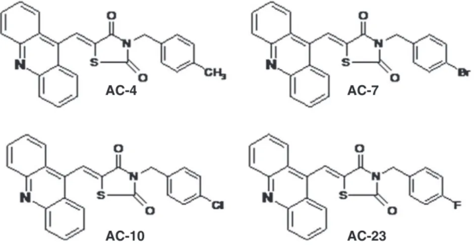
Thiazacridine derivatives (ATZD) are a novel class of cytotoxic agents that combine an acridine and thiazoli-dine nucleus. In this study, the cytotoxic action of four ATZD were tested in human colon carcinoma HCT-8 cells: (5Z)-5-acridin-9-ylmethylene-3-(4-methylbenzyl)-thiazolidine-2,4-dione—AC-4; (5ZE)-5-acridin-9-ylmethylene-3-(4-bromo-benzyl)-thiazolidine-2,4-dione—AC-7; (5Z)-5-(acridin-9-ylmethylene)-3-(4-chloro-benzyl)-1,3-thiazolidine-2,4-dione — AC-10; and (5ZE)-5-(acridin-9-ylmethylene)-3-(4-fl uoro-benzyl)-1,3-thiazolidine-2,4-dione — AC-23. All of the ATZD tested reduced the proliferation of HCT-8 cells in a concentration- and time-dependent manner. There were significant increases in internucleosomal DNA fragmentation without affecting membrane integrity. For morphological analyses, hematoxylin–eosin and acridine orange/ethidium bromide were used to stain HCT-8 cells treated with ATZD, which presented the typical hallmarks of apoptosis. ATZD also induced mitochondrial depolarisation and phosphatidylserine exposure and increased the activation of caspases 3/7 in HCT-8 cells, suggesting that this apoptotic cell death was caspase-dependent. In an assay usingSaccharomyces cerevisiaemutants with defects in DNA topoisomerases 1 and 3, the ATZD showed enhanced activity, suggesting an interaction between ATZD and DNA topoisomerase enzyme activity. In addition, ATZD inhibited DNA topoisomerase I action in a cell-free system. Interestingly, these ATZD did not cause genotoxicity or inhibit the telomerase activity in human lymphocyte cultures at the experimental levels tested. In conclusion, the ATZD inhibited the DNA topoisomerase I activity and induced tumour cell death through apoptotic pathways.
© 2013 Elsevier Inc. All rights reserved.
Introduction
Topoisomerases are enzymes that regulate the overwinding or underwinding of DNA. They relax DNA supercoiling and perform cat-alytic functions during replication and transcription. There are two types of topoisomerases: type I enzymes that cleave one strand of DNA; and type II enzymes that cleave both strands. Both types of topoisomerases are essential for mammalian cell survival. Therefore, DNA topoisomerases are important targets for the development of
cy-totoxic agents (Miao et al., 2007; Moukharskaya and Verschraegen,
2012; Pommier et al., 2010; Vos et al., 2011). Topoisomerases I and II
are important anticancer targets, and topoisomerase inhibitors such as camptothecin derivatives (e.g., topotecan and irinotecan), which are used clinically to inhibit the enzymatic activity of topoisomerase I (type I enzyme), and podophyllotoxin derivatives (e.g., etoposide and teniposide), which inhibit the enzymatic activity of topoisomerase II
(type II enzyme) (Hartmann and Lipp, 2006) are used to block cancer
growth.
Amsacrine (m-AMSA), an acridine derivative, was thefirst
syn-thetic topoisomerase inhibitor approved for clinical treatment.
Al-thoughm-AMSA is an intercalator and topoisomerase II inhibitor, its
metabolism has been associated with the production of free radicals,
which may cause serious harm to normal tissues (Belmont et al.,
2007; Blasiak et al., 2003; Ketron et al., 2012; Sebestik et al., 2007). A number of clinical and experimental studies have demonstrated that acridine and thiazolidine derivatives are promising cytotoxic
⁎ Corresponding authors. Fax: +55 85 3366 8333.
E-mail addresses:danielpbezerra@gmail.com(D.P. Bezerra),cpessoa@ufc.br
(C. Pessoa).
0041-008X/$–see front matter © 2013 Elsevier Inc. All rights reserved.
http://dx.doi.org/10.1016/j.taap.2013.01.010
Contents lists available atSciVerse ScienceDirect
Toxicology and Applied Pharmacology
agents. Recently, we described the synthesis of a novel class of cyto-toxic agents, thiazacridine derivatives (ATZD), that couple the
acridine and thiazolidine nucleus: (5Z
)-5-acridin-9-ylmethylene-3-(4-methylbenzyl)-thiazolidine-2,4-dione (AC-4); (5ZE
)-5-acridin-9-ylmethylene-3-(4-bromo-benzyl)-thiazolidine-2,4-dione (AC-7);
(5Z
)-5-(acridin-9-ylmethylene)-3-(4-chloro-benzyl)-1,3-thiazolidin-e-2,4-dione (AC-10); and (5ZE
)-5-(acridin-9-ylmethylene)-3-(4-fluoro-benzyl)-1,3-thiazolidine-2,4-dione (AC-23). The chemical
structures of these ATZD are illustrated inFig. 1; their ability to
inter-act with DNA was demonstrated using an electrochemical technique. These ATZD have demonstrated a solid tumour-selective cytotoxicity (Barros et al., 2012). Here, we study the mechanism of ATZD's
selec-tive cytotoxicity (AC-4,AC-7,AC-10andAC-23) in human colon
car-cinoma HCT-8 cells.
Material and methods
The synthesis of thiazacridine derivatives. The chemical data and
syn-thetic procedures for (5Z
)-5-acridin-9-ylmethylene-3-(4-methylbenzyl)-thiazolidine-2,4-dione (AC-4), (5ZE
)-5-acridin-9-ylmethylene-3-(4-bromo-benzyl)-thiazolidine-2,4-dione (AC-7), (5Z
)-5-(acridin-9-ylmeth-ylene)-3-(4-chloro-benzyl)-1,3-thiazolidine-2,4-dione (AC-10) and
(5ZE)-5-(acridin-9-ylmethylene)-3-(4-fl
uoro-benzyl)-1,3-thiazolidi-ne-2,4-dione (AC-23) are reported elsewhere (Barros et al., 2012;
Mourão et al., 2005; Silva et al., 2001). Thiazolidine-2,4-dione was
N-(3)-alkylated in the presence of potassium hydroxide, which
en-abled the thiazolidine potassium salt to react with the substituted benzylhalide in a hot alcohol medium. The thiazacridine derivatives were synthesised by the nucleophilic addition of substituted 3-benzyl-thiazolidine-diones on 3-acridin-9-yl-2-cyano-acrylic acid ethyl ester. The mechanisms of cytotoxic action for the thiazacridine
derivatives were studied as singleZ isomers forAC-4 and AC-10.
TheAC-7andAC-23compounds were studied as isomeric mixtures,
but theZisomer was the major stereoisomer.
Strains and media for the yeast assays. TheSaccharomyces cerevisiae
strains in this study were acquired from Euroscarf (European
Saccharo-myces cerevisiae Archive for Functional Analysis). The following S. cerevisiaegenotypes were used in this study: BY-4741 (MATa;his3Δ
1;leu2Δ0;met15Δ0;ura3Δ0);Top1Δ(YOL006c), same as BY4741 with YOL006c::kanMX4; Top3Δ (YLR234w), same as BY4741 with YLR234w::kanMX4. The media, solutions and buffers were prepared as
previously described (Burke et al., 2000). Complete medium (YPD),
containing 1% yeast extract, 2% peptone and 2% glucose was used for routine growth. The stationary-phase cultures were obtained by inocu-lating an isolated colony into liquid YPD medium and incubating the
culture at 28 °C for 72 h with shaking (for aeration). Cultures in the
ex-ponential phase were obtained by inoculating 5 × 10⁶cells/ml of the
stationary-phase YPD culture into fresh YPD medium at 28 °C for 2 h. The cell concentrations were determined in a Neubauer chamber using a light microscope (LO, Laboroptik GmbH, Bad Homburg, Hessen, Germany).
Cell lines and cell culture. The cytotoxicity of ATZD was evaluated using human colon carcinoma HCT-8 cells donated by the Children's Mercy Hospital, Kansas City, MO, USA. The cells were maintained in RPMI-1640 medium supplemented with 10% foetal bovine serum,
2 mM glutamine, 100μg/ml streptomycin and 100 U/ml penicillin.
The cells were kept in tissue-cultureflasks at 37 °C in a humidified
at-mosphere with 5% CO2and were harvested with a 0.15% trypsin–
0.08% EDTA, phosphate-buffered saline solution (PBS).
The following experiments were performed to determine ATZD's cy-totoxic mechanisms in HCT-8 cells. For all cell-based assays, the HCT-8
cells were seeded (0.7 × 10⁵cells/ml) and incubated overnight to
allow the cells to adhere to the plate surface. Then, the cells were
treat-ed for 12- and/or 24-h at concentrations of 2.5, 5 and/or 10μg/ml,
cor-responding to: 6.1, 12.2 and 24.4μM forAC-4; 5.3, 10.6 and 21.2μM for
AC-7; 5.8, 11.6 and 23.2μM forAC-10; 6.0, 12.1 and 24.1μM forAC-23, respectively. The trypan blue exclusion test was performed before each experiment described below to assess cell viability. The negative control was treated with the vehicle (0.1% DMSO) used for diluting the tested
substances. Amsacrine (m-AMSA, 0.3μg/ml [0.8μM], Sigma Chemical
Co. St Louis, MO, USA) or doxorubicin (0.3μg/ml [0.6μM], Sigma
Chem-ical Co. St Louis, MO, USA) was used as the positive control. The
con-centrations of ATZD used here were based on their IC50value in this
cell line (3.1μg/ml forAC-4, 5.3μg/ml forAC-7, 3.6μg/ml forAC-10
and 2.3μg/ml forAC-23) as previously described (Barros et al., 2012).
Trypan blue dye exclusion test. Cell proliferation was determined using the Trypan blue dye exclusion test. After each incubation peri-od, the cell proliferation was assessed. Cells that excluded trypan blue were counted using a Neubauer chamber.
BrdU incorporation assay. Twenty microliters of 5-bromo-20-deoxy-uridine (BrdU, 10 mM) was added to each well and incubated for 3 h at 37 °C before 24-h of drug exposure. To assess the amount of BrdU in-corporated into DNA, cells were harvested, transferred to cytospin slides (Shandon Southern Products Ltd., Sewickley Pennsylvania, USA) and allowed to dry for 2 h at room temperature. Cells that had incorporated BrdU were labelled by direct peroxidase immuno-cytochemistry using the chromogen diaminobenzidine. The slides were counterstained with hematoxylin, mounted and put under a
AC-4
AC-7
AC-10
AC-23
cover slip. A light microscopy (Olympus, Tokyo, Japan) was used to de-termine BrdU-positivity. Two hundred cells per sample were counted to determine the percent of BrdU-positive cells.
Morphological analyses using hematoxylin–eosin staining. Untreated or ATZD-treated HCT-8 cells were examined for morphological changes under a light microscopy (Metrimpex Hungary/PZO-Labimex Model Studar lab). To evaluate any alterations in morphology, cells from the cultures were harvested, transferred to a cytospin slide,
fixed with methanol for 30 s, and stained with hematoxylin–eosin.
Morphological analyses using afluorescence microscope. Cells were
pelleted and resuspended in 25μl of PBS. Then, 1μl of aqueous acridine
orange/ethidium bromide solution (AO/EB, 100μg/ml) was added and
the cells were observed under afluorescence microscope (Olympus,
Tokyo, Japan). Three hundred cells were counted per sample and
classi-fied as viable, apoptotic or necrotic (McGahon et al., 1995).
Cell membrane integrity. The integrity of the cell membrane was
evaluated using the exclusion of propidium iodide (2μg/ml, Sigma
Chemical Co. St Louis, MO, USA). Cellfluorescence was determined by
flow cytometry in a Guava EasyCyte Mini System cytometer using
CytoSoft 4.1 software (Guava Technologies, Hayward, California, USA). Five thousand events were evaluated per experiment and the cellular debris was omitted from the analysis.
Cell cycle distribution. The cells were harvested in a lysis solution
(citrate 0.1%, triton X-100 0.1% and propidium iodide 50μg/ml)
(Nicoletti et al., 1991), and the cellfluorescence was determined by
flow cytometry, as described above.
Measurement of the mitochondrial transmembrane potential. The mitochondrial transmembrane potential was determined by the
reten-tion of rhodamine 123 dye (Gorman et al., 1997; Sureda et al., 1997).
The cells were washed with PBS, incubated with rhodamine 123
(5μg/ml, Sigma Chemical Co. St Louis, MO, USA) at 37 °C for 15 min
in the dark and washed twice. The cells were then incubated again in
PBS at 37 °C for 30 min in the dark and theirfluorescence was
mea-sured byflow cytometry, as described above.
Annexin assay. Phosphatidylserine externalisation was analysed
byflow cytometry (Vermes et al., 1995). A Guava® Nexin Assay Kit
(Guava Technologies, Hayward, CA) determined which cells were ap-optotic (early apap-optotic + late apap-optotic). The cells were washed
twice with cold PBS and then re-suspended in 135μl of PBS with
5μl of 7-amino-actinomycin D (7-AAD) and 10μl of Annexin V–PE.
The cells were gently vortexed and incubated for 20 min at room
tem-perature (20–25 °C) in the dark. Afterwards, the cells were analysed
byflow cytometry, as described above.
Caspase 3/7 activation. Caspase 3/7 activity was analysed byflow cytometry using the Guava® EasyCyte Caspase 3/7 Kit (Guava Tech-nologies, Hayward, CA). The cells were incubated with Fluorescent Labelled Inhibitor of Caspases (FLICATM) and maintained for 1 h at
37 °C in a CO2incubator. After incubation, 80μl of wash buffer was
added and the cells were centrifuged at 2000 rpm for 5 min. The
resulting pellet was resuspended in 200μl of wash buffer and
centrifuged. The cells were then re-suspended in the working solution (propidium iodide and wash buffer) and analysed immediately using
flow cytometry, as described above.
Drop test assay to determine the sensitivity of mutant S. cerevisiae strains with defective topoisomerases. The drop test assay determined the
relative sensitivity of differentS. cerevisiaestrains to ATZD treatment.
The followingS. cerevisiaestrains were used: BY-4741, Top1Δ and
Top3Δ. Cells were treated with ATZD at concentrations of 50 and
100μg/ml and more, 4 dilutions 1:10 were performed. A suspension
of 2 × 10⁵cells/ml ofS. cerevisiaein the exponential phase was used.
An aliquot of 3μl of each dilution was added to plates containing
YEPD medium (YEL + agar). After 3–4 days of growth at 28 °C, the
plates were photographed.m-AMSA served as the positive control.
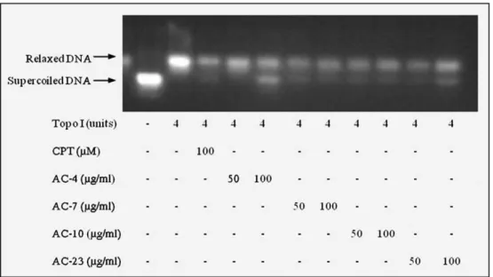
DNA relaxation assay. The inhibitory effects of ATZD on human DNA topoisomerase I were measured using a Topo I Drug Screening Kit (TopoGEN, Inc.). Supercoiled (Form I) plasmid DNA (250 ng) was in-cubated with human Topo I (4 units) at 37 °C for 30 min in relaxation buffer (10 mM Tris buffer pH 7.9, 1 mM EDTA, 0.15 M NaCl, 0.1% BSA, 0.1 mM spermidine and 5% glycerol) in the presence or absence of
ATZD (50 and 100μg/ml,final 20μl). The concentrations used were
based on the positive control indicated in this Kit. CPT (100μM)
served as the positive control. The reaction was terminated by the
addition of 10% SDS (2μl) and proteinase K (50μg/ml) and incubated
at 37 °C for 30 min. The DNA samples were added to the loading dyes
(2μl) and subjected to electrophoresis on a 1% agarose gel for 90 min
at room temperature and visualised with ethidium bromide.
Assessment of the genotoxic effect in human lymphocytes. A primary culture was obtained using a standard protocol and a Ficoll gradient. In addition, phytohemagglutinin (PHA) served as a mitogen to trigger cell division in T-lymphocytes. Peripheral blood was collected from
four (two women and two men) healthy donors, 19–30 years of age
with no history of smoking/drinking or chronic drug use. Venous blood (10 ml) was collected from each donor into heparinised vials. Lymphocytes were isolated with a Ficoll density gradient (Histopaque-1077; Sigma Diagnostics, Inc., St. Louis). The culture medium consisted of RPMI 1640 supplemented with 20% foetal bovine
serum, phytohemagglutinin (final concentration: 2%), 2 mM glutamine,
100 U/ml penicillin and 100μg/ml streptomycin at 37 °C with 5% CO2
(Berthold, 1981; Brown and Lawce, 1997; Hutchins and Steel, 1983). For all of the experiments, cell viability was performed using the Trypan Blue assay. Ninety percent of the cells had to be viable before starting the experiments.
Alkaline comet assay. The alkaline (pH> 13) version of the comet assay (Single Cell Gel Electrophoresis) was performed, as described by Singh et al. (1988) with minor modifications (Hartmann and Speit,
1997). The slides were prepared in duplicate and 100 cells were
screened per sample (50 cells from each duplicate slide) using afl
uores-cence microscope (Zeiss) equipped with a 515–560 nm excitationfilter,
a 590 nm barrierfilter, and a 40× objective. The cells were visually
scored and sorted intofive classes according to tail length: (1) class 0:
undamaged, without a tail; (2) class 1: with a tail shorter than the
diam-eter of the head (nucleus); (3) class 2: with a tail length 1–2× the
diam-eter of the head; (4) class 3: with a tail longer than 2× the diamdiam-eter of the head; and (5) class 4: comets with no heads. A value of damage index (DI) was assigned to each comet according to its class, using the
formula: DI=(0×n0) +(1×n1) +(2×n2) + (3×n3) + (4×n4), where
n=number of cells in each class analysed. The damage index ranged
from 0 (completely undamaged: 100 cells × 0) to 400 (with maximum damage: 100 cells× 4). DI was based on migration length and on the amount of DNA in the tail and was considered a sensitive measure of
DNA (Speit and Hartmann, 1999).
Chromosome aberration assay. We used naturally synchronised human peripheral blood lymphocytes with more than 95% of the cells
in the G0phase (Bender et al., 1988; Wojcik et al., 1996). Short-term
lymphocytes cultures, at a concentration of 0.3 × 10⁶cells/ml, were
ini-tiated according to a standard protocol (Preston et al., 1987). ATZD were
studied at different phases of the cell cycle based on the protocol
described byCavalcanti et al. (2008)with minor modifications.
Doxoru-bicin (0.3μg/ml) served as a positive control. In the experimental
stages were exposed, whilst it can be assumed that when ATZD was
added after 69 h, only cells in the G2stage were exposed. When ATZD
was added at the same time as the PHA stimulation (in culture start,
0 h), the cells were exposed in the G1stage. To obtain a sufficient
num-ber of analysable metaphases, colchicine was added at afinal
concen-tration of 0.0016%, 2 h prior to harvesting. The cells were harvested by centrifugation, treated with 0.075 M KCl at 37 °C for 20 min,
centrifuged andfixed in 1:3 (v/v) acetic acid:methanol. Finally, the
slides were prepared, air-dried and stained with a 3% Giemsa solution
(pH 6.8) for 8 min (Moorhead et al., 1960).
The slides were analysed with a light microscope; the structural and numerical CAs were examined during metaphase in the ATZD-treated cultures and the respective controls. The frequency of CAs (in 100 meta-phases per culture) and the mitotic index (MI, number of metameta-phases per 2.000 lymphocytes per culture) were determined.
Telomerase inhibition assay. The ability of ATZD to inhibit
telome-rase action was measured by determining telomere length usingfl
uo-rescence in situ hybridisation with probes to telomeric sequences
(TELO-FISH), as described byLansdorp (1995) and Lansdorp et al.
(1996). Short-term lymphocyte cultures were initiated according to
a standard protocol (Preston et al., 1987) and werefixed (methanol:
acetic acid, 3:1) on slides. The slides were hybridised with the pan telomeric Star FISH probe. The measurement of telomere length de-termined in each nucleus, was acquired using the image capturing software Applied Special Imaging analysis system. The images were
processed using the TFL-TELO software following the protocol (Poon
et al., 1999).
Statistical analysis. The data are presented as the means ± standard
error of the mean ofnexperiments. The differences among
experi-mental groups were compared using a one-way analysis of variance
(ANOVA) followed by a Newman–Keuls test (pb0.05). All analyses
were carried out using the GRAPHPAD programme (Intuitive Soft-ware for Science, San Diego, California, USA).
Results
Thiazacridine derivatives inhibit the proliferation of human colon carcinoma in HCT-8 cells
Human colon carcinoma HCT-8 cells were treated with 2.5, 5 and
10μg/ml of ATZD for 12- and/or 24-h and analysed in three different
assays (trypan blue dye exclusion, propidium iodide exclusion and
NC PC 2.5 5 10 2.5 5 10 2.5 5 10 2.5 5 10 0 5 10 15 20 25 30 35 AC-4
(µg/ml)
A
AC-7 AC-10 AC-23
*
*
*
C e lls num ber ( x 10 4 /m l)NC PC 2.5 5 10 2.5 5 10 2.5 5 10 2.5 5 10 0 5 10 15 20 25 30 35 AC-4
*
*
*
(µg/ml)
B
AC-7 AC-10 AC-23
*
*
*
*
*
*
*
*
*
*
C e lls num ber ( x 10 4/m l)NC PC 2.5 5 10 2.5 5 10 2.5 5 10 2.5 5 10 0 5 10 15 20 25 30 35 AC-4
(µg/ml)
C
AC-7 AC-10 AC-23
*
*
*
*
C e lls num ber ( x 10 4 /m l)NC PC 2.5 5 10 2.5 5 10 2.5 5 10 2.5 5 10 0 5 10 15 20 25 30 35 AC-4
*
*
*
(µg/ml)
D
AC-7 AC-10 AC-23
*
*
*
*
*
*
*
*
*
*
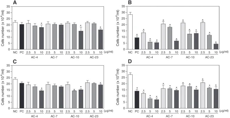
C e lls num b er ( x 10 4/m l)Fig. 2.The effect of thiazacridine derivatives on the proliferation of human colon carcinoma HCT-8 cells. A and B—the inhibition of cell proliferation was determined using the trypan blue dye exclusion method after 12- and 24-h incubations, respectively. C and D—the inhibition of cell proliferation was also determined usingflow cytometry and propidium iodide after 12- and 24-h incubations, respectively. The data are presented as the mean values ± S.E.M. from three independent experiments performed in duplicate. The negative control was treated with the vehicle (NC, 0.1% DMSO) that diluted the test substance. Amsacrine (PC,m-AMSA, 0.3μg/ml) was the positive control. For theflow cy-tometry analyses, 5000 events were analysed in each experiment. *,pb0.05 compared to the negative control using an ANOVA followed by a Student Newman–Keuls test.
NC PC 2.5 5 10 2.5 5 10 2.5 5 10 2.5 5 10 0 5 10 15 20 25 30 35 40 AC-4
*
*
*
(µg/ml)
AC-7 AC-10 AC-23
*
*
*
*
*
*
*
*
*
*
BrdU-positive cells (%)
BrdU incorporation). ATZD reduced the proliferation of HCT-8 cells in a concentration- and time-dependent manner.
After a 12-h incubation, cell proliferation was reduced at higher
concentration tested, which was confirmed by trypan blue dye
exclu-sion and propidium iodide excluexclu-sion (pb0.05, Figs. 2A, C). After a
24-h incubation, ATZD reduced cell number (pb0.05) at all
concen-trations tested using trypan blue dye exclusion (Fig. 2B), propidium
iodide exclusion (Fig. 2D) and BrdU incorporation (Fig. 3).m-AMSA,
the positive control, also reduced HCT-8 cell proliferation.
Thiazacridine derivatives preferentially caused human colon carcinoma HCT-8 cells to transition from the G2/M phase to DNA fragmentation
The effects that these ATZD had on cell cycle progression were
evaluated usingflow cytometry after 12- and 24-h. All DNA that was
sub-diploid in size (sub-G1) was considered to be caused by
inter-nucleosomal DNA fragmentation.Table 1indicates the cell cycle
distri-bution obtained. After a 12-h incubation, the ATZD treated withAC-4,
AC-7andAC-10(2.5μg/ml) caused a small increase in the number of
cells in the G2/M phase compared with the negative control (15.7%,
pb0.05). For the ATZD-treated cells, the percentage of cells in the
G2/M phase were 19.7%, 19.2% and 19.9%, forAC-4,AC-7andAC-10,
respectively. After a 24-h incubation, the cells in the G0/G1and S
phases remained mostly unchanged; however, there were fewer
cells in the G2/M phase. Additionally, all ATZD caused significant
internucleosomal DNA fragmentation at all of the concentrations
test-ed (pb0.05), which implies that ATZD preferentially caused cells from
the G2/M phase to transition into sub-G1. Cells treated withm-AMSA
served as the positive control, and had an increased number of cells
in the G2/M interval and a significant amount of internucleosomal
DNA fragmentation.
Thiazacridine derivatives induce apoptosis in human colon carcinoma HCT-8 cells
After 12- and 24-h incubations, the effects of ATZD were evaluated
based on cell morphology using hematoxylin–eosin and acridine
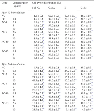
Table 1
The effect of thiazacridine derivatives on the cell cycle distribution on human colon HCT-8 cells.
Drug Concentration (μg/ml)
Cell cycle distribution (%)
Sub-G1 G1/G0 S G2/M After 12-h incubation
NC – 1.8 ± 0.1 62.8 ± 0.8 12.5 ± 0.3 15.7 ± 0.4 PC 0.3 1.5 ± 0.4 32.5 ± 1.7⁎ 20.3 ± 2.4⁎ 40.5 ± 2.1⁎ AC-4 2.5 3.8 ± 0.5⁎ 58.2 ± 1.7 13.8 ± 0.9 19.7 ± 0.8⁎ 5 4.4 ± 0.3⁎ 57.0 ± 1.1 15.2 ± 0.5 17.4 ± 1.3 10 9.9 ± 1.1⁎ 55.2 ± 2.7 14.5 ± 0.6 12.9 ± 0.9 AC-7 2.5 2.4 ± 0.4 58.3 ± 1.2 13.5 ± 0.6 19.2 ± 0.5⁎
5 5.0 ± 0.6⁎ 57.8 ± 1.3 15.3 ± 1.0 16.2 ± 0.4 10 8.4 ± 0.9⁎ 58.1 ± 1.6 15.3 ± 1.4 12.2 ± 0.6 AC-10 2.5 3.7 ± 0.5⁎ 57.6 ± 1.6 14.1 ± 0.5 19.9 ± 0.5⁎
5 5.3 ± 0.6⁎ 58.2 ± 1.2 14.4 ± 0.3 17.6 ± 0.7 10 6.9 ± 0.5⁎ 58.4 ± 1.3 13.5 ± 0.6 16.7 ± 0.9 AC-23 2.5 3.3 ± 0.8 58.4 ± 1.0 14.7 ± 1.0 16.6 ± 0.3 5 4.6 ± 1.0⁎ 57.6 ± 0.7 16.6 ± 1.1 13.9 ± 0.5 10 8.1 ± 0.8⁎ 60.5 ± 0.3 13.0 ± 0.8 11.9 ± 0.5⁎
After 24-h incubation
NC – 4.7 ± 0.4 59.6 ± 0.8 14.4 ± 0.8 16.0 ± 0,3 PC 0.3 8.1 ± 1.1⁎ 27.8 ± 1.6⁎ 24.0 ± 2.3⁎ 26.3 ± 1.0⁎
AC-4 2.5 13.9 ± 1.5⁎ 55.2 ± 0.8 15.1 ± 1.1 17.5 ± 0.9 5 24.7 ± 2.2⁎ 51.8 ± 0.9⁎ 15.1 ± 0.9 3.9 ± 0.8⁎
10 43.3 ± 1.5⁎ 44.0 ± 1.3⁎ 10.0 ± 0.5⁎ 3.7 ± 0.7⁎
AC-7 2.5 10.9 ± 0.8⁎ 55.7 ± 1.5 14.9 ± 0.4 11.6 ± 0.4⁎ 5 13.7 ± 1.4⁎ 54.9 ± 3.3⁎ 13.6 ± 0.7 9.8 ± 0.7⁎ 10 26.6 ± 3.6⁎ 54.6 ± 3.4⁎ 8.9 ± 0.5⁎ 5.2 ± 0.7⁎ AC-10 2.5 26.8 ± 0.7⁎ 52.6 ± 2.3⁎ 14.7 ± 0.8 10.8 ± 1.0⁎
5 27.1 ± 1.0⁎ 49.1 ± 3.1⁎ 14.3 ± 0.6 9.7 ± 1.1⁎
10 37.5 ± 1.4⁎ 48.8 ± 1.8⁎ 10.2 ± 0.5⁎ 4.7 ± 0.5⁎
AC-23 2.5 12.1 ± 2.0⁎ 56.3 ± 1.9 12.5 ± 0.5 10.8 ± 1.4⁎ 5 24.4 ± 2.1⁎ 55.8 ± 3.3 11.1 ± 0.7 9.8 ± 1.2⁎ 10 28.9 ± 2.0⁎ 52.2 ± 3.5⁎ 6.4 ± 0.6⁎ 4.5 ± 0.4⁎ The data are presented as the mean values±S.E.M. from three independent experiments performed in duplicate. The negative control was treated with the same vehicle (NC, 0.1% DMSO) that diluted the tested substance. Amsacrine (PC,m-AMSA) served as the positive control. Five thousand events were analysed for theflow cytometry analysis in each experiment.
⁎ pb0.05 compared to negative control by ANOVA followed by a Student Newman– Keuls test.
NC PC 2.5 5 10 2.5 5 10 2.5 5 10 2.5 5 10 0
20 40 60 80 100
AC-4
(µg/ml)
A
AC-7 AC-10 AC-23
*
*
*
*
*
*
*
*
Cells (%)
NC PC 2.5 5 10 2.5 5 10 2.5 5 10 2.5 5 10
0 20 40 60 80 100
AC-4
(µg/ml)
B
* *
* *
AC-7 AC-10 AC-23
*
*
*
* *
*
*
* *
*
*
* *
*
Cells (%)
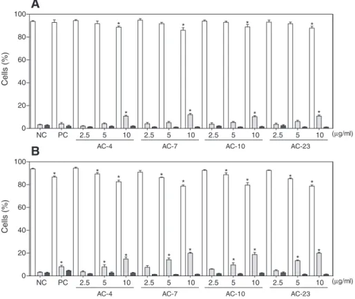
Fig. 5.The effect of thiazacridine derivatives on the viability of human colon carcinoma HCT-8 cells. A and B—cell viability (viable cells–white bar; apoptotic cells–grey bar; and necrotic cells–black bar) was determined byfluorescence microscopy using acridine orange/ethidium bromide after 12- and 24-h incubations, respectively. The data are presented as the mean values ± S.E.M. from three independent experiments performed in duplicate. The negative control was treated with the same vehicle (NC, 0.1% DMSO) that diluted the tested substance. Amsacrine (PC,m-AMSA, 0.3μg/ml) served as the positive control. *,pb0.05 compared to negative control by ANOVA followed by a Student Newman–Keuls test.
NC PC 2.5 5 10 2.5 5 10 2.5 5 10 2.5 5 10 0
20 40 60 80 100
AC-4
(µg/ml)
(µg/ml)
(µg/ml)
(µg/ml)
A
*
*
*
AC-7 AC-10 AC-23
*
Cell membrane integrity (%)
NC PC 2.5 5 10 2.5 5 10 2.5 5 10 2.5 5 10 0
20 40 60 80 100
AC-4
B
*
*
*
AC-7 AC-10 AC-23
*
*
*
*
*
Cell membrane integrity (%)
NC PC 2.5 5 10 2.5 5 10 2.5 5 10 2.5 5 10 0
5 10 15 20 25
AC-4
C
*
*
*
*
AC-7 AC-10 AC-23
Mitochondrial despolarisation (%) NC PC 2.5 5 10 2.5 5 10 2.5 5 10 2.5 5 10 0
5 10 15 20 25
AC-4
D
*
*
*
*
*
*
*
*
*
*
*
AC-7 AC-10 AC-23
Mitochondrial despolarisation (%)
orange/ethidium bromide staining. The integrity of the cell mem-brane and the mitochondrial memmem-brane potential were also
deter-mined by flow cytometry. Additionally, after a 24-h incubation,
phosphatidylserine externalisation and caspase 3/7 activation were
measured byflow cytometry.
After a 12-h incubation, HCT-8 cells either treated or untreated with ATZD, were tested at all concentrations and presented slight morpho-logical changes (data not shown). On the other hand, after a 24-h incu-bation, morphological examination of HCT-8 cells showed severe
drug-mediated changes. The hematoxylin–eosin stained HCT-8 cells
treated with ATZD presented a morphology consistent with apoptosis, including a reduction in cell volume, chromatin condensation and
nu-clei fragmentation (Fig. 4). The acridine orange/ethidium bromide
stained and treated cells also displayed a morphology consistent with
apoptosis, in a time- and concentration-dependent manner (pb0.05,
Fig. 5).m-AMSA, served as the positive control, which also induced morphological changes consistent with apoptosis.
The integrity of the cell membrane is a parameter of cell viability that differs between apoptotic and necrotic cells. After 12- or 24-h of exposure, ATZD induced a slight disruption in the plasmatic mem-brane, which was only observed at the higher concentrations tested (Figs. 6A, B). As cited above, the internucleosomal DNA
fragmenta-tion was markedly increased in ATZD-treated cells (pb0.05,Table 1).
Both of these modifications are characteristics of apoptotic cells. In
addition, ATZD induced mitochondrial depolarisation in a time- and
concentration-dependent manner (pb0.05,Figs. 6C, D).m-AMSA served
as the positive control, which also induced mitochondrial depolarisation and DNA fragmentation without affecting the membrane's integrity.
In addition, phosphatidylserine externalisation (AC-4andAC-10at
concentrations of 2.5 and 5μg/ml) and caspase 3/7 activation (AC-4,
AC-10andAC-23at concentrations of 5 and 10μg/ml) were measured in ATZD-treated cells after a 24-h incubation. Phosphatidylserine
exposure (pb0.05,Fig. 7A) and an increase in caspase 3/7 activation
(pb0.05, Fig. 7B) were also observed, suggesting that a
caspase-dependent apoptotic cell death had occurred. Doxorubicin served as the positive control and also induced phosphatidylserine exposure and increased caspase 3/7 activation.
Thiazacridine derivatives inhibits DNA topoisomerase I action
Because ATZD interact with DNA, they are potential topoisomer-ase inhibitors. The effect of ATZD on DNA topoisomertopoisomer-ase activity was evaluated in a yeast-based assay and in a cell-free assay.
First, the effects of ATZD were evaluated using a drop test assay in a
mutant strain ofS. cerevisiaethat was defective in topoisomerase type
I (Fig. 8). The type IB topoisomerases (topoisomerase 1 in yeast) relax both positively and negatively supercoiled DNA, whereas type IA topoisomerases (topoisomerase 3 in yeast) preferentially relax
nega-tively supercoiled DNA. At a concentration of 50μg/ml, the ATZD
were more resistant in yeast mutants that lacked topoisomerase 1
(Top1Δ) activity compared with the wild-type strain (BY-4741),
indi-cating that these molecules may induce lesions in topoisomerase 1. In
ATZD at higher concentration (100μg/ml), the Top1Δmutant was
more sensitive than the wild-type strain, which indicates that an addi-tional cytotoxicity mechanism (i.e., interaction with topoisomerase II) may be involved. Moreover, the strain without topoisomerase 3, but
with topoisomerase 1, (Top3Δ), was more sensitive to the ATZD,
with the exception ofAC-23.m-AMSA served as the positive control,
which showed similar effects.
In addition, the effect of ATZD on topoisomerase I activity was
evaluated in a cell-free system. Purified human DNA topoisomerase I
was incubated with ATZD (50 and 100μg/ml) in the presence of
supercoiled plasmid DNA; the products of this reaction were subjected to electrophoresis on agarose gels to separate the closed and open cir-cular DNAs. Relaxation of the DNA strand was inhibited in both of the
concentrations tested (Fig. 9). CPT served as the positive control
be-cause it also inhibits DNA topoisomerase I.
Thiazacridine derivatives do not cause genotoxicity or inhibit telomerase activity in human lymphocytes
The genotoxicity of ATZD (AC-4,AC-7, AC-10and AC-23) was
evaluated in human lymphocyte cultures using an alkaline comet
assay at concentrations of 2.5, 5 and 10μg/ml. The genotoxicity of
ATZD (AC-4andAC-10) was also evaluated in human lymphocyte
cultures using a chromosome aberration assay at concentrations of
2.5, 5 and 10μg/ml. The ability of ATZD (AC-4andAC-10) to inhibit
telomerase action was performed using a pan telomeric probe at a
concentration of 2.5μg/ml. None of the ATZD showed genotoxic
ac-tivity or anti-telomerase acac-tivity at any experimental concentrations tested (data not shown). Doxorubicin served as the positive control, and demonstrated potent genotoxic activity.
Discussion
The present work demonstrates the mechanism by which ATZD
(AC-4,AC-7,AC-10andAC-23) are cytotoxic in human colon
carcino-ma HCT-8 cells. As cited above, these agents were recently synthesised as a novel class of solid tumour-selective cytotoxic agents. These ATZD exhibit a relatively high cytotoxicity in colon carcinoma (HCT-8, HCT-15, SW-620 and COLO-205), prostate carcinoma (PC-3 and DU-145), ovarian carcinoma (OVCAR-8), melanoma (UACC-62 and MDA-MB-435) and glioblastoma (SF-295) tumour cell lines. However, these compounds were not active in leukaemia (HL-60, K-562 and
NC PC 2.5 5 2.5 5
0 20 40 60 80 100
*
*
*
*
AC-4 AC-10
(µg/ml)
A
Annexin V-positive cells (%)
NC PC 5 10 5 10 5 10
0 20 40 60
*
*
*
*
AC-4
(µg/ml)
B
*
*
*
AC-10 AC-23
Caspases 3/7-activated cells (%)
CEM), breast carcinoma (MDA-MB-231, HS-578-T and MX-1) or
nor-mal lymphoblast (PBMC) cells (Barros et al., 2012). Here, we
demon-strate the effects of ATZD on cell proliferation, cell cycle progress and apoptotic-induction using HCT-8 cells as a model. Studies in a yeast-based assay and a cell-free assay examine how ATZD interfere in topoisomerase I activity.
The ATZD inhibit human colon carcinoma HCT-8 cell proliferation in a concentration- and time-dependent manner, and their cytotoxic ac-tivity was assessed using different assays. Previously, we demonstrated that ATZD exhibited relatively high cytotoxicity against colon carcino-mas and that the highlight of these ATZD was their selectivity toward solid tumours because these ATZD were not active in leukaemias or
normal lymphoblasts (Barros et al., 2012). The pyrazoloacridines,
bisannulated acridines, aminoderivatives of azapyranoxanthenone
and pyranoisoflavones have also been cited as solid tumour-selective
cytotoxic agents (Gao et al., 2011; Kolokythas et al., 2006; Sebolt, et
al., 1987; Thale et al., 2002). Therefore, this feature is noteworthy but the mechanisms accounting for this selectivity are poorly understood.
The population of cells in the G2/M phase was shifted to the sub-G1
population in ATZD-treated HCT-8 cells, whilst few changes occurred
in the population of cells in the G0/G1or S phases. This indicates that
the ATZD preferentially guide cells from the G2/M phase into
apopto-sis. Manipulating the regulatory events at this checkpoint is a
promis-ing approach that will improve the efficiency of cytotoxic drugs and
overcome drug resistance (Links et al., 1998). In addition, HCT-8
cells treated with ATZD presented typical hallmarks of apoptosis. Se-lective apoptosis, the deletion of certain cells in tissues without
con-comitant inflammation, is advantageous in tissue homeostasis. The
induction of apoptosis is one of the main mechanisms that inhibit can-cer growth and proliferation and is used by several antitumor agents (Los et al., 2003; Schultz and Harrington, 2003). Moreover, ATZD
treatment induces mitochondrial depolarisation, phosphatidylserine exposure and an increase in caspase 3/7 activation, which suggests that ATZD treatment leads to a caspase-dependent apoptotic cell
death. Caspases play an essential role in apoptosis (Fan et al., 2005;
Kitazumi and Tsukahara, 2011): these caspases are responsible for the cleavage of cellular proteins, such as cytoskeletal components, which leads to the morphological changes previously observed in
the cells that undergo apoptosis (Kothakota et al., 1997).
The mechanism by which acridine and thiazolidine derivatives act has been continuously researched. Thiazolidine derivatives activate
peroxisome proliferator-activated receptors (Barros et al., 2010).
Mean-while, acridine derivatives used in cancer chemotherapy have biological targets, such as DNA topoisomerases I and/or II, telomerase/telomeres
and kinases (Castillo-González et al., 2009; Guo et al., 2009; Oppegard
et al., 2009). Our understanding of ATZD's cytotoxic mechanisms have been limited to results from double stranded-DNA biosensors and single stranded-DNA solutions, which show a positive interaction
with these ATZD that couple acridine and thiazolidine (Barros et
al., 2012). Here, we demonstrate that ATZD inhibit DNA topoisomer-ase I activity.
The cytotoxicity of DNA topoisomerase I inhibitors is caused by blocking DNA topoisomerase I cleavage complexes or by inhibiting DNA topoisomerase I catalytic activity. Then, DNA topoisomerase I inhibitors work by stabilising the DNA topoisomerase I cleavage
com-plexes, which cause DNA damage (Hsiang et al., 1989; Pommier et al.,
1998; Stewart et al., 1998). Because malignant cells often contain greater amounts of DNA topoisomerase I than normal cells, tumour cells should be more sensitive to the toxic effects of these inhibitors. The malignant cells that often contain great amounts of DNA topo-isomerase I include colon adenocarcinoma, several types of non-Hodgkin's lymphoma, leukaemias, melanoma and carcinomas of the Fig. 8.The sensitivity of a wild-type strain ofSaccharomyces cerevisiaeand mutants with defective topoisomerases. The sensitivity of topoisomerases type I to thiazacridine deriv-atives was determined by a drop test assay. A suspension ofS. cerevisiaecells in the exponential phase of growth was treated for 24-h in the absence or in the presence of thiazacridine derivatives at the indicated concentrations. The diluted cell cultures (107–103from left to right) were spotted on YPD agar plates. Amsacrine (m-AMSA) served as
stomach, breast and lung (Potmesil, 1994). This partially explains the selective cytotoxic effects of ATZD. However, the exact mechanism of this selective antitumor activity remains to be determined.
Previous studies have reported that some acridine and thiazolidine derivatives are somatic- and germ-cell mutagenic agents capable of inducing both numerical and structural chromosome aberrations in
vitro and in vivo (Attia, 2008; Attia, in press; Kao-Shan et al., 1984;
Nishi et al., 1989). These compounds are highly cytotoxic/genotoxic to normal lymphocyte cells. Therefore, to improve our understanding of the ATZD's cytotoxic actions, we assessed their genotoxic effects in human peripheral lymphocytes. Previously, the cytotoxicity of these
compounds was assessed against normal lymphocyte cells (Barros et
al., 2012); however, the genotoxicity had not been investigated. The genotoxic effects of ATZD were determined using an alkaline comet assay and a chromosome aberration assay; the anti-telomerase activ-ity was determined using a pan telomeric probe. In our studies, none of these ATZD agents showed genotoxicity and/or anti-telomerase ac-tivity in cultured human lymphocytes at the experimentally tested concentrations. Therefore, unlike the acridine and thiazolidine deriva-tives, ATZD did not cause cytotoxicity, genotoxicity and the inhibition of telomerase activity in human lymphocytes.
In this manuscript, we show that the ATZD are solid tumour-selective cytotoxic agents that inhibit DNA topoisomerase I activity and induce tumour cell death through caspase-dependent apoptosis pathways without causing genotoxicity in human lymphocytes.
These data confirm that these ATZD are promising anticancer drugs.
Conflicts of interest
The authors declare no conflicts of interest.
Acknowledgments
This study was supported by the Brazilian National Research Council, National Institute of Science and Technology for Pharmaceutical Innova-tion (CNPq/RENORBIO/INCT-IF) and INCT-Bioanalítica. The English was edited by American Journal Experts (key#354F-6EF9-BC4F-6B4A-E706).
References
Attia, S.M., 2008. Mutagenicity of some topoisomerase II-interactive agents. Saudi Pharm. J. 17, 1–24.
Attia, S.M., in press. Molecular cytogenetic evaluation of the mechanism of genotoxic potential of amsacrine and nocodazole in mouse bone marrow cells. J. Appl. Toxicol.http://dx.doi.org/10.1002/jat.1753.
Barros, C.D., Amato, A.A., de Oliveira, T.B., Iannini, K.B., Silva, A.L., Silva, T.G., Leite, E.S., Hernandes, M.Z., Lima, M.C.A., Galdino, S.L., Neves, F.A., Pitta, I.R., 2010. Synthesis and anti-inflammatory activity of new arylidene-thiazolidine-2,4-diones as PPAR ligands. Bioorg. Med. Chem. 18, 3805–3811.
Barros, F.W., Silva, T.G., Pitta, M.G.R., Bezerra, D.P., Costa-Lotufo, L.V., de Moraes, M.O., Pessoa, C., de Moura, M.A., de Abreu, F.C., de Lima, M.D., Galdino, S.L., Pitta, I.R., Goulart, M.O., 2012. Synthesis and cytotoxic activity of new acridine–thiazolidine derivatives. Bioorg. Med. Chem. 20, 3533–3539.
Belmont, P., Bosson, J., Godet, T., Tiano, M., 2007. Acridine and acridone derivatives, an-ticancer properties and synthetic methods: where are we now? Anan-ticancer Agents Med. Chem. 7, 139–169.
Bender, M.A., Awa, A.A., Brooks, A.L., Evans, H.J., Groer, P.G., Littlefield, L.G., Pereira, C., Preston, R.J., Wachholz, B.W., 1988. Current status of cytogenetic procedures to de-tect and quantify previous exposures to radiation. Mutat. Res. 196, 103–159. Berthold, F., 1981. Isolation of human monocytes byficoll density gradient
centrifuga-tion. Blut 3, 367–371.
Blasiak, J., Gloc, E., Drzewoski, J., Wozniak, K., Zadrozny, M., Skórski, T., Pertynski, T., 2003. Free radical scavengers can differentially modulate the genotoxicity of amsacrine in normal and cancer cells. Mutat. Res. 535, 25–34.
Brown, M.G., Lawce, H.J., 1997. Peripheral blood cytogenetic methods. In: Barch, M.J., Knutsen, T., Spurbeck, J.L. (Eds.), The AGT Cytogenetics Laboratory Manual. Lippincott-Raven Publishers, Philadelphia, pp. 77–171.
Burke, D., Dawson, D., Stearns, T., 2000. Methods in Yeast Genetics. Cold Spring Harbor, Laboratory Press, New York.
Castillo-González, D., Cabrera-Pérez, M.A., Pérez-González, M., Helguera, A.M., Durán-Martínez, A., 2009. Prediction of telomerase inhibitory activity for acridinic deriv-atives based on chemical structure. Eur. J. Med. Chem. 44, 4826–4840.
Cavalcanti, B.C., Sombra, C.M.L., Oliveira, J.H.H.L., Berlinck, R.G.S., Moraes, M.O., Pessoa, C., 2008. Cytotoxicity and genotoxicity of ingenamine G isolated from the Brazilian marine spongePachychalina alcaloidifera. Comp. Biochem. Physiol. C 147, 409–415. Fan, T.J., Han, L.H., Cong, R.S., Liang, J., 2005. Caspase family proteases and apoptosis.
Acta Biochim. Biophys. Sin. 37, 719–727.
Gao, S., Xu, Y.M., Valeriote, F.A., Gunatilaka, A.A., 2011. Pierreiones A–D, solid tumour selective pyranoisoflavones and other cytotoxic constituents fromAntheroporum pierrei. J. Nat. Prod. 74, 852–856.
Gorman, A.M., Samali, A., McGowan, A.J., Cotter, T.G., 1997. Use offlow cytometry tech-niques in studying mechanisms of apoptosis in leukemic cells. Cytometry 29, 97–105. Guo, C., Gasparian, A.V., Zhuang, Z., Bosykh, D.A., Komar, A.A., Gudkov, A.V., Gurova, K.V., 2009. 9-Aminoacridine-based anticancer drugs target the PI3K/AKT/mTOR, NF-kappaB and p53 pathways. Oncogene 28, 1151–1161.
Hartmann, J.T., Lipp, H.P., 2006. Camptothecin and podophyllotoxin derivatives: inhib-itors of topoisomerase I and II—mechanisms of action, pharmacokinetics and toxic-ity profile. Drug Saf. 29, 209–230.
Hartmann, A., Speit, G., 1997. The contribution of cytotoxicity to DNA effects in the sin-gle cell gel test (comet assay). Toxicol. Lett. 90, 183–188.
Hsiang, Y.H., Lihou, M.G., Liu, L.F., 1989. Arrest of replication forks by drug-stabilized topo-isomerase I-DNA cleavable complexes as a mechanism of cell killing by camptothecin. Cancer Res. 49, 5077–5082.
Hutchins, D., Steel, C.M., 1983. Phytohaemagglutinin-induced proliferation of human T lymphocytes: differences between neonate and adults in accessory cell require-ments. Clin. Exp. Immunol. 52, 355–364.
Kao-Shan, C.S., Micetich, K., Zwelling, L.A., Whang-Peng, J., 1984. Cytogenetic effects of amsacrine on human lymphocytesin vivo andin vitro. Cancer Treat. Rep. 68, 989–997.
Ketron, A.C., Denny, W.A., Graves, D.E., Osheroff, N., 2012. Amsacrine as a topoisomer-ase II poison: importance of drug-DNA interactions. Biochemistry 51, 1730–1739. Kitazumi, I., Tsukahara, M., 2011. Regulation of DNA fragmentation: the role of
caspases and phosphorylation. FEBS J. 278, 427–441.
Kolokythas, G., Pouli, N., Marakos, P., Pratsinis, H., Kletsas, D., 2006. Design, synthesis and antiproliferative activity of some new azapyranoxanthenone aminoderivatives. Eur. J. Med. Chem. 41, 71–79.
Kothakota, S., Azuma, T., Reinhard, C., Klippel, A., Tang, J., Chu, K., McGarry, T.J., Kirschner, M.W., Koths, K., Kwiatkowski, D.J., Williams, L.T., 1997. Caspase-3-generated fragment of gelsolin: effector of morphological change in apoptosis. Science 278, 294–298. Lansdorp, P.M., 1995. Telomere length and proliferation potential of hematopoietic
stem cells. J. Cell Sci. 108, 1–6.
Lansdorp, P.M., Verwoerd, N.P., van de Rijke, F.M., Dragowska, V., Little, M.T., Dirks, R.W., Raap, H.J., 1996. Heterogeneity in telomere length of human chromosomes. Hum. Mol. Genet. 5, 685–691.
Links, M., Ribeiro, J., Jackson, P., Friedlander, M., Russell, P.J., 1998. Regulation and de-regulation of G2checkpoint proteins with cisplatin. Anticancer Res. 18, 4057–4066.
Los, M., Burek, C.J., Stroh, C., Benedyk, K., Hug, H., Mackiewicz, A., 2003. Anticancer drugs of tomorrow: apoptotic pathways as target for drug design. Drug Discov. Today 8, 67–77.
McGahon, A.J., Martin, S.J., Bissonnette, R.P., Mahboubi, A., Shi, Y., Mogil, R.J., Nishioka, W.K., Green, D.R., 1995. The end of the (cell) line: methods for the study of apopto-sisin vitro. Methods Cell Biol. 46, 153–185.
Miao, Z.H., Player, A., Shankavaram, U., Wang, Y.H., Zimonjic, D.B., Lorenzi, P.L., Liao, Z.Y., Liu, H., Shimura, T., Zhang, H.L., Meng, L.H., Zhang, Y.W., Kawasaki, E.S., Popescu, N.C., Aladjem, M.I., Goldstein, D.J., Weinstein, J.N., Pommier, Y., 2007. Nonclassic functions of human topoisomerase I: genome-wide and pharmacologic analyses. Cancer Res. 67, 8752–8761.
Moorhead, P.S., Nomell, P.C., Mellman, W.J., Battips, D.M., Hungerford, D.A., 1960. Chro-mosome preparations of leukocytes cultured from human peripheral blood. Exp. Cell Res. 20, 613–616.
Moukharskaya, J., Verschraegen, C., 2012. Topoisomerase 1 inhibitors and cancer ther-apy. Hematol. Oncol. Clin. North Am. 26, 507–525.
Mourão, R.H., Silva, T.G., Soares, A.L., Vieira, E.S., Santos, J.N., Lima, M.C., Lima, V.L., Galdino, S.L., Barbe, J., Pitta, I.R., 2005. Synthesis and biological activity of novel acridinylidene and benzylidene thiazolidinediones. Eur. J. Med. Chem. 40, 1129–1133.
Nicoletti, I., Magliorati, G., Pagliacci, M.C., Grignani, F., Riccardi, C., 1991. A rapid and simple method for measuring thymocyte apoptosis by propidium iodide staining andflow cytometry. J. Immunol. Methods 139, 271–279.
Nishi, Y., Miyakawa, Y., Kato, K., 1989. Chromosome aberrations induced by pyrolysates of carbohydrates in Chinese hamster V79 cells. Mutat. Res. 227, 117–123. Oppegard, L.M., Ougolkov, A.V., Luchini, D.N., Schoon, R.A., Goodell, J.R., Kaur, H.,
Billadeau, D.D., Ferguson, D.M., Hiasa, H., 2009. Novel acridine-based compounds that exhibit an anti-pancreatic cancer activity are catalytic inhibitors of human topoisomerase II. Eur. J. Pharmacol. 602, 223–229.
Pommier, Y., Pourquier, P., Fan, Y., Strumberg, D., 1998. Mechanism of action of eukary-otic DNA topoisomerase I and drugs targeted to the enzyme. Biochim. Biophys. Acta 1400, 83–105.
Pommier, Y., Leo, E., Zhang, H., Marchand, C., 2010. DNA topoisomerases and their poi-soning by anticancer and antibacterial drugs. Chem. Biol. 17, 421–433. Poon, S.S., Martens, U.M., Ward, R.K., Lansdorp, P.M., 1999. Telomere length
measure-ments using digitalfluorescence microscopy. Cytometry 36, 267–278.
Potmesil, M., 1994. Camptothecins: from bench research to hospital wards. Cancer Res. 54, 1431–1439.
Preston, R.J., San Sebastian, J.R., McFee, A.F., 1987. Thein vitrohuman lymphocyte assay for assessing the clastogenicity of chemical agents. Mutat. Res. 189, 175–183. Schultz, D.R., Harrington, W.J., 2003. Apoptosis: programmed cell death at molecular
level. Semin. Arthritis Rheum. 32, 345–369.
Sebestik, J., Hlavacek, J., Stibor, I., 2007. A role of the 9-aminoacridines and their conju-gates in a life science. Curr. Protein Pept. Sci. 8, 471–483.
Sebolt, J.S., Scavone, S.V., Pinter, C.D., Hamelehle, K.L., Von Hoff, D.D., Jackson, R.C., 1987. Pyrazoloacridines, a new class of anticancer agents with selectivity against solid tumoursin vitro. Cancer Res. 47, 4299–4304.
Silva, T.G., Barbosa, F.S.V., Brandão, S.S.F., Lima, M.C.A., Galdino, S.L., Pitta, I.R., Barbe, J., 2001. Synthesis and structural elucidation of new benzylidene imidazolidines and acridinylidene thiazolidines. Heterocycl. Commun. 7, 523–528.
Singh, N.P., Mccoy, M.T., Tice, R.R., Schneider, E.L.A., 1988. Single technique for quanti-tation of low levels of DNA damage in individual cells. Exp. Cell Res. 175, 184–191. Speit, G., Hartmann, A., 1999. The comet assay (single-cell gel test). A sensitive genotoxicity test for the detection of DNA damage and repair. Methods Mol. Biol. 113, 203–212.
Stewart, L., Redinbo, M.R., Qiu, X., Hol, W.G., Champoux, J.J., 1998. A model for the mechanism of human topoisomerase I. Science 279, 1534–1541.
Sureda, F.X., Escubedo, E., Gabriel, C., Comas, J., Camarasa, J., Camins, A., 1997. Mito-chondrial membrane potential measurement in rat cerebellar neurons byflow cy-tometry. Cytometry 28, 74–80.
Thale, Z., Johnson, T., Tenney, K., Wenzel, P.J., Lobkovsky, E., Clardy, J., Media, J., Pietraszkiewicz, H., Valeriote, F.A., Crews, P., 2002. Structures and cytotoxic prop-erties of sponge-derived bisannulated acridines. J. Org. Chem. 67, 9384–9391. Vermes, I., Haanen, C., Steffens-Nakken, H., Reutelingsperger, C., 1995. A novel assay for
apoptosis. Flow cytometric detection of phosphatidylserine expression on early apoptotic cells usingfluorescein labelled Annexin V. J. Immunol. Methods 184, 39–51.
Vos, S.M., Tretter, E.M., Schmidt, B.H., Berger, J.M., 2011. All tangled up: how cells direct, manage and exploit topoisomerase function. Nat. Rev. Mol. Cell Biol. 12, 827–841.