The role of oxidative stress on the pathophysiology
of metabolic syndrome
FABIANE VALENTINI FRANCISQUETI1*, LIDIANA CAMARGO TALON CHIAVERINI2, KLINSMANN CAROLODOS SANTOS1, IGOR OTÁVIO MINATEL3,
CAROLINA BERCHIERI RONCHI4, ARTUR JUNIO TOGNERI FERRON5, ANA LÚCIA A. FERREIRA6, CAMILA RENATA CORRÊA7
1MSc, Department of Pathology, Faculdade de Medicina de Botucatu, Universidade Estadual Paulista "Júlio de Mesquita Filho" (Unesp), Botucatu, SP, Brazil 2PhD, Department of Pathology, Faculdade de Medicina de Botucatu, Unesp, Botucatu, SP, Brazil
3PhD, Department of Chemistry and Biochemistry, Instituto de Biociências de Botucatu, Unesp, Botucatu, SP, Brazil 4PhD, Department of Internal Medicine, Faculdade de Medicina de Botucatu, Unesp, Botucatu, SP, Brazil 5MSc, Department of Internal Medicine, Faculdade de Medicina de Botucatu, Unesp, Botucatu, SP, Brazil
6MD, Adjunct Professor of the Department of Internal Medicine, Faculdade de Medicina de Botucatu, Unesp, Botucatu, SP, Brazil
7Post-Doctorate, Lecturer in the Pathology Graduate Program, Department of Pathology, Faculdade de Medicina de Botucatu, Unesp, Botucatu, SP, Brazil
S
UMMARYStudy conducted at Faculdade de Medicina de Botucatu, Universidade Estadual Paulista "Júlio de Mesquita Filho" (Unesp), Botucatu, SP, Brazil
Article received: 3/9/2016
Accepted for publication: 6/20/2016
*Correspondence:
Address: Av. Prof. Montenegro Distrito de Rubião Junior, s/n Botucatu, SP – Brazil Postal code: 18618-970 [email protected]
http://dx.doi.org/10.1590/1806-9282.63.01.85
Metabolic syndrome (MetS) has a high prevalence around the world. Considering the components used to classify MetS, it is clear that it is closely related to obe-sity. These two conditions begin with an increase in abdominal adipose tissue, which is metabolically more active, containing a greater amount of resident macrophages compared to other fat deposits. Abdominal adiposity promotes inlammation and oxidative stress, which are precursors of various complications involving MetS components, namely insulin resistance, hypertension and hy-perlipidemia. One way to block the effects of oxidative stress would be through the antioxidant defense system, which offsets the excess free radicals. It is known that individuals with metabolic syndrome and obesity have high consumption of fats and sugars originated from processed foods containing high levels of sodium as well as low intake of fruits and vegetables, thus maintaining a state of oxidative stress, that can speed up the onset of MetS. Healthy eating habits could prevent or delay MetS by adding antioxidant-rich foods into the diet.
Keywords: oxidative stress, metabolic syndrome, obesity.
I
NTRODUCTIONMetabolic syndrome (MetS), also known as syndrome X or insulin resistance syndrome, is characterized by the clustering of cardiovascular risk factors such as hyperten-sion, insulin resistance, central obesity, and atherogenic dyslipidemia (high LDL-cholesterol, high triglycerides, and low HDL-cholesterol).1 MetS is a major health issue
of westernized modern societies1 and it already appears
as one of the main challenges of current clinical practice. In general, the International Diabetes Federation (IDF) estimates that one-quarter of the world’s adult population has MetS and the observed prevalence of MetS in Na-tional Health and Nutrition Examination Survey (NHANES) was 5% among the subjects of normal weight, 22% among the overweight, and 60% among the obese.2
For the diagnosis of MetS, there are at least three cri-teria based on ive components: waist circumference, blood
pressure, blood glucose, triglycerides, and HDL-cholesterol. The National Cholesterol Education Program – Adult Treat-ment Pannel III (NCEP – ATP III)3 adopts at least three
com ponents for diagnosis of MetS. The IDF4 considers the
abdominal circumference and two more components, and the World Health Organization (WHO)5 uses the waist/hip
ratio, presence of type 2 diabetes mellitus (DM) or insulin resistance, microalbuminuria, hypertension and triglycerides. Observing the components that classify the indi-vidual as having metabolic syndrome, it can be noted that they are all complications that commonly affect obese individuals, which shows that there is a direct link between these two diseases.6 In general, these two conditions begin
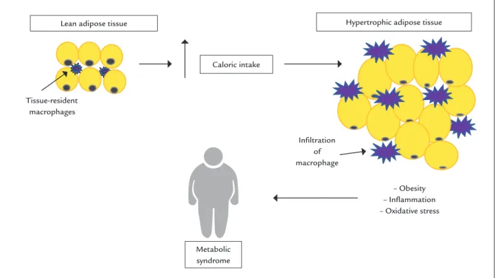
to increase in abdominal adipose tissue which is more metabolically active, containing a higher amount of res-ident macrophages compared to other fat deposits.7
stress, which are precursors of various complications involving MetS components, namely insulin resistance, hypertension, and hyperlipidemia (Figure 1).8,9 Because
MetS is associated with cardiovascular complications, the main cause of death worldwide, it is important to under-stand the factors that are involved in this disorder. Thus, this review aims to present the involvement of oxidative stress in the onset of metabolic syndrome.
M
ETABOLIC SYNDROME AND OXIDATIVE STRESSInlammation and oxidative stress occur when the energy supply begins to exceed the storage capacity of adipocytes and, as a result, hypertrophy occurs.10 This hypertrophy
leads to a higher release of adipokines as proinlamma-tory cytokines such as interleukin-1 (IL-1), interleukin-6 (IL-6) and tumor-necrosis factor alpha (TNF-α), resulting in low-grade chronic inlammation, which begins in adi-pose tissue and eventually reaches the circulation and other organs.11,12 One of the irst consequences of
inlam-mation is insulin resistance, since TNF-α prevents the phosphorylation of insulin receptors, interfering in their cascade action and preventing their functioning.13 Insulin
resistance and type 2 diabetes mellitus are classically char-acterized by dyslipidemia with hypertriglyceridemia, low HDL-cholesterol and LDL-cholesterol appearance.14
In-sulin resistance decreases inIn-sulin function, leading to a change in storage lipids that is a mechanism dependent on this hormone.15
Another cause of inlammation is oxidative stress, which can be triggered by adipocytes. When fat mass in-creases, insuficient irrigation can lead to lack of oxygen and, thus, to cell necrosis. The process of phagocytosis to eliminate these dead cells results in increased inlamma-tory iniltration and also oxidative stress by liberation of free radicals such as nitric oxide and hydrogen peroxide,16,17
which may negatively impact components of MetS.18
Oxidative stress is classically deined as an event re-sulting from the magnitude of imbalance between oxidant and antioxidant substances,19,20 generated in a setting of
oxidation-reduction reactions. Since the generation and the action of these substances depend on this oxidation--reduction system, authors now use the term “imbalance of redox system” to refer to the oxidative stress.21,22
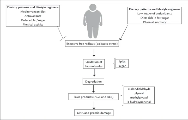
Com-monly known as free radicals, oxidants include reactive oxygen and nitrogen species, which perform the oxidation of lipids (lipoxidation) and glucose (glycation), substanc-es found in excsubstanc-ess in obsubstanc-esity. Excsubstanc-essive food intake in-creases the amount of energy and nutrients in the blood stream.23 Lipoxidation products include malondialdehyde,
glyoxal, acrolein, 4-hydroxy-nonenal (HNE), while the
– Obesity – Inlammation – Oxidative stress Lean adipose tissue
Caloric intake
Metabolic syndrome Tissue-resident
macrophages
Iniltration of macrophage
Hypertrophic adipose tissue
products generated from glycation include glyoxal and methyl glyoxal. These compounds bind to the amino grouping of amino acids, resulting in advanced glycation products (AGEs) and advanced lipoxidation end--products (ALEs),24 which are highly reactive and participate
in the development of other components of MetS. Clinical studies in patients with hypertension showed that systolic and diastolic blood pressure are positively correlated with biomarkers of oxidative stress and nega-tively correlated with the levels of antioxidants.25-27 This
fact is attributed to endothelial dysfunction caused by oxidative stress and inlammation, producing imbalance of vasoconstrictor and vasodilator products. This is evi-denced by an inverse association between factors that trigger vasodilation, plasma levels of malondialdehyde and positive association with antioxidants.28
Oxidative stress plays an important role on the patho-genesis of insulin resistance by disrupting the release of adipokines by adipose tissue such as TNF-α and IL-6, which can trigger inlammation, a mechanism already described above.29-31 Thus, it seems that obesity and MetS
are factors associated to inlammation and oxidative stress.
A
NTIOXIDANT DEFENSEOxidative stress is controlled by the endogenous antioxi-dant defense system, which includes antioxiantioxi-dant enzymes such as superoxide dismutase, catalase, glutathione per-oxidase, glutathione reductase; and nonenzymatic com-pounds such as ferritin, transferrin, bilirubin, ceruloplas-min, and even albumin carrier low molecular weight, such as uric acid and lipoic acid.32 Exogenous antioxidants from
fruits and vegetables, including hydrophilic as vitamin C and lavonoids and lipophilic as vitamin E and carotenoids, are also included. Carotenoids are divided into a group of pigments that give yellow and orange colors to plants, animals, and microorganisms. More than 700 carotenoids have been identiied; however, lutein, zeaxanthin, crypto-xanthin, alpha-carotene, beta-carotene, and lycopene rep-resent 95% of the carotenoids in human plasma.33
Antioxidants are able to trap free radicals generated by cellular metabolism or exogenous sources through the donation of hydrogen atoms of these molecules, breaking the chain reaction, which prevents attack on lipids, ami-no acids in proteins, double bond of the polyunsaturated fatty acids, and DNA bases, avoiding formation of lesions and loss of cell integrity.34 Another role of antioxidants
is the protection mechanism, which acts in the repair of damage caused by free radicals, a process related to the removal of the DNA molecule of damage and restoration of damaged cell membranes.35
The literature reports that a diet rich in fruits, vege-tables and grains prevents various diseases, such as car-diovascular diseases and cancer.36,37 Other intervening
factor in antioxidant response and the manifestations of MetS is the association between dietary adequacy and physical exercise.38 This is due to the exogenous and
en-dogenous antioxidants acting in synergy in combating free radicals.25 However, it is important to note that this
intake needs to be steady and orderly and that the intake of vitamins in supplement form may result in pro-oxidant effects called stress antioxidative.39
B
IOMARKERS OF OXIDATIVE STRESSThe reactive species are very unstable and have a very short half-life, which makes it a major challenge to perform an accurate assessment of these species. For this reason, meth-ods have been developed for measuring products resulting from the redox markers in biological samples, which are oxidation products of lipids, DNA and proteins.40 Among
the most common are the products of lipid peroxidation because of polyunsaturated fatty acids (such as phospho-lipids and glycophospho-lipids). When these phospho-lipids are oxidized, two products classically measured in biological samples, malo-ndialdehyde (MDA) and isoprostan, are formed.40,41
MDA is formed by the peroxidation of polyunsatu-rated fatty acids and can interact with proteins. MDA can be detected by the thiobarbituric acid (TBA) using a colorimetric method based on MDA TBA reaction and form a pink color, so gauging MDA and all species react-ing with this acid.42 The MDA can be speciically measured
by high performance liquid chromatography (HPLC). The same reaction occurs between MDA and TBA, but due to the apparatus of the luorescence detector, only the MDA is identiied, making this more speciic test.43
The isoprostane is a stable product of lipid peroxida-tion, and can be measured both in the tissues and in bio-logical luids including urine, plasma, and cerebrospinal luid. The level of this compound in plasma and urine correlates with the levels of reactive oxygen species and oxidative stress in experimental studies in humans.44
However, in healthy individuals at risk for obesity and hyperlipidemia their levels are increased, suggesting it as a good marker for cardiovascular risk.41
Total antioxidant capacity can be considered a mark-er of oxidative stress, since it measures the state of anti-oxidant capacity in biological luids. This method gives deeper insight into the involvement of oxidative stress in several pathophysiological conditions, but also monitors the effectiveness of antioxidant interventions.44 In this
quanti-ied by comparing the area under the curve (AUC) on the oxidation kinetics of BODIPY (4,4-diluoro-1,3,5,7,8-pentamethyl-4-bora-3a, 4a-diaza-s-indacene), a luorescent lipophilic oxidizable compound, radical initiator opposite 2.20 azobis- (2-amidinopropane) dihydrochloride (AAPH) in relation to the oxidation of phosphatidylcholine used as a reference lipid matrix.45
Another approach to assess the antioxidant capacity is to measure antioxidants individually. However, as there are many, this would require time and a variety of ana-lytical techniques, instruments, and procedures. In addi-tion, this approach lacks information about the possible synergy and cooperation between the hydrophilic and lipophilic antioxidants.45
Protein and DNA molecules are also highly suscep-tible to modiication by changes in redox state.46 The
protein oxidation occurs when proteins of amino acids (proline, arginine, threonine, lysine, histidine and cysteine) bind to glycoxidation and lipoxidation products, forming carbonyl groups. This reaction called carbonylation may be irreversible and lead to changes in their biological function; the detection of these toxic products (carbonyl) can be made by the mass spectrometer.24
As for DNA damage, comet assay can be performed, a cell microgel electrophoresis technique, very useful and widely used to assess damage and DNA repair in indi-vidual cells. Its basic principle is the lysis of cell membranes, followed by induction of DNA released from the electro-phoretic migration on agarose matrix. When viewed un-der a microscope, the migrated cell takes the apparent form of a comet, with a head, the nuclear region, and a tail, which contains fragments or DNA strands that have migrated towards the anode. The analysis of comets is based on the degree of DNA fragmentation and migration by microelectrophoresis.47 Measures such as the total
length of the “tail” and the DNA density provide indirect information about the state of the sample DNA. To detect oxidative damage, endonuclease III (ENDOIII) and phos-phatidylinositol-pyrimidine DNA glycosylase (FPG) are used to repair enzyme thus detecting oxidation bases in the pyrimidine and purine.48
D
IETARY INTAKE,
PHYSICAL ACTIVITY,
OXIDATIVE STRESS,
AND METABOLIC SYNDROMEAccording to what has been previously described, it is clear that when there is an imbalance within a large sup-ply of nutrients and a low antioxidant intake, obesity carries a picture of oxidative stress promoting metabolic syndrome.23 Corroborating this fact, the literature
indi-cates that individuals with metabolic syndrome and
obe-sity have a high consumption of fat and sugars derived from processed foods with high sodium content,49-51 as
well as low antioxidant intake.
Diets with high antioxidant content, such as the well--known Mediterranean diet, which consists of olive oil,
fruits and vegetables, cereals, nuts, and a small amount of red meat and foods high in sugar, are also ways to manage oxidative stress and inlammation.52,53
Research-ers suggest that individuals with MetS and obesity delayed and attenuated complications, such as insulin resistance, hypertension, and hyperlipidemia, when they had an intervention and began to consume this type of diet. One of the arrangements set out for this improvement was the reduction of oxidative stress and inlammation.53-57
Among these studies, Mitjavila et al.58 observed a
de-crease of some markers of oxidative stress after one year of dietary intervention. In this same study, subjects with MetS who consumed the Mediterranean diet were com-pared to a group that consumed a diet with only low levels of fat. It showed that a diet richer in antioxidants resulted in improvement in markers of oxidative stress and decreased DNA damage. This shows the importance of diet quality and the consistency and effectiveness of antioxidants in its composition.
Another factor associated to MetS is reduced daily physical activity in healthy young adults, which leads to negative metabolic consequences such as decreased insu-lin sensitivity and increased abdominal fat.59 Therefore,
increased physical activity is likely to be the evolutionary favored pathway to prevent the development of insulin resistance during metabolic derangements. The impact of exercise on insulin sensitivity is evident for 24 to 48 hours and disappears within 3 to 5 days, so continuous practice is essential.2 Besides, exercise increases the
pro-duction of oxidative stress. However, these increases seem to be necessary in order to allow for an upregulation in endogenous antioxidant defenses, thus providing benei-cial effects to the individual engaged in chronic exercise.60
A combination of resistance and aerobic exercise is the best, but any activity is better than none.
The association between good eating habits and exer-cise practice is also important. A recent study showed that food adequacy (intake of fruits and vegetables) associated with physical exercise for 20 weeks resulted in higher car-diorespiratory itness in residents of the city of Botucatu, SP. Moreover, reduction in visceral adiposity (waist circum-ference) was observed, reducing the prevalence of MetS and mainly increasing signiicantly glutathione concentra-tion and total antioxidant protecconcentra-tion (TAP) of the plasma.38
an-aerobic exercise, and resistance training in association with weight loss has been shown to be advantageous in amelio-rating oxidative stress and alleviating inlammation in obesity. Studies report that healthy obese patients doing exercise showed decreased lipid peroxidation indicator.61-63
Thus, it is important to consider strategies that increase the antioxidant defense capacity of the body, since the same part not only of detoxiication of free radicals, but are also closely related to modulation of pathophysiologic pro-cesses present in the MetS (Figure 2).
F
INAL CONSIDERATIONSThis review's approach can highlight the involvement of inlammation, and especially oxidative stress, in the pathogenesis of MetS. Given that obesity may be a key event in the development of this syndrome, treatment strategies are necessary to control and attenuate oxida-tive stress so that the body does not develop complications leading to MetS.
One of the main factors triggering these two pathol-ogies is food imbalance. In recent decades, there has been an increase in the intake of sugars and fats parallel to a reduction of the consumption of fruit and vegetables;
this alone promotes oxidative stress and has been the root cause in the growing epidemic of chronic diseases that affect developed and developing countries. A decrease in food intake coupled with physical activity would be a determining factor in the reduction of oxidative stress. The excess calories from sugar and fat intake, combined with a sedentary lifestyle, cause the body to manage the excess energy that must be metabolized. Other macronu-trients undergo oxidation within the mitochondria, pro-moting an increase in production of free radicals, which has been proposed as a unifying mechanism linking exces-sive intake of nutrients, insulin resistance, metabolic syndrome, and diabetes. Therefore, what could prevent or delay the onset of MetS would be the maintenance of healthy eating habits, with the inclusion of foods rich in antioxidants and physical activity.
A
CKNOWLEDGMENTThomas Patrick Wisniewski, Krupp Foundation Research Fellow, Harvard University.
C
ONFLICT OF INTERESTThe authors declare no conlict of interest.
Dietary patterns and lifestyle regimens
Mediterranean diet Antioxidants Reduced fat/sugar
Physical activity
Excessive free radicals (oxidative stress)
Oxidation of biomolecules
Degradation
DNA and protein damage Toxic products (AGE and ALE)
lipids sugar
malondialdehyde glyoxal methylglyoxal 4-hydroxynonenal
Dietary patterns and lifestyle regimens
Low intake of antioxidants Diets rich in fat/sugar
Physical inactivity
FIGURE 2 Biochemical way of oxidative stress.
R
ESUMOO papel do estresse oxidativo na isiopatologia da sín-drome metabólica
A síndrome metabólica apresenta elevada prevalência na população mundial. De acordo com os componentes que a classiicam, nota-se que está intimamente relacionada com a obesidade. Essas duas condições se iniciam com o aumento do tecido adiposo abdominal, o qual é metabo-licamente mais ativo, contendo uma quantidade maior de macrófagos residentes em comparação com outros depósitos de gordura. Essa situação favorece a inlamação e o estresse oxidativo, ambos precursores de diversas com-plicações, mas principalmente as envolvidas na síndrome metabólica, como resistência à insulina, hipertensão ar-terial e hiperlipidemia. Uma maneira de bloquear os efei-tos do estresse oxidativo seria pelo sistema de defesa an-tioxidante, o qual anula os radicais livres em excesso. É sabido que indivíduos portadores de síndrome metabó-lica e obesos apresentam um alto consumo de gorduras, açúcares oriundos de alimentos industrializados com alto teor de sódio e uma baixa ingestão de frutas e verduras, apresentando uma condição de estresse oxidativo contí-nuo. A manutenção de hábitos alimentares saudáveis, com a inclusão de alimentos ricos em antioxidantes, po-deria prevenir ou retardar o surgimento da SM.
Palavras-chave: estresse oxidativo, síndrome metabóli-ca, obesidade.
R
EFERENCES1. Steckhan N, Hohmann CD, Kessler C, Dobos G, Michalsen A, Cramer H. Effects of different dietary approaches on inlammatory markers in patientes with metabolic syndrome: a systematic review and meta-analysis. Nutrition. 2016; 32(3):338-48.
2. Kaur J. A comprehensive review on metabolic syndrome. Cardiol Res Pract. 2014; 2014:943162.
3. Expert Panel on Detection, Evaluation, and Treatment of High Blood Cholesterol in Adults. Executive Summary of The Third Report of The National Cholesterol Education Program (NCEP) Expert Panel on Detection, Evaluation, And Treatment of High Blood Cholesterol In Adults (Adult Treatment Panel III). JAMA. 2001; 285(19):2486-97.
4. Alberti KG, Zimmet P, Shaw J; IDF Epidemiology Task Force Consensus Group. The metabolic syndrome – a new worldwide deinition. Lancet. 2005; 366(9491):1059-62.
5. World Health Organization (WHO). Diet, nutrition and the prevention of chronic diseases. Geneva: WHO/FAO Expert consultation on diet, nutrition and the prevention of chronic diseases; 2002.
6. Dupuy AM, Jaussent I, Lacroux A, Durant R, Cristol JP, Delcourt C. Waist circumference adds to the variance in plasma C-reactive protein levels in elderly patients with metabolic syndrome. Gerontology. 2007; 53(6):329-39. 7. Luna-Luna M, Medina-Urrutia A, Alarcón G, Coss-Rovirosa F,
Vargas-Barrón J, Pérez-Méndez O. Adipose tissue in metabolic syndrome: onset and progression of atherosclerosis. Arch Med Res. 2015; 46(5):392-407.
8. Yao L, Herlea-Pana O, Heuser-Baker J, Chen Y, Barlic-Dicen J. Roles of the chemokine system in development of obesity, insulin resistance, and cardiovascular disease. J Immunol Res. 2014; 2014:181450.
9. Francisqueti FV, Nascimento AF, Corrêa CR. Obesidade, inlamação e complicações metabólicas. Nutrire. 2015; 40(1):81-9.
10. Klöting N, Blüher M. Adipocyte dysfunction, inlammation and metabolic syndrome. Rev Endocr Metab Disord. 2014; 15(4):277-87.
11. Cotillard A, Poitou C, Torcivia A, Bouillot JL, Dietrich A, Klöting N, et al. Adipocyte size threshold matters: link with risk of type 2 diabetes and improved insulin resistance after gastric bypass. J Clin Endocrinol Metab. 2014; 99(8):E1466-70.
12. Skurk T, Alberti-Huber C, Herder C, Hauner H. Relationship between adipocyte size and adipokine expression and secretion. J Clin Endocrinol Metab. 2007; 92(3):1023-33.
13. Andrade-Oliveira V, Câmara NOS, Moraes-Vieira PM. Adipokines as drug targets in diabetes and underlying disturbances. J Diabetes Res. 2015; 2015:681612.
14. Grundy SM. Hypertriglyceridemia, atherogenic dyslipidemia, and the metabolic syndrome. Am J Cardiol. 1998; 81(4A):18B-25B.
15. Borer KT. Counterregulation of insulin by leptin as key component of autonomic regulation of body weight. World J Diabetes. 2014; 5(5):606-29. 16. Bhattacharya I, Domínguez AP, Drägert K, Humar R, Haas E, Battegay EJ.
Hypoxia potentiates tumor necrosis factor-alpha induced expression of inducible nitric oxide synthase and cyclooxygenase-2 in white and brown adipocytes. Biochem Biophys Res Commun. 2015; 461(2):287-92. 17. Kosacka J, Kern M, Klöting N, Paeschke S, Rudich A, Haim Y, et al. Autophagy
in adipose tissue of patients with obesity and type 2 diabetes. Mol Cell Endocrinol. 2015; 409:21-32.
18. Netzer N, Gatterer H, Faulhaber M, Burtscher M, Pramsohler S, Pesta D. Hypoxia, oxidative stress and fat. Biomolecules. 2015; 5(2):1143-50. 19. Ferreira AL, Yeum KJ, Matsubara LS, Matsubara BB, Correa CR, Pereira EJ, et
al. Doxorubicin as an antioxidant: maintenance of myocardial levels of lycopene under doxorubicin treatment. Free Radic Biol Med. 2007; 43(5):740-51. 20. Yeum KJ, Russell RM, Krinsky NI, Aldini G. Biomarkers of antioxidant
capacity in the hydrophilic and lipophilic compartments of human plasma. Arch Biochem Biophys. 2004; 430(1):97-103.
21. Grant CM. Metabolic reconiguration is a regulated response to oxidative stress. J Biol. 2008;7(1):1.
22. Poli G, Schaur RJ, Siems WG, Leonarduzzi G. 4-hydroxynonenal: a mem-brane lipid oxidation product of medicinal interest. Med Res Rev. 2008; 28(4):569-631.
23. Dandona P, Ghanim H, Chaudhuri A, Dhindsa S, Kim SS. Macronutrient intake induces oxidative and inlammatory stress: potential relevance to atherosclerosis and insulin resistance. Exp Mol Med. 2010; 42(4):245-53. 24. Aldini G, Dalle-Donne I, Facino RM, Milzani A, Carini M. Intervention
strategies to inhibit protein carbonylation by lipoxidation-derived reactive carbonyls. Med Res Rev. 2007; 27(6):817-68.
25. Montezano AC, Dulak-Lis M, Tsiropoulou S, Harvey A, Briones AM, Touyz RM. Oxidative stress and human hypertension: vascular mechanisms, biomarkers, and novel therapies. Can J Cardiol. 2015; 31(5):631-41. 26. Wong WT, Tian XY, Huang Y. Endothelial dysfunction in diabetes and
hypertension: cross talk in RAS, BMP4, and ROS-dependent COX-2-derived prostanoids. J Cardiovasc Pharmacol. 2013; 61(3):204-14.
27. Hernanz R, Briones AM, Salaices M, Alonso MJ. New roles for old pathways? A circuitous relationship between reactive oxygen species and cyclo-oxygenase in hypertension. Clin Sci (Lond). 2014; 126(2):111-21.
28. Ward NC, Hodgson JM, Puddey IB, Mori TA, Beilin LJ, Croft KD. Oxidative stress in human hypertension: association with antihypertensive treatment, gender, nutrition, and lifestyle. Free Radic Biol Med. 2004; 36(2):226-32. 29. Tangvarasittichai S. Oxidative stress, insulin resistance, dyslipidemia and
type 2 diabetes mellitus. World J Diabetes. 2015; 6(3):456-80.
30. Houstis N, Rosen ED, Lander ES. Reactive oxygen species have a causal role in multiple forms of insulin resistance. Nature. 2006; 440:944-8. 31. Hotamisligil GS. Inlammation and metabolic disorders. Nature. 2006;
444(7121):860-7.
32. Poljsak B, Šuput D, Milisav I. Achieving the balance between ROS and antioxidants: when to use the synthetic antioxidants. Oxid Med Cell Longev. 2013; 2013:956792.
bioavailability and their protective role in humans. Mol Nutr Food Res. 2009; 53(Suppl 2):S194-218.
34. Sies H. Strategies of antioxidant defense. Eur J Biochem 1993; 215(2):213-9. 35. Sies H. Oxidative stress: from basic research to clinical application. Am J
Med. 1991; 91(3C):31S-8S.
36. Mangge H, Becker K, Fuchs D, Gostner JM. Antioxidants, inlammation and cardiovascular disease. World J Cardiol. 2014; 6(6):462-77.
37. Fiedor J, Burda K. Potential role of carotenoids as antioxidants in human health and disease. Nutrients. 2014; 6(2):466-88.
38. Moreto F, Kano HT, Torezan GA, de Oliveira EP, Manda RM, Teixeira O, et al. Changes in malondialdehyde and C-reactive protein concentrations after lifestyle modiication are related to different metabolic syndrome-associated pathophysiological processes. Diabetes Metab Syndr. 2015; 9(4):218-22.
39. Niki E. Biomarkers of lipid peroxidation in clinical material. Biochim Biophys Acta. 2014; 1840(2):809-17.
40. Petramala L, Pignatelli P, Carnevale R, Zinnamosca L, Marinelli C, Settevendemmie A, et al. Oxidative stress in patients affected by primary aldosteronism. J Hypertens. 2014; 32(10):2022-9; discussion 2029. 41. Basu S. Bioactive eicosanoids: role of prostaglandin F(2alpha) and
F(2)-isoprostanes in inlammation and oxidative stress related pathology. Mol Cells.; 30(5):383-91.
42. Reis GS, Augusto VS, Silveira AP, Jordão AA Jr, Baddini-Martinez J, Poli Neto O, et al. Oxidative-stress biomarkers in patients with pulmonary hypertension. Pulm Circ. 2013; 3(4):856-61.
43. Davies SS, Roberts LJ 2nd. F2-isoprostanes as an indicator and risk factor for coronary heart disease. Free Radic Biol Med. 2011; 50(5):559-66. 44. Fraga CG, Oteiza PI, Galleano M. In vitro measurements and interpretation
of total antioxidant capacity. Biochim Biophys Acta. 2014; 1840(2):931-4. 45. Beretta G, Aldini G, Facino RM, Russell RM, Krinsky NI, Yeum KJ. Total
antioxidant performance: a validated luorescence assay for the measurement of plasma oxidizability. Anal Biochem. 2006; 354(2):290-8.
46. Shacter E. Quantiication and signiicance of protein oxidation in biological samples. Drug Metab Ver. 2000; 32(3-4):307-26.
47. Prado RP, dos Santos BF, Pinto CL, de Assis KR, Salvadori DM, Ladeira MS. Inluence of diet on oxidative DNA damage, uracil misincorporation and DNA repair capability. Mutagenesis. 2010; 25(5):483-7.
48. Collins AR, Gedik CM, Olmedilla B, Southon S, Bellizzi M. Oxidative DNA damage measured in human lymphocytes: large differences between sexes and between countries, and correlations with heart disease mortality rates. FASEB J. 1998; 12(13):1397-400.
49. Novak EM, Keller BO, Innis SM. Dietary lipid quality and long-term outcome. Nestle Nutr Workshop Ser Pediatr Program. 2011; 68:17-27; discussion 27-32.
50. Johnson RJ, Nakagawa T, Sanchez-Lozada LG, Shaiu M, Sundaram S, Le M, et al. Sugar, uric acid, and the etiology of diabetes and obesity. Diabetes. 2013; 62(10):3307-15.
51. Keller U. Dietary proteins in obesity and in diabetes. Int J Vitam Nutr Res. 2011; 81(2-3):125-33.
52. Castro-Quezada I, Román-Viñas B, Serra-Majem L. The Mediterranean diet and nutritional adequacy: a review. Nutrients. 2014; 6(1):231-48. 53. Casas R, Sacanella E, Estruch R. The immune protective effect of the
Mediterranean diet against chronic low-grade inlammatory diseases. Endocr Metab Immune Disord Drug Targets. 2014; 14(4):245-54.
54. Kastorini CM, Milionis HJ, Esposito K, Giugliano D, Goudevenos JA, Panagiotakos DB. The effect of Mediterranean diet on metabolic syndrome and its components: a meta-analysis of 50 studies and 534,906 individuals. J Am Coll Cardiol. 2011; 57(11):1299-313.
55. Salas-Salvadó J, Bulló M, Babio N, Martínez-González MÁ, Ibarrola-Jurado N, Basora J, et al. Reduction in the incidence of type 2 diabetes with the Mediterranean diet: results of the PREDIMED-Reus nutrition intervention randomized trial. Diabetes Care. 2011; 34(1):14-9.
56. Doménech M, Roman P, Lapetra J, García de la Corte FJ, Sala-Vila A, de la Torre R, et al. Mediterranean diet reduces 24-hour ambulatory blood pressure, blood glucose, and lipids: one-year randomized, clinical trial. Hypertension. 2014; 64(1):69-76.
57. Vincent-Baudry S, Defoort C, Gerber M, Bernard MC, Verger P, Helal O, et al. The Medi-RIVAGE study: reduction of cardiovascular disease risk factors after a 3-mo intervention with a Mediterranean-type diet or a low-fat diet. Am J Clin Nutr. 2005; 82(5):964-71.
58. Mitjavila MT, Fandos M, Salas-Salvadó J, Covas MI, Borrego S, Estruch R, et al. The Mediterranean diet improves the systemic lipid and DNA oxidative damage in metabolic syndrome individuals. A randomized, controlled, trial. Clin Nutr. 2013; 32(2):172-8.
59. Golbidi S, Mesdaghinia A, Laher I. Exercise in the metabolic syndrome. Oxid Med Cell Longev. 2012; 2012:349710.
60. Huang CJ, McAllister MJ, Slusher AL, Webb HE, Mock JT, Acevedo EO. Obesity-related oxidative stress: the impact of physical activity and diet manipulation. Sports Med Open. 2015; 1:32.
61. Phillips MD, Patrizi RM, Cheek DJ, Wooten JS, Barbee JJ, Mitchell JB. Resistance training reduces subclinical inlammation in obese, postmenopausal women. Med Sci Sports Exerc. 2012; 44(11):2099-110.
62. Oh S, Tanaka K, Warabi E, Shoda J. Exercise reduces inlammation and oxidative stress in obesity-related liver diseases. Med Sci Sports Exerc. 2013; 45(12):2214-22.