(Annals of the Brazilian Academy of Sciences) ISSN 0001-3765
www.scielo.br/aabc
Chemical constituents of
Piptadenia gonoacantha
(Mart.)
J.F. Macbr (pau jacaré)
MÁRIO G. DE CARVALHO1, MARITZA A.R. CARDOZO1, FRANCISCO E.A. CATUNDA JUNIOR1 and ACÁCIO G. DE CARVALHO2
1Departamento de Química, ICE, Universidade Federal Rural do Rio de Janeiro
BR 465, km 07, 23890-000 Serópedica, RJ, Brasil
2Departamento de Produtos Florestais, Instituto de Florestas, Universidade Federal Rural do Rio de Janeiro
BR 465, km 07, 23890-000 Seropédica, RJ, Brasil
Manuscript received on February 4, 2009; accepted for publication on November 24, 2009
ABSTRACT
The phytochemical investigation of Piptadenia gonoacantha (Mart.) J.F. Macbr. (Leguminosae-Mimosoideae), commonly known as “pau jacaré” (alligator stick), afforded sitosterol, campesterol, stigmasterol, the N-benzoyl-phenylalanine-2-benzoylamide-3-phenylpropyl ester, known as asperphenamate, sitosterol-3-O-β-D-glucopyranoside,
besides three flavonoids, apigenin, 5-O-methylapigenin and 7,4′-dihydroxy-3′,5-dimethoxyflavone from its branches.
From its leaves, the methyl gallate and two flavonoids, vitexin and isovitexin, were isolated. From its bark, a mix-ture of sitosterol, campesterol, and stigmasterol, besides a mixmix-ture of cycloartenone, cycloartan-25-en-3-one, and 24-methylene-cycloartenone, and the pure triterpenes 24-methylenecycloartanol, friedelin, lupeol and lupenone, were isolated. Their structures were established on the basis of spectral analysis, comparison with literature data and GC-MS analysis of the mixtures. The ester, flavonoids and the cycloartanes are been identified for first time in the genus
Piptadenia.
Key words:Leguminoseae,Piptadenia gonoacantha, terpenoids, asperphenamate, flavonoids, “pau jacaré”.
INTRODUCTION
The Piptadeniagenus belong to Mimosoideae (Legu-minosae) and have about 80 tropical species frequently occurring in South America. ThePiptadeniaspecies are known in Brazil as angico, and as cebil in Argentina and Paraguay. These species have been used in tannery due to the tannins, in building due to the hard and heavy wood and in the recovery of forests because they can grow in poor and degraded soil (Lorenzi 1998, Correa 1984). The scientific interest onPiptadeniaspecies is motivated by their use in snuff preparation, such asP. peregrina that causes humans euphoria due to the indole alkaloid from its seeds (Stromberg 1954). More frequently, in-dole alkaloids, such as bufotenine and derivatives, have
Correspondence to: Mário Geraldo de Carvalho E-mail: [email protected]
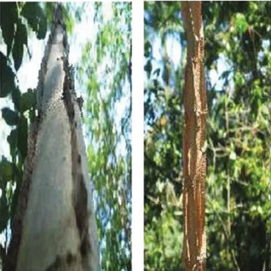
due to its salience in the bark like lamina, and owed to it the tree is named as “icarapé”, “caniveteiro”, “casco-de-jacaré” and mainly as “pau jacaré” (alligator stick) (Fig. 1). This is the first phytochemical study of P. gonoacantha in which we describe the presence of three cicloartenones, cicloartanol, three steroids, sitos-terol-3-O-β-D-glycopiranoside, three pentaciclic
triter-penes, methyl gallate, the ester asperphenamate, and five flavonoids, apigenin, apigenin-5-methyl ether, 7,4′
-di-hydroxy-3′,5-dimethoxyflavone, vitexin and isovitexin
(Fig. 2).
Fig. 1 – Stalk aspect ofPiptadenia gonoacantha, “pau-jacaré”.
MATERIALS AND METHODS
GENERALEXPERIMENTALPROCEDURE
Melting points have not been corrected. IR spectra were recorded on a Perkin-Elmer 1605 FT-IT spectrophoto-meter using KBr for solids and film for liquid samples (range 4000-600 cm−1). 1H and13C NMR spectra
(in-cluding 1D and 2D specials techniques) were recorded on a Brüker AC-200 (1H: 200 and 13C: 50 MHz) of
UFRRJ, and Brücker DRX-500 (1H: 500 and13C: 125
MHz) of UFC. DMSO-d6, CD3OD or CDCl3with TMS
as internal standard were used as solvents. Bruker Ac-200 was used in the NOEDIFF experiments. LRMS were recorded on Varian saturn 2000 instrument with ion trap at 70eV and electron ionization. The Chromatography
columns were packed with silica gel (Vetec and Aldrich 0.05-0.20 mm) and Sephadex LH-20 (Sigma, USA); sil-ica gel F254 G (Vetec) was used for preparative TLC; aluminum backed (Sorbent) silica gel plates W/UV254 were used for analytical TLC, with visualization un-der UV (254 and 366 nm), with AlCl3-ETOH (1%),
Lieberman-Burchard and/or Godin reagents, or expo-sure to iodine vapor.
PLANTMATERIAL
The branches and leaves of Piptadenia gonoacantha (Mart.) J.F. Macbr (Fig. 1) were collected in UFRRJ Campus, Seropédica, Rio de Janeiro, Brazil, in 2005 by Professor Acácio Geraldo de Carvalho. A voucher spec-imen (RBR 6939) has been deposited at RBR Herbar-ium, Instituto de Biologia, UFRRJ.
EXTRACTION ANDISOLATION
The powdered branches (1448 g) and leaves (560 g) of Piptadenia gonoacanthawere extracted with methanol at room temperature. The solvent was removed under vac-uum to yield the residuesPGBrM(46.4 g) andPGLM
(19.7 g), respectively. The bark (650.0 g) was extracted with dichlorometane and methanol, and the residues PG-BaD(5.0 g) and PGBaM (70 g) were obtained. The residuePGBrM (40.4 g) was partitioned into CHCl3,
ethyl acetate, and methanol:H2O (9:1) to yield fractions PGBrMC (4.0 g), PGBrMA (4.5 g), and PGBrMM
(24.3 g), respectively. FractionPGBrMCwas chroma-tographed on a silica gel column eluting initially with CHCl3and gradually increasing the polarity with MeOH
to give 35 subfractions. The fractionsPGBrMC-6-7, af-ter recrystallization from MeOH, afforded a solid com-posed by the mixture of 4, 5 and6. The subfraction
PGBrMC-2-11 was further purified by CC eluted with CHCl3100% to obtain10(31.0 mg). Subfraction PGBr-MC-16-20 was further purified by crystallization from methanol to afford 4a(37.2 mg). Fraction PGBrMA
was subjected to silica gel CC eluting with CHCl3:MeOH
and increasing the polarity with MeOH (100%) to ob-tain 33 subfractions. FractionsPGBrMA-6-7 was pu-rified in silica gel CC eluting with CHCl3:MeOH (9:1)
R
1=R
3=H, R
2= glu
R
1=Me, R
3=H, R
2= glu
R
1=R
2=H, R
3= glu,
R=H
R=CH
3R
1=OH, R
2=H
R
1=OCH
3, R
2=H
R
1=R
2=OCH
3O OCH3 RO RO OR 2 3 4 5 6 7 1 10
O
R
1 3 4 6 8 9 11 12 13 14 16 17 18 19 20 21 28 2930 4 29
6 3 10
RO
R
1 1 8 9 11 12 13 14 16 17 18 19 20 21R=H, R
1=
, R=glu
R=H, R
1=
R=H, R
1=
22 23 24 25 26 27 22 22 23 2 3 24 24 25 25 26 27 27 28 28 28 29 26
R=
R=
R=
with 3-OH
22 23 24 25 26 27 31 22 22 23 23 24 24 25 25 26 27 27O
OH
R
2O
R
1HO
2 3 4 5 6 7 8 91' 2' 3'
4' 5' 6'
N
O
O
N
O
O
H
H
H
H
H
H
H
H
1 2 3 4 5 6 7 8 9 10 11 12 13 14 15 16 1' 2' 3' 4' 5' 6' 7' 8' 9' 10' 11' 12' 13' 14' 15' 16'
O
OR
1O
OR
1R
1O
R
3R
2 2 3 4 5 6 7 8 9 1' 2' 3' 4' 5' 6' 29 23 20 10 O 1 3 4 6 8 9 11 12 13 14 16 17 19 20 2124 25 26 30 27 29 28
R
1,R
2=O
R
1=OH, R
2=H
10 1 3 4 6 8 9 11 12 13 14 16 17 19 21 23 24 25 26
R
1R
2 27 28 18 30Fraction PGGMA-12 was further purified by TLC (CHCl3:AcOEt:MeOH, 7:2.5:0.5) to give12(5.0 mg).
Fraction PGBrMA-21 was subjected to silica gel CC eluting with CHCl3:MeOH and increasing the polarity
with methanol to obtain 8 subfractions; fraction PGBr-MA-21/6 was purified by TLC (CHCl3:MeOH, 9:1) to
give13(6.5 mg).
The residue PGLM (15.0 g) was extracted with CHCl3 to obtain the fractions PGLMC (3.9 g) and PGLMM(10.4 g), respectively. The fraction obtained with chloroform had a mixture of hydrocarbons and steroids. The residue from the methanol fraction PGL-MM (10.0 g) was chromatographed over silica gel, eluted with CHCl3:MeOH (8:2) as eluent and
increas-ing the polarity until MeOH 100%. Eleven fractions were collected. FractionPGLMM-2 was subjected to silica gel CC eluting with CHCl3:MeOH (9:1) to obtain
5 fractions, including thePGLMM-2/2-3 with16(112.0 mg). FractionPGLMM-2/4 was subjected to silica gel CC eluting with CHCl3:MeOH (8:2) to afford 6
frac-tions. Fraction PGLMM-2/4-4 afforded14(24.0 mg) and fractionPGLMM-2/4-5 was applied to a Sephadex LH-20 gel column eluting with CHCl3:MeOH (7:3) and
furnished15(25.0 mg).
The dichlorometane extract from the bark ( PG-BaD, 4.0 g) was fractionated on a silica gel column using hexane as the initial eluent and increasing the polarity with chloroform and methanol until methanol (100%). Sixty fractions of 25 ml were collected. The solid ma-terial obtained from the fractions 7-10 yielded 1 + 2 + 3 (54.7 mg). Fractions 11-14 yielded a solid 7 (53.4 mg). Fractions 23-25 afforded a solid 3a (99.8 mg), and fractions 47-49 were crystallized from methanol to yield the mixture4 + 5 + 6(53.9 mg). The extract PG-BaM, (70.0 g) was dissolved in methanol:water (8:2) and partitioned with dichlorometane, ethyl acetate and buthanol. The residues PGBaMD(2.0 g), PGBaMA
(5.8 g),PGBaMB(4.9 g) andPGBaMM(50.3 g) were obtained from the respective solutions. PGBaMD(1.5 g) was fractionated on a silica gel column using chloro-form as the initial eluent and increasing the polarity with methanol until methanol (100%). Thirty fractions of 25 ml were collected and analyzed by TLC plate. Frac-tions 15-20 (340 mg) were submitted to flash silica gel column using hexane and methanol mixture to methanol
100%. Twenty fractions of 15 ml were collected and an-alyzed by TLC. FractionsPGBaMD-15-20/3-5 yielded a solid after crystallization from methanol, which was identified as8(82.2 mg). FractionsPGBaMD -15-20/9-12 were crystallized from dimethylketone affording 9
(86.9 mg).
Tri-O-methylvitexin (5,7,4′
-trimethoxy-flavone-8-C-glucopiranoside,14a): 1H NMR (200 MHz, DMSO-d6)δH: 8.09 (d, J=8.0Hz, H-2′,6′), 7.0 (d, J=8Hz, H),
6.60 (s, 2H, H-3 and H-6), 4.70 (d,J=10 Hz, H-1′′), 3.92,
3.88, 3.83 (s, 3H each), 3.9-3.2 (m).
Methyl-gallate (16): 1H NMR (200 MHz,
DMSO-d6)δH: 9.5 (HO), 6.96 (s, 2H), 3.72 (s, 3H);13C-NMR
(50.3 MHz, DMSO-d6): δC166.7 (C-7), 145.9 (C-3,5),
138.8 (C-4), 119.7 (C-1), 108.9 (C-2,6), 51.9 (OCH3);
Methyl trimethyl-gallate:1H NMR (200 MHz,
DMSO-d6)δH: 7.21 (s, H-2,6), 3.82, 3.81, 3.81, 3.72 (s, OCH3
×4).
RESULTS AND DISCUSSION
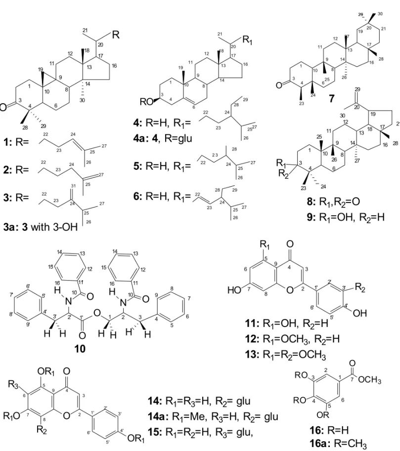
The phytochemical investigation of the extracts from the leaves, branches and bark of Piptadenia gonoacantha allow the identification of four cycloartane triterpenes, cycloartenone (1), cycloartan-25-26-en-3-one (2), 24-methylene-cycloartanone (3) and 24-methylenecycloar-tanol (3a), three steroids, sitosterol (4), campesterol (5), and stigmasterol (6), a saponin, sitosterol-3-O-β
-D-glu-copyranoside (4a), three pentaciclic triterpenes, fried-elin (7), lupenone (8), and lupeol (9), the N-benzoyl-phenylalanine-2-benzoylamide-3-phenylpropyl ester (asperphenamate,10), five flavonoids, apigenin (5,7,4′
-trihydroxyflavone,11), 5-methylapigenin (12), 7, 4′
-di-hydroxy-3′, 5-dimethoxyflavone (13), vitexin
(8-C-glu-copyranosyl-5, 7, 4′-trihydroxyflavone,14), and
isovite-xin (6-C-glucopyranosyl-5,7,4′-trihydroxyflavone, 15),
and methyl gallate (16), Figure 2. Their structures were established on the basis of spectral analysis, comparison with literature data and GC-MS analysis of steroids and cycloartenones mixtures.
The identification of compounds1-3, 3a,4-6and
7-9was achieved by the analysis of IR, NMR and GC-EIMS spectra and comparison with literature data. The
1H and 13C NMR spectra and the use of the Olea and
identifica-tion of the respective series of1-3a (cycloartane), 4-6
(steroids) and 7-9 (pentacyclic triterpenes). Detailed analysis of13C NMR (BBD and DEPT), and
compar-ison with literature data allowed the identification of the cycloartenones (1-3) and 24-methylene cycloartenol
3a(Silva et al. 2005, Davies et al. 1992, Silveira and Pessoa 2005), friedelane (7), lupenes (8, 9) (Davies et al. 1992, Carvalho et al. 1995, Mahato and Kundu 1994) and steroids 4-6 (Dutra et al. 1992, Kojima et al. 1990, Chaurasia and Wichtl 1987). These structures were defined by the GC-MS analysis that allow the iden-tification of three compounds in the fractions group con-taining the cycloartenones: cycloartenone1 (Rt 14.29 min, M+
∙424), cycloartan-25-26-en-3-one (2: Rt 14.29
min, M+
∙424), 24-methylene-cycloartanone (3: Rt 15.61
min, M+
∙426); the pure compound
24-methylenecyclo-artanol (3a, Rt 16.14, M+
∙ 426]; the steroids in
mix-ture: campesterol (5: Rt 13.20 min, M+∙400), sitosterol
(4: Rt 14.81, M+∙414) and stigmasterol (6: Rt 16.66
min, M+∙412). The glycoside4a(sitosterol-3-O-β
-D-glucopyranoside) was identified mainly by1H and13C NMR (BBD and DEPT) data analysis and by compar-ison with literature data (Chaurasia and Wichtl 1987). The number of C, CH, CH2 and CH3 and comparison
of the values with those from the literature (Olea and Roque 1990, Davies et al. 1992, Carvalho et al. 1995, Mahato and Kundu 1994) for7-9allowed to confirm the structure of the triterpenes, friedelin (7), lupenone (8) and lupeol (9).
The ester10, a solid (MP 184-186◦C), was
identi-fied by IR, NMR (1D and 2D) and mass spectra analy-sis. The IR spectrum of10showed absorption bands of N-H (νNH 3310 cm−1),νC=O (1750 cm−1),νCO (1640
cm−1), besides bands of,ν
C-Oand bands characteristics
of aromatic rings. The1H and13C NMR and 2D cor-related NMR techniques, [1H-1H- COSY and 1H-13 C-COSY-n
JC H (n=1, HMQC; n=2 and 3, HMBC)] were
used to identify this substance and make the complete proton and carbon-13 chemical shift assignments. The analysis of1H NMR,1H-1H- COSY and1H-13 C-COSY-1J
C Hspectra allow the identification of signals of
hydro-gens in aromatic rings (δH7.66-7.15) that were
compat-ible with four mono substituted benzene rings, signals atδH 4.85-2.8 of five methylene groups and two
me-tine [δH 2.85/2.93 (dd, 1H each);δH 3.17/3.22 (dd, 1H
each),δH 3.96/4.47 (dd, 1H) andδH 4.84 (t) and 4.53
(m)] connected to carbonsδC H2: 37.03, 37.20, 65.41,
and withδC H: 54.50 and 50.21, respectively. Besides
the signals ofnJ
C H detected in HMBC spectrum, the
values of hydrogen and carbon-13 chemical shift of10
were compared with those of ester described by Cata-lan et al. (2003), named N-benzoylphenylaCata-lanine-2- N-benzoylphenylalanine-2-benzoylamide-3-phenylpropyl ester, isolated from Cro-ton hieronymi(Catalan et al. 2003). The mass spectrum shows peaks at m/z(%): 355 (10), 328(50), 238 (70), 146 (100), 118 (60), 91(70) that were used to confirm the structure of10. This ester was isolated fromZeyhera digitalis(Bignoniaceae) (Faccione et al. 2004), Piper aurantiacatum(Piperaceae) (Banerji and Ray 1981), and Medicargo polymorpha(Leguminosae) (Poi and Adity-achoudhury 1986). This compound has been isolated from fungus species, such asAspergillus flavipes(Clark et al. 1977), Anaphalis subumbellata(Talapatra et al. 1983),Penicilliumspecies (McCorkindale et al. 1978, Bird and Campbell 1982, Nozawa et al. 1989), and it has been named as asperphenamate.
The flavones11-13were identified by comparison of these1H and13C NMR data (including NOEDIFF
ex-periments of12and13) and mass-spectra, and compari-son with literature data. These compounds show positive test for flavonoids using AlCl3/EtOH in TLC plate. 1H
NMR spectra of flavone11show two broad singlets atδH
6.44 (1H), 6.20 (1H), one singlet atδH 6.68 (1H), two
doublets atδH 7.92 (J=8.0 Hz, 2H), and 6.90 (J=8.0
Hz, 2H), besides a singlet at 13.01 of quelated hidroxyl group (5-OH). These data were compared with those of 5,7,4′-trihydroxyflavone and confirmed the structure of
11 that is known as apigenin (Miyazawa and Hisama 2003). 1H NMR spectrum of12was similar to that one of 11only with an additional signal atδH 3.78 of the
methoxyl group. Besides the analysis of13C NMR and
1H-1H-COSY data, the spectra obtained by NOEDIFF
experiment show only one signal of NOE (4%) atδH
6.37 (H-6) by irradiation atδC H33.78, and NOE (14%)
atδH 7.84 (H-2′,6′) by irradiation atδH 6.50 (H-3). The 13C NMR data were identical to those of
5-O-methyl-apigenin (Wagner et al. 1976). The spectra of13show signal atδH6.31 (brs), 6.47 (brs), 6.57 (s), 7.44 (brs, 2H)
and 6.88 (d, J=8 Hz, 1H), and two singlets of OCH3at
with12were made with13and allow the identification of NOE atδH6.31 (H-6) and 7.44 (H-2′), confirming the
methoxyl group at 5 and 3′ positions. These data and
analysis of 1H×1H-COSY, besides the LREIMS
spec-trum [m/z(%): 314 (1), 180 (100), 163 (50), 147 (10), 137 (50), 124 (20), 109(10)], allow the identification of
13as 7,4′-dihydroxy-5,3′-dimethoxyflavone.
The 1H NMR spectrum of flavonoids 14 and 15
shows signals of a flavone moiety containing four groups: three hydroxyl group and one sugar unit in both14and
15 as indicated by the following signals: 14: δH 8.0
(d, J=8Hz, 2H)/6.88 (d, J=8Hz, 2H) (AA′BB′system),
6.77 (H-3)/6.26(H-6), 4.68(d, J=10 Hz, 1H), multiplet between 3.8-3.0 and singlet at 13.2;157.90(d, J=8 Hz, 2H)/6.92 (d, J=8Hz, 2H) (AA′BB′ system),
6.75(H-3)/6.53(H-8), 4.58(d, J=10 Hz, 1H), multiplet between
δH 4.5-3.0 and 13.6(s). Comparison of the13C-NMR
(BBD and DEPT) data showed that all the carbon chem-ical shifts were similar, but small differences wereδC H
93.7, δC 79.0, 108.5 in 15. These data and
compar-ison with 1H and 13C NMR literature data, allow the identification of 14as vitexin (Zhou et al. 2005), and
15 as isovitexin (Pedras et al. 2003). NOEDIFF ex-periments confirmed these identifications. Irradiation of 14 at δH O-5 (13.2) shows NOE at δH 6.78 (H-6),
and irradiation onδH-3shows NOE atδH 8.0 (H-2′.6′).
The same experiments were made with 15and the ob-tained results were according with its identification as isovitexin. Methylation of 14 (in methanol) with di-azomethane ether solution yielded 14a, which is addi-tional data to confirm the identification of14.
The IR,1H and13C NMR spectra of16were
ana-lyzed and compared with literature data to identify this compound as methyl gallate (Scott 1972). The tri-O-methyl derivative obtained by the treatment of 16with diazomethane ether solution yielded 16a (see experi-mental) and confirmed its identification.
ACKNOWLEDGMENTS
The authors are grateful to Conselho Nacional de De-senvolvimento Científico e Tecnólogico (CNPq), Fun-dação Carlos Chagas Filho de Amparo à Pesquisa do Estado do Rio de Janeiro (FAPERJ), Coordenação de Aperfeiçoamento de Pessoal de Nível Superior (CAPES) for grants and fellowships, and thank CENAUEMN,
UFC, Fortaleza-CE, Brazil, for the 500 MHz NMR spectra.
RESUMO
O estudo fitoquímico de galhos dePiptadenia gonoacantha
(Mart.) J.F. Macbr. (Leguminosae-Mimosoideae), comumente conhecida como “pau jacaré”, forneceu sitosterol, estigmas-terol, o éster N-benzoilfenilalaninato de 2-N-benzoil-3-fenil-propila, conhecido como asperfenamato, 3-O-β
-D-glicopira-nosil-sitosterol, além de três flavonóides, apigenina (5,7,4′
-triidroxiflavona), apigenina-5-O-metil éter e 7,4′-dihidroxi-3′,
5-dimetoxiflavona. Das folhas isolaram-se galato de metila e dois flavonóides, 8-C-glicopiranosil-5,7,4′-trihidroxiflavona e
6-C-glicopiranosil-5,7,4′-trihidroxiflavona, conhecidas como
vitexina e isovitexina. Das cascas desta planta isolaram-se uma mistura de sitosterol, campesterol e estigmasterol; mistura de cicloartenona, cicloartan-25,26-en-3-ona e 24-metileno-ciclo-artanona, além dos triterpenos, 24-metilenocicloartenol, fri-delina, lupeol e lupenona. As estruturas foram estabelecidas através de análise de espectros de IV, RMN1H e13C e massas, além de análise com CG-EM para identificar os componentes das misturas de cicloartanos e esteróides. O éster conhecido como asperfenamato, os flavonóides e os cicloartanos estão sendo registrados pela primeira vez emPiptadenia.
Palavras-chave: Leguminoseae, Piptadenia gonoacantha, terpenóides, asperfenamato, flavonóides, pau jacaré.
REFERENCES
BANERJIAANDRAYR. 1981. Auranamide, a new pheny-lalanine derivative isolated fromPiper aurantiacumWall.
Ind J Chem B 20B: 597–598.
BIRDBAANDCAMPBELLLM. 1982. Disposition of myco-phenolic, brevianamide A, asperphenamate, and ergoste-rol in solid cultures ofPenicillium brevicompactum. Appl Environ Microbiol 43: 345–348.
CARVALHO MG DE, ALMEIDA MEL DE, HAUPTLI MB
AND MELEIRO LAC. 1995. Triterpenos Isolados de Eschweilera rabelilanaMori (Lecythidaceae). Rev Univ Rural Ser Cienc Ex e da Ter 17(1-2): 33–36.
CATALANCAN, HELUANICS, KOTOWICZC, GEDRISTE
ANDHERZW. 2003. A linear sesterterpene, two
squa-lene derivatives and two peptide derivates from Croton hieronymi. Phytochemistry 64: 625–629.
CHAURASIANANDWICHTLM. 1987. Sterols and steryl
CLARKAM, HUFFORDCD ANDROBERTSONLW. 1977. Two metabolites fromAspergillus flavipes. J Nat Prod (Loydia) 40: 146–151.
CORREAMP. 1984. Dicionário de plantas úteis do Brasil, e das exóticas cultivadas, Rio de Janeiro, RJ, Editora Minis-tério da Agricultura, Instituto Brasileiro de Desenvolvi-mento Florestal, 4329 p.
DAVIESNW, MILLERJM, NAIDURANDSOTHEESWARAN
S. 1992. Triterpenoids in bud exudates of FijianGardenia
species. Phytochemistry 31: 159–162.
DUTRA NN, ALVES H DE M, CARVALHO MG DE AND
BRAZ-FILHOR. 1992. Constituintes Químicos de Sima-ba obovata. Quim Nova 15: 10–14.
FACCIONEM, FERREIRADT, BRAZ-FIHORANDPOMINI
AM. 2004. Synthesis of asperphenamate and auranti-amide benzoate for structural revision. Rev Latinoamer Quim 32: 7–14.
GIESBRECHTAM. 1960. Bufotenine occurrence in Piptade-nia falcateseeds. An da Assoc Bras de Quim 19: 117–119.
KOJIMAH, SATO N, HATANO A ANDOGURA H. 1990.
Sterol Glucosides from Prunella vulgaris. Phytochem-istry 29: 2351–2355.
LEGLER G ANDTSCHESCHER R. 1963. The isolation of N-methyltriptamine, 5-methoxy-N-methyltriptamine and 5-methoxy-N,N-dimethyltryptamine from the bark of
Piptadenia peregrine. Naturwissenschaften 50: 94–95. LORENZI H. 1998. Árvores Brasileiras: Manual de
iden-tificação e cultivo de plantas arbóreas nativas do Brasil, 2aed., Nova Odessa, SP, Editora Plantarum, 357 p.
MAHATOSBANDKUNDUAP. 1994. 13C-NMR spectra of
pentacyclic triterpenoids – a compilation and some salien-te features. Phytochemistry 37: 1517–1575.
MCCORKINDALENJ, BAXTERRL, ROYTP, SHIELDSHS, STEWARTRMANDHUTCHINSON SA. 1978.
Synthe-sis and chemistry of N-benzoyl-O-[N′
-benzoyl-L-pheny-lalanyl]-L-phenylalaninol, the major mycelial metabolite ofPenicillium canadense. Tetrahedron 34: 2791–2795.
MIYAUCHIY, YOSHIMOTOT ANDMINAMIK. 1976. Ex-tractives of hardwood, IX, ExEx-tractives from heartwood of
Piptadeniasp. Mokuzai gakkaishi 22: 47–50.
MIYAZAWAMANDHISAMAM. 2003. Antimutagenic activ-ity of flavonoids fromChysantemum morifolium. Biosc Biotecnol Biochem 67: 2091–2099.
NASCIMENTOIA, GOMES MS, CARVALHO MG DE AND
CARVALHOAGDE. 2003. Deslocamentos químicos de 1H e13C de 5-H-flavanona e 5-H-flavonol isolados de Leguminosae. Rev Univ Rural Ser Cienc Ex e da Ter 22 (1,2): 81–87.
NOZAWA K, UDAGAWAD S, NAKAJIMA S AND KAMAI
KS. 1989. A dioxopiperazine derivative fromPenicillium megasporum. Phytochemistry 28: 929–931.
OLEARSCANDROQUENF. 1990. Análise de Misturas de
Triterpenos por RMN de13C. Quim Nova 13: 171–175. PATCHERIJ, ZACHARIUSDEANDRIBEIROO. 1959. Índole
alkaloids ofAcer saccharinum(silver maple),Dictyoloma incanescens, Piptadenia columbrine, andMimosa hosti-litis. J Org Chem 24: 1285–1287.
PEDRASMSC, CHUMALAPBANDSUCHYM. 2003. Phy-toalexins from Thlaspi arvensea wild crucifer resistant
virulent Leptosphaeria maculans: structures, syntheses and antifungal activity. Phytochemistry 64: 949–956. PIACENTES, BALDERRAMAL,DETOMASSILH, MORA
-LESL, VARAGASLANDPIZAC. 1999. Anadanthoside:
a flavanol-3-O-β-D-xilopyranoside fromAnadenanthera macrocarpa. Phytochemistry 51: 709–711.
POIRANDADITYACHOUDHURYN. 1986. Occurrence of
two rare amides inMedicago polymorpha. Ind J Chem B 25B: 1245–1246.
SCOTTKN. 1972. Carbon-13 nuclear magnetic resonance of biologically important aromatic acids I. Chemical shifts of benzoic acid and derivatives. J Am Chem Soc 29: 8564– 8568.
SILVAMSS, CITÓAMGL, CHAVESMHANDLOPESJAD.
2005. Triterpenos tipo cicloartano de Terezina-PI. Quim Nova 28: 801–804.
SILVEIRAERANDPESSOAODL. 2005. Constituintes mi-cromoleculares de plantas do Nordeste com potencial far-macológico: com dados de RMN13C. Fortaleza expressão Gráfica e Editora, 216 p.
STROMBERG VL. 1954. The isolation of bufotenine from
Piptadenia peregrina. J Am Chem Soc 76: 1707.
TALAPATRASK, PAL JK, MALLIKAK ANDTALAPATRA
B. 1983. Structure and synthesis of (-)-anabellamide. A new phenylalanine derivative ester amide fromAnaphlis subumbellataoccurrence of 4′-hydroxydehydrokawain. J
Nat Prod 46: 140–143.
WAGNERHW, CHARIVMANDSONNENBICHLERJ. 1976. Carbon-13 NMR spectra of naturally occurring flavo-noids. Tetrahedron Lett 21: 1799–1802.
ZHOUX, JINYONGP, FANGANDWUY. 2005. Isolation and purification of flavonoid glycosides from Trollius ledebouri using high-speed counter-current