UM inho | 20 14 Ana Mar garida F err eir a da Cunha P ain, emo
tion and cognition in lef
t and right sided peripheral neuropat
hies: hemispheric-specific in
vol
vement of dopamine
Universidade do Minho
Escola de Ciencias da Saúde
Ana Margarida Ferreira da Cunha
Pain, emotion and cognition in left and
right sided peripheral neuropathies:
hemispheric-specific involvement of dopamine
Ana Margarida Ferreira da Cunha
Pain, emotion and cognition in left and
right sided peripheral neuropathies:
hemispheric-specific involvement of dopamine
Outubro de 2014
Dissertação de Mestrado
Mestrado em Ciências da Saúde
Trabalho Efetuado sob a orientação do
Professor Doutor Hugo Almeida
Universidade do Minho
Escola de Ciencias da Saúde
ii
DECLARAÇÃO
Nome: Ana Margarida Ferreira da Cunha
Endereço electrónico: [email protected] Número do Bilhete de Identidade: 13830531
Título da dissertação: Pain, emotion and cognition in left and right sided peripheral neuropathies: hemispheric-specific involvement of dopamine
Orientador: Professor Doutor Hugo Almeida Ano de conclusão: 2014
Ramo de conhecimento do mestrado: Mestrado em Ciências da Saúde
É AUTORIZADA A REPRODUÇÃO INTEGRAL DESTA TESE/TRABALHO APENAS PARA EFEITOS DE INVESTIGAÇÃO, MEDIANTE DECLARAÇÃO ESCRITA DO INTERESSADO, QUE A TAL SE COMPROMETE;
Universidade do Minho, Outubro de 2014 Assinatura:
iii
Para a minha avó
v
Agradecimentos
Ao Hugo, pelo entusiasmo contagiante, por me incentivar a pensar e a olhar criticamente para todos os factos, resultados e teorias, pelas nossas discussões. Por acreditar em mim (e particularmente na minha capacidade de escrever a dita cuja revisão) muitas das vezes mais do que eu própria e por fazer questão de me ir lembrando para ser uma voz e não um eco.
Ao Marco, pelo companheirismo, pelo apoio, por partilhares comigo os altos e baixos deste ano. Pela tua alegria constante e pelas tuas mentirinhas irritantes, sem as quais já não te conheço.
Aos dois, pela ajuda, pelas horas partilhadas no biotério ou fora dele, por tudo o que me ensinaram. Esta tese, muito mais do que minha, é nossa.
À Ana e à Madalena também pela ajuda, pelo trabalho e pelas palavras de força.
A todos os NeRD’s que direta ou indiretamente me ajudaram com este trabalho, por estarem sempre disponíveis a partilhar o vosso tempo, conhecimento e reagentes e materiais comigo, pelas discussões e colaborações, pelo apoio, pelas palavras amigas, pela animação constante.
Ao gang da cantina, cuja companhia tornou a hora de almoço num tempo de alegria e recuperação de forças.
Às tratadoras, principalmente à Susana e à Celina pela paciência, por me ajudarem com tudo o que precisava, mesmo quando lhes pedia em cima da hora, por fazerem o seu trabalho com simpatia e prontidão.
Aos meus pais, por estarem sempre lá para mim, por compreenderem as minhas ausências, pelo orgulho, pelo amor e pela dedicação. Ao meu irmão, pelo carinho e pela
vi
companhia apesar dos jogos perdidos, apesar dos fins de semana que não pude estar com ele, apesar de toda a falta de atenção que lhe dei este ano.
Ao tio Domingos por assegurar a minha formação, pelo apoio e confiança constantes. À Ana, por ter sido um apoio incontornável ao longo deste ano, pelos desabafos, pelas partilhas, pela amizade.
Ao Nélson, por me ouvir constantemente a falar de dopamina, das minhas experiências ou de ciência em geral, por ter sempre as palavras (e as músicas) certas para cada ocasião e por me ensinar que tudo, incluindo o meu trabalho, pode (e deve) ser uma expressão de amor.
Ao Fábio, ao Miguel Ângelo, ao Mile, à Fabiana, ao Carlos, à Ana Luísa, à Tatiana e a todos os meus amigos pelas conversas, pelos lanches e jantares, por estarem lá para mim.
vii
Pain, emotion and cognition in left and right sided peripheral neuropathies: hemispheric-specific involvement of dopamine
Pain is a complex sensation with a physiological protective role. However, in some poorly understood and individual-specific conditions, pain can chronify becoming highly debilitating and comorbid with emotional and cognitive impairments. Studies in animal models of chronic pain indicate that pain duration, left/right location and age are critically associated with the manifestation of emotional and cognitive behavioral outcomes. Paradoxically, these studies also suggest some degree of dissociation between pain and its comorbidities. Dopamine (DA) is in this context an interesting research target as it is a neurotransmitter associated with emotion, cognition and pain and its hemispheric asymmetries and age-related variations have been thoroughly demonstrated.
This experimental work aims to elucidate the relation between pain and its comorbidities and to study the involvement of DA in these lateralized phenomena. First, the impact of left- and right-sided peripheral neuropathies on behavior was studied in painful and non-painful conditions. The spared nerve injury (SNI) model was installed in the left (SNI-L) or right (SNI-R) side and, 1 month later, behavior was analyzed in a battery of behavioral paradigms. SNI-L rats presented an anxiety-like phenotype in the dark/light and spontaneous burrowing behavior paradigms. Also, SNI-R rats presented increased impulsivity in variable delay-to-signal test. In both cases, side-specific effects were in accordance with previous observations of the group. Moreover, these behaviors manifested independently of the presence of allodynia, a hallmark of neuropathic pain. Secondly, D1 and D2 DA receptors’ mRNA was quantified by qPCR in the medial prefrontal cortex, orbitofrontal cortex, dorsal striatum and nucleus accumbens in both hemispheres. Results revealed an increase in D1 and D2 expression in the nucleus accumbens contralateral to the SNI lesion. Furthermore, ablation of cortical and subcortical DA efferences resulted in heightened impulsivity in right-sided lesions suggesting a causal relation between the side of the lesion and the behavioral impairments.
In conclusion, we demonstrated that the impact of a peripheral neuropathic lesion on emotional and cognitive behavior is to a certain extent independent of pain manifestations, suggesting that the nerve injury alone (i.e. in the absence of pain) can trigger central plastic events. Further studies should nevertheless be performed to clarify
viii
the relation between the peripheral nerve injury and the central events in the lateralized bias observed.
ix
Dor, emoção e cognição em neuropatias periféricas esquerdas e direitas: envolvimento lateralizado da dopamina
A dor é uma sensação complexa que possui uma função protetora. No entanto, em alguns indivíduos, por razões ainda não entendidas, esta pode tornar-se crónica sendo frequentemente acompanhada por problemas cognitivos e emocionais. Estudos em modelos animais de dor crónica indicam que a duração da dor, a localização à esquerda/direita e a idade estão intimamente associados com a manifestação de comportamentos emocionais e cognitivos. Paradoxalmente, estes estudos também sugerem um certo grau de dissociação entre a dor e as suas comorbilidades. A Dopamina (DA) é neste contexto um alvo de interesse, uma vez que é um neurotransmissor associado com emoção, cognição e dor. Além disso, assimetrias hemisféricas e variações relacionadas com a idade também já foram demonstradas.
Este trabalho experimental tem como objetivo elucidar a relação entre a dor e as suas comorbilidades e estudar o envolvimento da DA neste fenómeno lateralizado. Inicialmente foi estudado o impacto de neuropatias periféricas esquerdas e direitas nos comportamentos emocionais e cognitivos em condições de dor e não dor. O modelo Spared Nerve Injury (SNI) foi realizado no lado esquerdo (SNI-L) ou direito (SNI-R) e 1 mês mais tarde, o comportamento foi analisado numa bateria de paradigmas comportamentais. Os ratos SNI-L demostraram um fenótipo ansioso nos testes dark/light e spontaneous burrowing. Além disso, os ratos SNI-R apresentaram um aumento de impulsividade. Nos dois casos, os efeitos do lado estão de acordo com as observações prévias do grupo. Mais ainda, estes efeitos manifestam-se independentemente da presença ou não de alodinia, uma característica da dor crónica. No segundo conjunto de experiências foi realizada a quantificação do mRNA dos receptores de DA D1 e D2 no córtex prefrontal medial, no córtex orbitofrontal, no estriado dorsal e no núcleo accumbens dos dois hemisférios. Os resultados revelaram uma expressão aumentada dos receptores no lado contralateral à lesão SNI no núcleo accumbens. Para além disso, a ablação dos eferentes corticais e subcorticais de DA resultou numa impulsividade acrescida nos animais com lesão direita sugerindo uma relação causal entre o lado da lesão e os défices comportamentais.
x
Em conclusão, demonstramos que o impacto de uma lesão neuropática periférica é, até certa extensão, independente da manifestação da dor, sugerindo que a lesão do nervo por si só (i.e. na ausência de dor) pode despoletar eventos plásticos centrais. Estudos futuros deverão no entanto ser realizados para esclarecer a relação entre lesões periféricas do nervo, os eventos centrais e o viés de lateralidade observado.
xi
TABLE OF CONTENTS
1. INTRODUCTION 1
1.1PAIN 3
1.1.1FROM THE NOCICEPTIVE STIMULUS TO PAIN 3
1.1.2PAIN MODULATION 5
1.1.3CATEGORIZATION OF PAI N 7
1.1.4CHRONIC PAIN COMORBIDITIES 10
1.2.DOPAMINE 12
1.2.1DOPAMINE AS A NEUROTRANSMITTER 12
1.2.2DOPAMINE AND ACUTE PAIN 13
1.2.3DOPAMINE AND CHRONIC PAIN 14
1.3RATIONAL AND AIMS 15
2. MATERIALS AND METHODS 17
2.1BEHAVIORAL PERFORMANCE IN PAINFUL AND NON-PAINFUL NEUROPATHIC CONDITIONS 19
2.1.1STUDY-DESIGN 19
2.1.2ANIMALS 19
2.1.3SPARED NERVE INJURY (SNI) 20
2.1.4PAIN BEHAVIOR ASSESSMENT 20
2.1.5BEHAVIORAL TESTS 21
2.1.6EUTHANASIA 23
2.2DOPAMINERGIC SYSTEM IN CHRONIC PAIN CONDITIONS 23
2.2.1ANIMALS 23
2.2.2SNI LESION AND PAIN ASSESSMENT 24
2.2.5EUTHANASIA 24
2.2.6D1R AND D2R MRNA QUANTIFICATION 24
2.3HABIT AND IMPULSIVITY IN 6-OHDA LESIONED ANIMALS 25
2.3.1ANIMALS 25 2.3.26-OHDALESIONS 25 2.3.3HABIT BEHAVIOR 26 2.3.4VDS 26 2.3.5EUTHANASIA 27 2.4STATISTICAL AN ALYSIS 27 3. RESULTS 29
3.1BEHAVIORAL PERFORMANCE IN PAINFUL AND NON-PAINFUL NEUROPATHIC CONDITIONS 31
xii
4. DISCUSSION 43
4.1GENERAL 45
4.2EXPERIMENTAL CONSIDERATIONS 46
4.3BEHAVIORAL PERFORMANCE IN PAINFUL AND NON-PAINFUL NEUROPATHIC CONDITIONS 48
4.4DOPAMINERGIC SYSTEM IN CHRONIC PAIN CONDITIONS 49
4.5IMPACT OF LATERALIZED DOPAMINE DEPLETION IN DECISION-MAKING 51
4.4CONCLUSIONS 52
xiii
Abbreviations
5-HT- 5-hydroxytryptamine (5-HTa) 6-OHDA- 6-Hydroxydopamine ACC- Anterior Cingulated Cortex
ADHD- Attention Deficit and Hyperactivity Disorder
ALL-L- Allodynic-left ALL-R- Allodynic-right AMY- Amygdala
BLA- Basolateral Nucleus of the Amygadala BP- Binding Potential
COMT- Catechol-O-methyl transferase CPP- Conditioned Place Preference CRF- Continuous Reinforcement Schedule CRPS- Complex Regional Pain Syndrome D/L- Dark/Light Test
D1-5R- Dopamine receptor 1 to 5
DA- Dopamine DH- Dorsal Horn
DNA- Deoxyribonucleic acid DRG- Dorsal Root Ganglia
EDTA- Ethylenediamine Tetraacetic acid EPM- Elevated Plus Maze
ERK- Neuronal Extracellular Signal-regulated Kinase
FC- Fear Conditioning
fMRI- functional Magnetic Resonance Image FST- Forced Swimming test
IASP- International Association for Study of Pain
DAT-1- Dopamine transporter 1 IL- Interleukin
L-DOPA - L-3,4-dihydroxyphenylalanine MAO-A- Monoamine Oxidase-A
mPFC- medial Prefrontal Cortex NAcc- Nucleus Accumbens N-All-L- Non-Allodynic-Left N-All-R- Non-Allodynic.Right NA- Noradrenaline
OFC- Orbitofrontal Cortex PAG– Periaqueductal Gray PD- Parkinson’s Disease
PET- Positron Emission Tomography PFA- Paraformaldehyde
PFC- Prefrontal Cortex
qPCR- quantification Polymerase Chain Reaction
RAIC- Rostral Agranular Insular Cortex RNA- Ribonucleic acid
Rpm- Rotations per minute RR- Random Ratio
RT- Room Temperature
RVM- Rostral Ventromedial Medulla SBB-Spontaneous Burrowing Behavior SNI-L- Spared Nerve Injury-Left SNI-R- Spared Nerve Injury-Left SNI- Spared Nerve Injury SNL- Spinal Nerve Ligation SN- Substantia Nigra
xv
SPF- Spontaneous Paw Flicks SS- Somatosensory
STR- Striatum
STT- Spinothalamic Tract TH- Tyrosine Hydroxylase TNF- Tumor Necrosis Factor VDS- Variable Delay-to-Signal VF- Von Frey monofilaments test VO- Ventral Orbitofrontal Cortex VTA- Ventral Tegmental Area WM- Working Memory
xv
Figures Index
Figure 1- Ascending and descending pain circuits. 4 Figure 2- Dopaminergic Pathways. 12 Figure 3- Experimental design of the behavioral performance in painful and non-painful
conditions. 19
Figure 4- Habit-behavior paradigm schematics. 26 Figure 5- Animals distribution by experiemtnal group. 32 Figure 6- Mechanical and spontaneous allodynia. 34 Figure 7- Anxious-like behavior in the Dark/Light test. 34 Figure 8- Total distance walked during Dark/Light test. 34 Figure 9- Percentage of gravel burrowed in Spontaneous Burrowing Behavior test. 35 Figure 10- Depressive-like behavior evaluated by swimming, struggling, immobility and latency
time in forced swimming test. 36
Figure 11- Learning and impulsivity evaluated by the Variable Delay to Signal test. 38 Figure 12- Mechanical allodynia by the VF test 37 Figure 13- Laterality Index of D1R and D2R mRNA expression in mPFC, NAcc, OFC and STR. 38
Figure 14- D1R expression in left OFC and mechanical allodynia. 39
Figure 15-Habit-behavior test 40
1
3
1.1 Pain
The International Association for the Study of Pain (IASP) defines pain as “an unpleasant sensory and emotional experience associated with actual or potential tissue damage, or described in terms of such damage” (Merskey & Bogduk, 1994) It is a complex experience that involves a sensory/discriminative dimension related with pain intensity and localization, an affective/motivational dimension associated with our capacity to interpret pain as an unlikable sensation and to modulate it according to our current status and a cognitive/evaluative dimension mostly related with our culture, education and memories (Casey, 1968).
The experience of pain starts at the peripheral primary afferent neurons - the nociceptor - that convey the signal to the spinal dorsal horn (DH) neurons, which, in turn, project supraspinally to multiple areas of the Brainstem, Thalamus and cortex. Activation of the nociceptor, i.e. nociception, and pain are autonomous, though related phenomena and can occur independently. For instance, tetraplegic patients do not feel evoked pain after a noxious stimulus in the limbs but reflexes are maintained.
Pain is an indispensable sensation since it activates withdrawal reflex, for instance in case of a burning, decreases the contact with the damaged parts, facilitating healing, and alert us that something is wrong. A dramatic evidence of this statement comes from individuals with congenital insensitivity to pain, which have no perception of the majority of painful stimuli without any other sensory deficit. These individuals present severe physical injuries including extensive burns and fractured bones and have a reduced lifespan when compared with the general population (Nagasako, et al., 2003).
On the opposite extreme, pain that persists for long periods of time without any evident biological gain is debilitating and frequently comorbid with increased anxiety, depression and cognitive impairments. The endogenous control pain system – which provides inhibitory and facilitatory control over the nociceptive stimulus – in a healthy individual has to be therefore under tight regulation at multiple stations of the neuroaxis.
1.1.1 From the nociceptive stimulus to pain
As stated in the previous section, pain is initiated by the stimulation of nociceptive afferent neurons. The cell-bodies of these neurons are located at the Dorsal Root Ganglia (DRG). From these, a single neurite ramifies projecting simultaneously to the periphery (skin, internal organs, bones and muscles) and to the DH of the spinal cord (Basbaum, et al., 2009) (Fig. 1). At the periphery, stimuli of different modalities (mechanical, thermal and chemical) can activate specific
4
afferent neurons, depending on the repertoire of the receptors expressed at their terminals (Harriott & Gold, 2009). Afferent neurons are categorized according to the caliber of their axons as Aβ, Aδ and C. Aβ are large myelinated fibers and
are therefore the fastest conducting axons of the three. They are mostly implicated in proprioception and light touch but some reports also indicate nociceptive-related activity (Djouhri & Lawson, 2004). On the other hand, Aδ and C fibers are intrinsically involved in nociception. Small myelinated Aδ fibers have a faster conduct velocity than unmyelinated C fibers and are responsible for the first sharp pain sensation; C fibers, on the contrary, are associated with a dull, less precise, secondary pain (Marchand, 2008).
The central terminals of afferent neurons synapse with secondary neurons at the DH of the spinal cord (Fig. 1) with a precise topographical organization. Fibers from specific body parts project to specific levels of DH and consequently to precise supraspinal loci, which allows pain localization. Even at the same level, different afferent fibers synapse at diverse locations: Aδ and peptidergic C fibers terminate mostly in the exterior part of dorsal horn (laminae I and II0 of Rexed laminae),
non peptidergic C fibers terminate at III and Aβ
synapse even more deep in laminae III to V (Ossipov, 2012). Some studies show that this specific organization goes even further by
revealing specific synapse loci for fiber subpopulations based on its expressed receptors (Braz, et al., 2014; Brown, et al., 1995). Secondary neurons project to supraspinal centers organized in different pathways, being the Spinothalamic tract (STT) one of the most characterized. The STT is classically divided into the lateral STT and the central STT based on their terminations on the
Figure 1- Ascending and descending pain circuits. Nociceptors stimulated in the periphery synapse in the dorsal horn (DH) of the spinal cord, which in turn sends inputs to supraspinal sites, namely to the thalamus. Pain modulation from brain nuclei goes through PAG and RVM, which project to the DH, facilitating or inhibiting pain. DRt=Dorsal Reticular nucleus, RVM= Rostral Ventromedial Medulla, LC= Locus coerulueus, PAG= Periaqueductal Gray área, LA=Lateral Amygdala, BLA=Basolateral Amygdala, CeA= Cental Nucleus of Amygdala
5
lateral nuclei of Thalamus and in the medial nuclei of Thalamus, respectively. The lateral STT projects further to the primary and secondary somatosensory (SS) cortices. These projections and SS cortices maintain a high level of somatotopy (spatial body representation) and therefore lateral STT is associated with the sensory and discriminative dimension of pain (Ploner, et al., 1999). On the other hand, neurons from central STT project to limbic system nuclei, being therefore precluded as part of the affective and motivational components of pain (Ploner, et al., 1999; Rainville, et al., 1997). Other tracts project to areas like Brainstem, Amygdala (AMY) or Hypothalamus and are involved in the discriminative and motivational components of pain but also in pain modulation, autonomic responses, sleep alterations and endocrine responses (see (Almeida, et al., 2006; Almeida, et al., 2004; Millan, 1999) for revision).
While most of the tract definition studies were performed in animal models, the involvement of specific brain areas in human pain response starts to be more easily studied with the development of non-invasive image techniques. I and II SS cortices, Anterior Cingulate Cortex (ACC), Insula, Thalamus and Prefrontal cortex (PFC) are areas described to be activated after a nociceptive stimulus in most of the image studies and consequently accepted to be closely involved in pain. Some studies also report activation of the Brain stem, AMY, Cerebellum and Striatum (STR) (Apkarian, et al., 2005). As mentioned before, activation of these distinct areas will give rise to the complex experience of pain in all its dimensions.
1.1.2 Pain Modulation
Pain is a subjective and individual experience. The amount of stimulation upon the nociceptors is not proportional to the amount of pain experienced and neither is the activation of the different brain nuclei (Bingel & Tracey, 2008). Between different individuals and even in the same person similar lesions can elicit different pain perceptions depending on the physiological condition and context. As stated previously, in the most extreme cases pain can even be experienced in the absence of any detectable lesion. For instance, in a functional Magnetic Resonance Image (fMRI) study by Osborne and Derbyshire (2010) it was reported that the observation of images containing painful conditions elicited similar patterns of activity as evoked by actual nociceptive experiences; these individuals also reported somatization upon the observation of the images. Modulation of pain is an important feature of the system because it enables a decrease or even a suppression of pain in survival threatening situations (Beecher, 1946). It also explains why anxiety, expectation and attention can result in an increase of
6
perceived pain or, on the other hand, why distraction and excitation can have the opposite result (Bingel, et al., 2011; Villemure & Bushnell, 2002).
Pain modulation operates via descending pathways, which act upon the afferent pathways in the DH of the spinal cord. The Periaqueductal Gray (PAG) area in the Mesencephalon was the first nuclei described to be involved in pain modulation. In a seminal study, Reynolds stimulated the PAG with a small current and the powerful anesthetic effect elicited allowed the execution of an invasive abdominal surgery in rats (Reynolds, 1969). Similar effects were also observed in humans (Richardson & Akil, 1977). PAG anesthetic effect is mediated by the Rostral Ventromedial Medulla (RVM), which in turn projects to the DH (Fig. 1). RVM receives inputs from the PAG but also from other areas that can modulate pain negatively like ACC and PFC, positively as Hypothalamus and Dorsal Reticular nucleus (DRt) or in both ways like AMY (Hardy & Haigler, 1985; Heinricher, et al., 2009; Lima & Almeida, 2002; Lorenz, et al., 2003; Martenson, et al., 2009; Strobel, et al., 2014). In that way, RVM is considered a critical relay in pain modulation. Studies in rodents demonstrated that the RVM presents bifunctional effects in pain sensation: it can inhibit pain when electrically stimulated with high intensity current (50-200 µA) or glutamate quantity (2.5 nmol) and facilitate when low current (5-25 µA) or glutamate quantity (0.25 nmol) is applied (Calejesan, et al., 2000; Gebhart, 2004; Zhuo & Gebhart, 1997). Very likely, this is possibly due to the presence of three different types of cells, not anatomically independent: On, Off and Neutral cells. In electrophysiological recordings On-cells increase firing upon the application of a noxious stimulus, just before a retraction movement is elicited (e.g. tail flick). Off-cells, on the other hand, maintain in normal conditions a tonic activity, which is interrupted upon the application of a noxious stimulus. These cells have been hypothesized to be involved in pain facilitation and inhibition, respectively. Finally Neutral cells present no alterations in their firing rate elicited by noxious stimulation (Fields, et al., 1983).
It is believed that overall pain perception results from the balance between facilitatory and inhibitory supraspinal stimulus from the RVM and other areas. These stimuli reach DH by different pathways including dorsolateral and ventrolateral funiculi. The interruption of these pathways has been show to block supraspinal inhibitory and facilitatory pain influences, respectively (Porreca, 2002; Zhuo & Gebhart, 2002). The involvement of different neurotransmitters and receptors in the process has also been demonstrated (Millan, 2002; Ossipov, et al., 2010).
7
Opioid receptors are one of the most studied. Their stimulation by opioids like morphine or endogenous opioid peptides like endorphins has a potent inhibitory effect on pain. Injection of opioids in spinal cord, AMY, PAG and RVM decrease pain perception (Fang, et al., 1989; Heinricher, et al., 1994; Helmstetter, et al, 1998; Sohn, et al., 2000) and administration of opioid’ antagonists systemically, in the PAG or in the RVM, has the opposite effect (Oliveras, et al., 1977; Sohn, et al., 2000). Opioids analgesic effect results in part from activation of Off-cells and inhibition of On-cells in RVM (Fang, et al., 1989; Fields, et al., 1983). Despite its side effects (constipation, nausea, respiratory depression, addiction) opioids are still very used in the clinic, especially in cancer-related pain and some hard to treat neuropathies (Ahlbeck, 2011; Ashburn & Staats, 1999).
Monoaminergic neurotransmitters like Noradrenaline (NA), a catecholamine involved in autonomic responses responsible for increased alertness, and Serotonin (5-HT:5-hydroxytryptamine), usually associated with feelings of happiness and well being, also play a role in pain modulation (Ossipov, et al., 2010; Pertovaara, 2006; Sommer, 2004). Locus Coerulueus, which is connected with PAG and RVM, and nucleus Raphe Magnus, a RVM’s nuclus, are the main afferent brain nucleus of NA and 5-HT, respectively, and both project to DH. Stimulation of PAG and RVM, which leads to antinociception, induces the release of NA and 5-HT in the spinal cord and the antinociceptive effect of PAG stimulation is blocked by serotonergic and NAergic antagonists administration, directly implicating these neurotransmitters in pain blockage (Cui, et al., 1999; Hammond & Yaksh, 1984; Hentall, et al., 2006). Central NA release has an antinociceptive effect, mostly mediated by α2 receptors (Pertovaara, 2006) and 5-HT induces pain facilitation and inhibition, depending of the stimulated receptors. Stimulation of 5-HT3, 5-HT4
and 5-HT2a receptors increases pain perception while stimulation of 5-HT1a, 5-HT1b, 5-HT2c and
5-HT7 is inhibitory (Dogrul, et al., 2009; Viguier, et al., 2013). Dopamine (DA), other
monoaminergic neurotransmitter, is also involved in pain modulation, and its role will be discussed further.
1.1.3 Categorization of pain 1.1.3.1 Stimulus
Pain can be classified according to the triggering stimulus as nociceptive, inflammatory and neuropathic (Treede, et al., 2008; Woolf, 2010, Merskey & Bogduk, 1994).
Nociceptive pain results from the activation of the nociceptors by noxious stimuli. This type of pain is short-lasting and it is not associated with lesions in the tissues (Woolf, 2010). On the
8
other hand, inflammatory pain can be both the result of a continued inflammation, like in the case of appendicitis, or be involved in the healing of damage tissues by discouraging m ovement and/or physical contact (Woolf, 2010; Xu & Yaksh, 2011). Inflammatory pain involves the presence of inflammatory modulators, like cytokines, which induce tissue sensitization that leads to hyperalgesia (increased pain response due to a usually painful stimulus) and allodynia (pain due to a usually innocuous stimulus) (Xu & Yaksh, 2011).
Finally, neuropathic pain arises from damage to the somatosensory system caused by a lesion or disease (Treede, et al., 2008). Neuropathic pain is the most frequent pathological pain and is associated with a number of clinical conditions such as diabetic neuropathy, trigeminal neuralgia, postherpetic neuralgia, fibromyalgia, neuropathic back pain and complex regional pain syndrome (CRPS) (Jay & Barkin, 2014; Taylor, 2006). Neuropathic pain patients complain about intense continuous or episodic pain sensations as stabbing, burn ing, shocking, itching and numbness, which are much of the times debilitating (Jay & Barkin, 2014; Woolf & Mannion, 1999). Moreover, treatment efficiency varies drastically from person to person (the best drugs benefit only 30 to 60% of the patients) and between chronic pain types, which implies that prescriptions are based in trial and error and even efficient therapies do not eliminate pain permanently (Finnerup, et al., 2005; Sindrup & Jensen, 1999).
1.1.3.1 Duration
Pain can also be classified according to its duration as acute - lasts from days to weeks (usually no more than 1 month) -or chronic- lasts more than 3 months and can last all life. Acute pain can refer to the first pain response associated with nociceptive pain and which does not last more than a few hours or to a secondary pain related with inflammatory pain and the healing process and that last from days to weeks.
This classification is based on pain duration, however, in fact, acute and chronic pain distinguish protective from pathological pain. Acute pain does not last more than a few weeks because it disappears as soon as the cause is gone or even before the healing process terminates (Xu & Yaksh, 2011). On the contrary, chronic pain remains long before the cause is gone and can even appear without any apparent reason. Nowadays, chronic pain is considered itself a disease and although it can have different etiologies, affect different body parts and respond differently to treatment, constant pain is the element in common.
9
A study from World Health Organization in 15 countries all over the world reveal that chronic pain affects in average 22% of people and Azevedo and colleagues show that it reaches 37% of Portuguese population (Azevedo, et al., 2012; Gureje & Korff, 1998). It is also important to consider that it not only impacts patients’ daily life but also has a huge economic impact, directly by the amount of money spent in the treatments and health care and indirectly by the costs of lost productivity since the permanent pain precludes patients to work. A recent study by Rasu and colleagues reports that only in United States $ 17.8 spent per year in medication prescribed for pain (Rasu, et al., 2014).
The mechanisms underlying arise and maintenance of chronic pain are not fully understood. However, studies in chronic pain patients and in experimental models, particularly concerning neuropathic pain, have been revealing important information at anatomical, functional and biochemical levels.
First, it is well accepted that chronic pain patients and neuropathic pain animal models show spontaneous ectopic activity generated in sensory nerve axons or in their cell bodies in DRG (Han, et al., 2000; Study & Kral, 1996), which arises between 12h and 2 days after injury and decreases over time (Han, et al., 2000; Sun, et al., 2005). Suppression of spontaneous activity, by nerve blockage, before or 3 to 5 days after injury reduces or totally eliminates pain behavior (Chaplan, et al., 2003; Xie, et al., 2005), implicating this mechanism in chronic pain initiation.
Other explored mechanism for chronic pain is central glia activation and releasing of proinflammatory cytokines. In animal models of neuropathic pain, microglia inhibition by administration of minocycline decreased mechanical hyperalgesia and allodynia (Hains & Waxman, 2006; Raghavendra, et al., 2003), directly implicating inflammation in chronic pain maintenance. This effect seems to be through the pro-inflammatory cytokines Tumor Necrosis Factor (TNF)-α and Interleukin 1(IL-1) release (Sommer, et al., 1999; Sorkin & Doom, 2000, Quintão, et al., 2006; Xu, et al., 2006).
Also, it was observed in humans and monkeys that after a nerve injury, cortical space formerly represented by the lesioned body part was functionally occupied by neighbor body parts (Elbert, et al., 1994; Merzenich, et al., 1984; Pons, et al., 1991; Wrigley, et al., 2009). Increased cortical reorganization is related with increased magnitude of pain in chronic back pain, complex regional pain syndrome and spinal cord injury patients (Flor, et al., 1997; Maihöfner, et al., 2003; Wrigley, et al., 2009). Nevertheless, it remains unknown if cortical reorganization is behind chronic pain appearance or if it is a response to pain maintenance.
10
Brain imaging studies showed also alterations in the grey matter volumes in several chronic pain conditions including chronic back pain (Apkarian, et al., 2004), fibromyalgia (Kuchinad, et al., 2007; Lutz, et al., 2008), trigeminal neuropathy (Obermann et al., 2013), CRPS pain (Geha et al., 2008) and chronic tension type headache (Schmidt-Wilcke et al., 2006) (see for review (May, 2008)). Cingulate, Orbitofrontal (OFC) and Insular cortices repeatedly present decreased grey matter volumes (Apkarian, et al., 2004; Emerson et al., 2014) which have been shown to revert upon analgesic treatment (Seminowicz et al., 2011). Similar observations were also made in an experimental model of neuropathic pain (Seminowicz et al., 2009). Of notice, these frontal brain areas, particularly the medial PFC (mPFC) and the OFC are critical elements of the networks involved in emotional and executive function (Kesner & Churchwell, 2011; Kouneiher, et al., 2009), which suggests a relation between chronic pain and the associated comorbidities.
1.1.4 Chronic pain comorbidities
Chronic pain is frequently comorbid with emotional and cognitive deficits (Bair & Robinson, 2003; Hart, et al., 2000; Moriarty, et al., 2011; Simons, et al., 2014). Chronic pain patients are 3 to 5 times more likely to develop depression than healthy subjects and its prevalence increases with pain severity (Bair & Robinson, 2003; DeVeaugh-Geiss et al., 2010). On the other hand, depressive patients complain more about pain and it has been shown that depressive symptoms can predict chronic pain appearance (Bair & Robinson, 2003). Chronic pain patients also have higher prevalence of anxiety (Jordan & Okifuji, 2011) and sleep disorders than healthy people (Menefee, et al., 2000; “Self-reported sleep and mood disturbance in chronic pain patients.,” n.d.) and present a suicide risk two times higher than healthy subjects (Tang & Crane, 2006).
In animal models of chronic pain, albeit some contradictory results, depression and anxiety-like behaviors have also been demonstrated ((Gonçalves, et al., 2008; Matsuzawa-Yanagida, et al., 2008; Wang, et al., 2011) confront with (Gonçalves, et al., 2008; Hasnie, et al., 2007; Kontinen, et also., 1999)). Interestingly, it has also been shown that basal anxiety traits are predictors of heightened pain after the installation of a neuropathy (Geerse, et al., 2006) suggesting, like in humans, a bidirectional relation between emotional factors and the sensory abnormalities.
The emergence of anxiety- and depression-like behaviors in the animal model is intrinsically related with the temporal factor, the former emerging 2-4 weeks after pain onset and the later 6-8 weeks (Suzuki, et al., 2007; Yalcin, et al., 2011); such suggests a well defined
11
kinetic of events between the onset of pain and the manifestation of behavioral abnormalities, which are not entirely understood. In addition, it has been demonstrated that the age of the individual plays a critical role as anxiety-like behavior in Spared Nerve Injury (SNI) rats increases with age and depressive-like behavior is only potentiated by the lesion in mid-aged animals (Leite-Almeida, et al., 2009). Furthermore, lesion side also impacts behavioral outcomes as rats lesioned in the left side spend less time in open arms when compared with Sham and right injured animals, in Elevated Plus Maze (EPM) test (Leite-Almeida, et al., 2012).
Concerning cognition, studies show that chronic pain patients have impairments in memory, particularly working-memory (WM) (Berryman, et al., 2013; Dick & Rashiq, 2007; Ling, et al., 2007; Luerding, et al., 2008), attention (Grisart & Plaghki, 1999) and decision-making in a Iowa Gambling Task (Apkarian, et al., 2004; Walteros, et al., 2011). Also, these cognitive deficits seem to be independent of emotional impairments (Grisart & Plaghki, 1999; Reyes Del Paso, et al., 2012).
Once again, similar deficits have been observed in animal models. For instance, Low and colleagues demonstrate that chronic neuropathic pain rats spend less time exploring a new object than Sham animals, revealing attention or memory impairments (Low, et al., 2012). In a chronic inflammatory pain rat model (intra-articular injection of Complete Freund's Adjuvant), impaired performance in a test analogous to Iowa Gambling Task revealed deficits in emotional decision-making as these showed a preference for high-risk options (Pais –Vieira, et al., 2009). In contrast, Grégoire and colleagues did not find differences in spatial recognition when comparing neuropathic and Sham rats (Grégoire, et al., 2012). Thus, as in the emotional studies, cognitive impairments depend of experimental conditions. Recent studies demonstrate the influence of lesion side and animals’ age in the effect of chronic pain in cognition. Right-side injured animals, but not left-side, show impairments in attentional set-shifting, WM and delay-to-signal impulsivity tasks in young animals (Leite-Almeida, et al., 2012, 2014). Left-sided pain, in turn, resulted in WM and reversal learning deficits but only in mid-aged rats (Leite-Almeida, et al., 2009).
Altogether, the animal studies suggest that the emergence of behavioural impairments in chronic pain conditions results from the interplay of numerous factors like pain duration, side and the age of the individual. Such most probably explains the apparent discrepancy observed between groups in the behavioural studies.
12
1.2. Dopamine
1.2.1 Dopamine as a neurotransmitter
Catecholamines are Tyrosine-derived chemicals which function both as hormones and neurotransmitters and include DA and NA (Eisenhofer, et al., 2004). L –tyrosine is converted into L-3,4-dihydroxyphenylalanine (L-DOPA) by Tyrosine Hydroxylase (TH), from which L-DOPA decarboxylase originates DA (3-hydroxytyramine). DA can further be converted in to NA by DA β-hydroxylase. Catecholaminergic neurons were identified by histochemical fluorescence and classified into groups according to their
localization. Areas A1 to A7 represent groups of noradrenergic neurons while areas A8 to A14 correspond to dopaminergic neurons (Dahlstroem & Fuxe, 1964; Felten & Sladek, 1983). Areas A9 and A10 correspond respectively to the Substantia Nigra (SN) and Ventral Tegmental Area (VTA), the major dopaminergic efferences to the cortical and subcortical areas. Though corresponding to only 1% of the total neurons in the human brain, this system
is of critical importance for motor and cognitive processes (Chinta & Andersen, 2005; Iversen & Iversen, 2007; Robbins & Roberts, 2007). Neurons from SN project to the STR via the Nigrostriatal pathway (Fig. 2), which is involved in motor control and coordination. Degeneration of these neurons, which happens for instance in Parkinson’s disease (PD), leads to motor impairments and other disabilities (Mazzoni, et al., 2012; Pavese, 2012). On the other hand, neurons from VTA project to the frontal cortex, as part of the Mesocortical pathway (Fig. 2), or to Nucleus Accumbens (NAcc), AMY and Hippocampus through Mesolimbic pathway. These pathways are involved in more emotional functions like motivation, reward and attention (Arias-Carrión, et al., 2010; Landau, et al., 2009; Wise, 2004). There are evidences that schizophrenia, a mental disorder characterized by the presence of hallucinations, impaired social behavior and paranoia, and attention deficit and hyperactivity disorder (ADHD),
Figure 2- Dopaminergic Pathways. Nigrostriatal pathway projects from SN to Sriatum. Mesolimbic and Mesocortic pathways project from VTA to Nucleus Accumbens and Prefrontal cortex, respectively. The first is more involved in motor coordination while the last two are related with motivation and reward. VTA=Ventral Tegmental Area, SNc= Substantia Nigra.
13
a disorder which symptoms are attention deficits, higher impulsivity and hyperactivity, are the result of alterations in these pathways (Genro, et al., 2010; Wu, et al., 2012).
Not long after the discovery of DA as a neurotransmitter, two G protein-coupled receptors were identified as DA receptors. Their distinction, D1R and D2R, was based, respectively, in the
stimulation or not of adenylyl cyclase. Three different receptors -D3R to D5R- were later identified.
Considering their similarities with D1R or D2R receptors, now D1R and D5R are known as the D1
R-like family and D2R, D3R and D4R as D2R-like family. Both families show differences in structure,
pharmacological properties, distribution and function (Missale, et al., 1998). 1.2.2 Dopamine and Acute Pain
A significant number of studies have been exploring the importance of DA in the context of pain. Pathologies characterized by substantial alterations of dopamine availability and/or processing have been providing interesting information in this respect. PD patients, for instance, although having normal ascending conduction of the nociceptive signal, have inferior pain thresholds and considerably more pain than general population (Beiske, et al., 2009) (Schestatsky, et al., 2007), which is diminished by administration of L-DOPA (Brefel-Courbon et al., 2005; Schestatsky et al., 2007). The alterations in muscular tonus might be a contributing factor for painful manifestations in these patients. However, the decreased availability of DA alone seems to be detrimental as PD patients with pain have a lack of habituation of sympathetic sudomotor responses to repetitive laser-induced pain stimuli, which reveals an abnormal control of the effects of pain inputs on autonomic centers.
In the opposite side, schizophrenia patients, which according to DA hypothesis have an overactivation of D2R (Seeman, 1987), present a diminished response to pain (Dworkin, 1994;
Potvin & Marchand, 2008), which is independent of their medication (Potvin & Marchand, 2008) and disease side-effects, since it is also present in schizophrenia patients’ families (Hooley & Delgado, 2001).
The role of DA in pain modulation and/or perception has been confirmed by other studies in healthy human subjects. For instance, the injection of the DA receptors agonist apomorphine, decrease the amount of pain experience during a noxious cold stimuli when applied simultaneously with a heat noxious stimulus in other body part, implicating DA in pain inhibition (Treister, et al., 2013). Moreover, Positron Emission Tomography (PET) studies show that D2/D3 Binding Potential (BP), in STR, decreases after pain induction (Scott, et al., 2007) and that this decrease correlates with pain threshold and low modulatory capacity (Hagelberg, et al., 2002;
14
Pertovaara, et al., 2004; Scott, et al., 2007). Which suggests i) an increase in DA release after pain stimulation and ii) that DA plays an inhibitory role in pain modulation. Also, genetic studies reveal that pain sensitivity is related with specific polymorphisms in the DA transporter 1 (DAT-1), DA degradation enzymes Monoamine Oxidase -A (MAO-A) and Catechol-O-methyl transferase (COMT) and DA receptors D3R and D4R genes (Diatchenko, et al., 2005; Potvin, et
al., 2009; Treister, et al., 2009). More specifically, it appears that people with polymorphisms which lead to lower DA levels, present higher sensitivity to pain.
Animal studies have also helped clearing the role of DA in pain modulation. Prolonged tail pinch stimulation in rats induce an increase of at least 25% in NAcc DA (Louilot, et al., 1986) indicating, like in humans, a DA release after pain stimuli. Similarly, apomorphine intrathecal administration in rats lead to a dose-dependent increase of nociceptive behavior responses latency in the hot plate and acetic acid tests, which was inhibited by the previous administration of DA but not NA, 5-HT or opioid antagonists (Jensen & Yaksh, 1984). The same authors, however, observed no differences in tail flick latencies in any of the tested doses, which suggest that DA antinociceptive effect bares some specificity.
Antinociception in formalin-induced responses was produced after chemical inhibition of DA (and NA) reuptake (i.e. increased synaptic DA availability) either systemically (Basile & Janowsky, 2007) or directly in the Rostral Agranular Insular cortex (RAIC) (Burkey, et al., 1999), reinforcing once again the importance of dopamine release in nociceptive modulation. Finally, 6-Hydroxydopamine (6-OHDA) lesions in SN and/or VTA, which completely depletes DAergic (and NAergic) inputs to the frontal circuitry, decrease latency to nociceptive behavior in rats and increase mechanical and thermal allodynia (Chudler & Lu, 2008; Dieb, et al., 2014; Saadé, et al., 1997; Zengin-Toktas, et al., 2013).
Both, human and animals studies, reveal that DA involvement in pain modulation is dependent of its receptors as the observed antinociceptive effects are mainly mediated via D2
R-like receptors and D1R-like agonists and antagonists apparently do not have an impact in the
animals nociceptive responses (Magnusson & Fisher, 2000; Mansikka, et al., 2005; Sheng, et al., 2009). DA receptors distribution in the brain can, for that reason, help clear the incongruities found in respect to the antinociceptive role of DA.
1.2.3 Dopamine and Chronic Pain
Besides its clear involvement in acute pain modulation, there are evidences of dysregulation in DAergic system in chronic pain patients and animals models. PET studies in
15
chronic pain patients show decreased presynaptic uptake of 6-[18F] fluorodopa and higher D2R
BP suggesting therefore reduced levels of DA (Jääskeläinen et al., 2001; Wood et al., 2007). Indeed, DA or its metabolites are diminished in the urine and Cerebrospinal Fluid (CSF) of chronic pain patients when compared with healthy controls (Legangneux et al., 2001; Riva, et al., 2012).
DA release after a painful stimulus, which correlates with perceived pain in healthy subjects, is altered in chronic pain conditions (Wood, et al., 2007) revealing impairments in DAergic reaction to pain stimuli. Furthermore, administration of Bupropion, a NA-DA reuptake inhibitor with antidepressive effects, decreased pain in chronic neuropathic pain patients (Moreira, 2011; Shah & Moradimehr, 2010).
In animals, injections of DA in ACC lead to a dose-dependent decrease in autotomy behavior in a rat model of chronic pain (López-Avila, et al., 2004). As in the acute pain, D2R seem
to play a major role in chronic pain since activation of this receptors in RAIC and STR induce antinociception in a neuropathic model, and in contrast, its inhibition was nociceptive (Ansah, et al., 2007; Coffeen, et al., 2008). In inflammatory and neuropathic pain animal models, a decrease in the nociceptive threshold was, just like in humans, observed after administration of Bupropion (Basile & Janowsky, 2007).
1.3 Rational and aims
Nowadays, it is accepted that the emergence of emotional and cognitive alterations in chronic pain patients and animal models is the result of continued pain. However, the literature suggests that pain alone, might not entirely explain the appearance of these comordibities. For instance, Dimitrov and colleagues recently reported the presence of emotional deficits in mice in the absence of pain manifestations. In this study, the cuff model was applied in the left sciatic nerve of C57BL/6J mice. When the cuff was removed 3 weeks later in a subgroup of animals, pain resolved but anxiety and depressive-like behaviors manifested at the same levels as the cuff group (Dimitrov, et al., 2014). Also, Ren and colleagues reported that right-sided SNI animals presented WM deficits whether or not allodynia manifested (Ren, et al., 2011). Secondly, as stated before, there is a temporal mismatch between the manifestation of pain, which starts immediately after the neuropathic lesion and the behavioral alterations, which take at least 2 weeks to emerge, suggesting a segregation between the sensory and the emotional/cognitive deficits (Suzuki, et al., 2007; Yalcin, et al., 2011). Furthermore, chronic pain in the right or in the left side of the body is associated with different emotional outcomes, although pain scores do not
16
differ. Men with left-sided pain show higher levels of anxiety and depression and lower quality of life than men with right-sided pain (Wasan, et al., 2010) and in an animal model of neuropathic pain, rats with lesion in the left paw show emotional deficits but no alterations in cognition and animals with lesion in the right paw display only cognitive problems (Leite-Almeida, et al., 2012). To test the hypothesis that pain is not needed to the establishment of emotional and cognitive deficits we decided, in a first moment, to study the behavior of SNI animals with and without pain.
The second part of the thesis, in turn, will focus on the biochemical alterations underlying the initiation and maintenance of chronic pain and its comorbidities. DA appears here as an appealing target since, as explained in the previous section, DAergic system is dysregulated in chronic pain conditions. Besides that, it has a pivotal role in WM, attention and decision-making, i.e. in behavioral domains affected by chronic pain, and it is involved in depression (Brown & Gershon, 1993; Cools & D’Esposito, 2011; de Wit, et al., 2012; Rogers, 2011; Savitz, et al., 2006). Finally, there are evidences of lateralization of the DAergic system in humans and rats (Larisch, et al., 1998; Molochnikov & Cohen, 2014; Nowak, 1989.; Schneider, et al., 1982; Tomer, et al., 2014; Vernaleken, et al., 2007).
Previous unpublished results from our lab showed that after habit induction rats lesioned in the left side are incapable of recognizing reward devaluation, indicating an impaired transition from habit-based to goal-directed behaviors in these animals. Once again, it is very likely that DA plays a role in this behavior.
Considering all of these, this master thesis intends to:
- Evaluate the emotional and cognitive behavior of right- and left-sided injured rats in painful and non-painful conditions
- Assess the state of the DAergic system in chronic pain conditions, considering brain hemisphere and lesion side.
17
19
2.1 Behavioral performance in painful and non-painful neuropathic conditions
2.1.1 Study-design
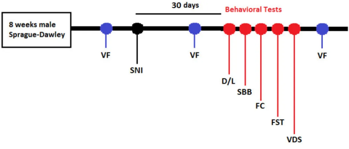
Two independent experiments were executed according to the study-design present in Fig. 3. A battery of behavioral tests (see 2.1.5) was performed 30 days after the installation of the SNI neuropathic lesion (see 2.1.3) and pain was measured with Von Frey (VF) monofilaments in three time points. The order of the behavioral tests was design to minimize possible cross negative impacts between tests on subsequent tests. The VDS was performed at the end of the battery as it requires food deprivation.
Figure 3. Schematic organization of the study 1 timeline. Behavioral tests started 30 days after SNI installation. The manifestation of allodynia was assessed 3 times: prior to SNI; after SNI pre- and post-behavior. VF=Von frey test, SNI= Spared Nerve Injury, D/L= Dark/Light test, SBB= Spontaneous Burrowing Behavior, FC= Fear conditioned, FST=Forced Swimming Test, VDS= Variable Delay-to-Signal
2.1.2 Animals
Two months-old male Sprague-Dawley rats were used. All of the procedures were made according to the guidelines of the European Communities Council Directive 2010/63/EU. Animals were housed in groups of three in a 12h light/dark cycle (lights on at 8 am) with controlled temperature (21±1°C) and humidity (50-60%). Rats had food (4RF21; Mucedola SRL, Settimo Milanese, Italy) and water ad libitum, except during the Variable Delay-to-Signal (VDS) test, in which food availability/consumption was restricted to the first hour of the circadian light cycle. Handling was done at least once a week until the start of behavior tests, beginning 2 weeks before surgery. Body weight was controlled along the experience.
20
2.1.3 Spared Nerve Injury (SNI)
Chronic neuropathic pain was induced using the SNI model, in the right (SNI-R) or left (SNI-L) hind paw, as described by Decosterd and Woolf (Decosterd & Woolf, 2000). Rats were anesthetized via intraperitoneal administration of 1:1.5 mix (1 ml/kg) of Dorbene® Medetomidine Hydrochloride) and Imalgene® (Ketamine), respectively. A blunt incision was then performed to expose the three branches of sciatic nerve: common peroneal, tibial and sural nerves. The first two nerves were individually ligated with a 4-0 suture (Silk Suture Thread, FST, Heidelberg, Germany) and distally cut; great care was taken to avoid lesions to sural nerve (spared nerve). In sham animals the nerves of right or left paw were exposed but left intact. After surgery muscle and skin were sutured separately in two layers, with 5-0 and 2-0 sutures (Coated vicryl braided absorbable suture, Johnson & Johnson, Inc., New Jersey, USA), respectively. 150µl of Antisedan® (Atipamezole Hydrochloride) were injected subcutaneously to reverse anesthesia and animals were then left to recover in their cages. One animal of each group (SNI-L, SNI-R and Sham) was present in all cages. Animals were monitored in the following days for open wounds and signs of inflammation. No major problem was observed in the study.
2.1.4 Pain behavior assessment
Measurement of allodynia was done using the up-and-down method with VF monofilaments test (Chaplan, et al., 1994). On the day before the first measurement, animals were habituated to the experimental setting, which consisted in an elevated mesh wire (approximately 50 cm).During habituation rats were left in the mesh for 3 minutes with their movement restricted by a plastic box (25 L x 15 W x 15 H) perforated to allow air exchange. In the test days, each animal was left alone for at least 30 seconds in the mesh. After that, spontaneous paw flicks (SPF) were scored for 2 minutes, without any interference. In the end, VF was done, as described by Chaplan and colleagues (Chaplan, et al., 1994). In this experiment, 8 monofilaments ranging from 0.4 to 15 g were used. The test started with the stimulation of the lesioned paw with the central monofilament (2 g). If the animal withdrawed the paw, a weaker monofilament was used, if not, a stronger one was applied. Stimulation was done until the animal responded to the 0.4 g, did not respond to 15 g monofilaments or after a total of 6 measures around threshold first crossing. 50% threshold was calculated using the formula:
50% g threshold =
21
in which Xf is the value (in log units) of the final monofilament used, K is the tabular value for the pattern of positive/negative responses and δ is the mean difference between stimuli (in this case it is equal to 0.224).
2.1.5 Behavioral tests
Behavioral tests started 4 weeks after SNI lesion and were done during the dark phase of the circadian cycle.
i) Dark/Light (D/L)
The D/L apparatus consists in a square arena (43.2x43.2 cm) in which half is brightly illuminated and the other half consists in a dark compartment (MedAssociates Inc. model ENV-515). Animals in the D/L face a conflict between staying in the close and safe compartment (dark) and the innate drive to explore a new and exposed environment (Crawley & Goodwin, 1980). Anxiety-like behavior in this paradigm manifests in higher D/L ratios. In the beginning of the test animals were placed in the illuminated area and left to explore the arena for 10 minutes. Infrared beams at the floor level allow to automatically monitor animal’s position. Time and distance spent in each area were calculated using Activity Monitor software (MedAssociates, Inc.). After each animal the arena was cleaned with 10% ethanol.
ii) Spontaneous Burrowing Behavior (SBB)
SBB evaluates the general well-being of rodents and it was also described as a good correlate of ongoing pain (Andrews, et al., 2012). It is based on the assumption that burrow is an ethologically relevant behavior in rodents and therefore alterations in SBB could signal disease and/or pain. The SBB protocol employed was the same described by Deacon (Deacon, 2006), except for some minor alterations as described. The SBB test was done in rats housing cages (without bedding) reserved for this sole propose. Each rat performed the test in the same location and in the same apparatus along the testing days. In the first day of habituation, empty PVC tubes (diameter: 90 mm, length: 210 mm) with one end closed were left in the rats’ home cages for a period of 24 hours. In the second day rats were putted in pairs in the SBB cages for 2 hours (1h with each cage mate); in this case the open end of the tubes were elevated 70 mm above the cage floor and each tube was filled with approximately 2 kg of gravel. On days 3, 4 and on test day each animal was placed alone in SBB cages for 2h after which amount of displaced (burrowed) gravel was weighted. Results are shown as the percentage of
22
initial gravel burrowed by the animals in test day. During the sessions no food or water was available in the SBB cages.
iii) Forced Swimming Test (FST)
To evaluate learned helplessness behavior the FST was done. Animals that spend more time immobile during the test are described as having a more depressive-like behavior (Porsolt, et al., 1977). Rats were placed in a glass cylinder filled with water (21-23°C) 30 cm deep, from which they cannot escape, and left there for 5 minutes. Two sessions in subsequent days were performed and the second was recorded for posterior behavioral scoring by a blind researcher using Etholog V 2.2 software (Ottoni, 2000). Immobility, struggling, swimming and latency times were calculated.
iv) Fear Conditioning (FC)
FC was done in a startle response box (SR-LABTM, San Diego Instruments, San Diego, CA,
USA) with a non-restrictive Plexiglas cylinder (diameter 8.8 cm, length 22.2 cm) and a steel grid through which an electric current could be passed.
FC protocol lasted for three consecutive days. In the first day (habituation) each animal was placed in the FC box for 11 minutes. In the second day (conditioning) each rat was left in the box for 11 minutes divided in 3 minutes without the stimulus and 8 minutes with 6 pairs of light/ shock (0.4±0.1 mA), with an inter-stimulus interval of 60 seconds. Light was on for 20 seconds and the shock was given immediately after light was turned off. In the last day (test day) the protocol was equal to the day before but no shock was given to the animal. Videos were recorded and freezing time analyzed in Etholog V 2.2 software (Ottoni, 2000). Freezing was defined as the complete absence of voluntary movements, except for respiratory movements.
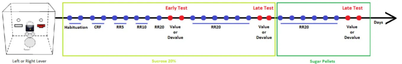
v) Variable Delay to Signal (VDS)
VDS was done in a 5-hole box (TSE-systems, Bad Homburg, Germany) as described by Leite-Almeida and colleagues (Leite-almeida, et al., 2013). The apparatus consists in a box with five holes in one side and a food magazine connected to a pellet dispenser on the opposite side. A total of 15 sessions were performed, two per night, including habituation, shaping and the VDS session, as explained below.
23
In the first night, rats were left exploring the box for 30 minutes. All the holes were closed and lights off. Sugared pellets (dustless precision pellets ® 45 mg, Bio-Serv) were left in the food magazine. In the second night of habituation, animals explored the box for 15 minutes. In these 2 sessions the central hole was open and 2-3 sugared pellets were also presented there. Food magazine, central hole and house lights were on during the entire session. 10 training sessions then followed. Each session started with the delivery of a sugared pellet in the food magazine and with the house-light on. Then, after a 3 seconds delay, the light in the central hole was switched on (for 60 seconds). If the animal nosepoked in that time (correct nosepoke) it was rewarded with a pellet. But if the animal nosepoked during the 3 seconds delay period when the light was off (premature response) or if it did not nosepok (omission) it was punished with 5 seconds in complete darkness and no reward was delivered. Each session ended after 100 complete trials or 30 minutes. Premature responses, correct nosepokes, omissions and perseverant responses were recorded. Correct nosepokes during the shaping protocol allow the study of learning capacity. In the VDS test proper (15th session), a total of 120 trials were
performed, consisting of 25 trials of 3 seconds delay in the beginning and in the end flanking 70 trials of 6 or 12 seconds delay (randomly distributed by the computer). Contrary to the shaping session, during the VDS animals are allowed to perform premature responses; these are registered but not punished. Prematurity rate – number of impulsive responses per time of available delay – was used to measure impulsivity.
2.1.6 Euthanasia
After behavior tests rats were euthanized. First they were anesthetized with an intraperotoneal injection of Eutasil ® (Pentobarbital). Then the sciatic nerve from both back paws was removed and stored at -80°C. Following perfusion with paraformaldeyde (PFA) 3% in PBS, brain and spinal medulla were removed, submersed in Richard-Allan Scientific™ Neg-50™ Frozen Section Medium (Thermo Fisher Scientific, Inc., USA) and stored for posterior analyzes.
2.2 Dopaminergic system in chronic pain conditions
2.2.1 Animals
Two months-old male Wistar-Han rats were used in this experimental set. Husbandry conditions were as described in 2.1.1.
24
2.2.2 SNI lesion and pain assessment
SNI lesion and pain assessment were performed following the procedures described in sections 2.1.3 and 2.1.4, respectively. Except for handling 1-2 times a week animals were left undisturbed during 1 month starting at SNI installation.
2.2.5 Euthanasia
Thirty days after SNI animals were euthanized. Euthanasia was done in a room of the animal facilities to decrease the time required for the procedure and minimize possible stress-related neurotransmitter fluctuations. A single cage at a time was brought to the room and between cages, sham, SNI-L and SNI-R animals were sacrificed by decapitation in alternate sequences to avoid biases, in a total time not superior to 2 minutes. The skull was then submerged in liquid nitrogen during 8 seconds and the brain immediately removed and placed in a cold brain slicer. 1 or 2 mm slices, depending in the areas of interest, were obtained. These were placed in a clean and cold Petri dish and mPFC, OFC, STR and NAcc from both hemispheres were macrodissected and immediately stored at -80°C.
2.2.6 D1R and D2R mRNA quantification
i) Ribonucleic acid (RNA) extraction and quantification
RNA extraction was done using Trizol® Reagent (Life technologies) according to the manufacturer instructions. 1 mL of Trizol reagent was added to the samples (in ice) and homogenization was done with a 23G needle. After homogenization, 200 µl of clorophorm was added, the tubes were manually shaken for 15 seconds and the mixture was kept 2-3 minutes at room temperature (RT). After that period, samples were centrifuged 15 minutes at 8000 rotation per minute (rpm) at 4°C. The upper phase containing the RNA was then removed to a new eppendorf and RNA was precipitated with 500 µl of isopropanol. After 10 minutes of incubation at RT, samples were centrifuged at 9000 rpm during 10 minutes at 4°C. Then, the supernatant was removed and the pellet was washed with 100 µl of ethanol 70%. Samples were centrifuged for 7 minutes at 5000 rpm at 4°C. Finally, ethanol was removed and the pellet was eluted in milliQ H2O. After RNA extraction, RNA in each sample was quantified using a nanodrop
spectrometer (Nanodrop Technologies, Inc., ThermoScientific, Wilmington, USA). 260/280 ratio of all the samples was higher than 1.8, ensuring its purity.
25
ii) DNase treatment and cDNA synthesis
In order to eliminate genomic DNA from the samples a treatment with DNase was done before cDNA synthesis. 1 µl of DNase and other of DNase buffer were added to 1 µg of RNA from each sample diluted in 10 µl of RNase free water. Samples were left 30 minutes at 37°C, the ideal temperature for DNase activity. 1 µl of EDTA was then added to stabilize RNA and samples were incubated for 10 minutes at 65°C, to inactivate DNase. cDNA was synthesized with iScript ® kit (Biorad) according to the manufacturer instructions. 4 µl of 5x iScript ® reaction mix and 1 µl of iScript ® Reverse Transcriptase were added to each sample and submitted 5 minutes at 25°C, 60 minutes at 42°C and 5 minutes at 85°C in a thermal cycler (MWG Biotech Inc. Primus 96 Thermal Cycler). cDNA was stored at -20°C.
iii) Quantification Polymerase Chain Reaction (qPCR)
D1R (Primers: Forward- 5- TCC TTC AAG AGG GAG ACG AA -3; Reverse- 5- CCA CAC AAA
CAC ATC GAA GG -3) and D2R (Primers: Forward- 5- CAT TGT CTG GGT CCT GTC CT-3; Reverse-
5- GAC CAG CAG AGT GAC GAT GA-3) gene expression was assessed in qPCR using Evagreen ® (Bio-Rad) reagent. GAPDH (Primers: Forward- 5- AGC CTC GTC TCA TAG ACA AGA TGG T -3; Reverse- 5- AGG TGA GCC CCA GCC TTC TCC -3) was used as the control gene. qPCR reaction ( 1 minutes 95°C and 40 cycles of 15 seconds at 95°C, 20 seconds at 60°C and 20s at 72°C) was done using the CFX96™Real-Time System (Bio-Rad). Transcript levels were calculated, by comparison, using the formula 2(−ΔΔCt). A laterality index (2(−ΔΔCt) left - 2(−ΔΔCt) right/ (2(−ΔΔCt) left + 2(−ΔΔCt) right) was used to evaluate the relative expression of the receptors.
2.3 Habit and impulsivity in 6-OHDA lesioned animals
2.3.1 Animals
A pilot study was conducted using 8 male Sprague-Dawley rats with 6 months. Husbandry conditions were as described in 2.1.1 except for food that was only available in the last hour of the light phase of the cycle. Body weight was controlled along the experience.
2.3.2 6-OHDA Lesions
Rats were anesthetized by intraperitoneal administration of a 2:3 mix (1 ml/kg) of Dorbene® (Medetomidine Hydrochloride) and Imalgene® (Ketamine), respectively. Then they were fixed in a stereotaxic frame and 2 µl of 6-OHDA (at 2 or 4 µg/µl) were unilaterally injected