Ana Filipa Martins Oliveira
Licenciada em Biologia
Microglial clearance function:
dependence on phenotypes
Dissertação para obtenção do Grau de Mestre em
Genética Molecular e Biomedicina
Orientador: Dora Maria Tuna de Oliveira Brites,
Investigadora Coordenadora e
Professora Catedrática Convidada,
Faculdade de Farmácia, Universidade de Lisboa
Co-orientador: Ana Sofia Iria Azevedo Falcão de Jesus,
Bolsa Pós-doutoramento,
Faculdade de Farmácia, Universidade de Lisboa
Júri:
Presidente: Prof. Doutor José Paulo Nunes de Sousa Sampaio
Arguente: Prof. Doutor Luís Filipe Ferreira Moita
Vogal: Prof. Doutora Dora Maria Tuna de Oliveira Brites
Microglial clearance function: dependence on phenotypes
Copyright Ana Filipa Martins Oliveira, FCT/UNL, UNL
Part of the results discussed in this thesis was presented in the following meeting:
Dora Brites, Sandra L. Silva, Ana Oliveira, Ana R. Vaz, Maria J. Diógenes, Nico van Rooijen, Ana M. Sebastião, Adelaide Fernandes, Cláudio Gomes, A. S. Falcão, Rui F.M. Silva. Microglia faces
AGRADECIMENTOS
O meu primeiro agradecimento é dirigido à Profª. Doutora Dora Brites pela oportunidade de realizar este projecto, dando assim início ao meu percurso neste mundo da ciência. Os seus conhecimentos e rigor científico assim como o seu espírito crítico foram qualidades essenciais para a concretização deste trabalho. Agradeço a orientação e as horas dispensadas na discussão desta tese, principalmente na fase final da sua elaboração.
Gostaria de agradecer de seguida à pessoa que orientou e seguiu de mais perto o meu trabalho, a Sofia. Um obrigado por todos os conhecimentos transmitidos durante este período e pela disponibilidade para discutir e ajudar em vários aspectos do meu trabalho. Agradeço ainda a tua sempre boa disposição que permitiram que rapidamente me sentisse que pertencia a este grupo.
Agradeço também ao Prof. Rui Silva e à Profª. Alexandra Brito por sempre se mostrarem disponíveis para discutir questões relativas ao meu trabalho. Adelaide, um muito obrigado pelas sugestões e pelo teu interesse neste trabalho. És sem dúvida uma inspiração, um exemplo de profissional que nunca vê limite para o conhecimento e que sempre procura fazer mais e melhor.
Quero também agradecer às restantes meninas que partilham comigo o dia-a-dia e que me receberam de abraços abertos. Conseguiram fazer-me acreditar que é possível trabalhar entre mulheres apenas! Obrigado a todas pela vossa preciosa ajuda e também pelos “raspanetes”…afinal também fazem parte! Desejo-vos a maior sorte a todos os níveis.
Às minhas companheiras de mestrado quero dizer que adorei partilhar com vocês esta fase da minha vida. Foram uma ajuda preciosa!
Cátia, apesar de não ter partilhado contigo muitos momentos fora do CPM, estiveste sempre perto para o que fosse preciso e sempre preocupada para que nada me faltasse. Desejo-te que consigas alcançar tudo aquilo que desejas porque mereces.
Inês, este percurso não teria sido o mesmo se não o tivesse feito na tua companhia. Agradeço-te principalmente por seres como és, amiga, sempre disponível para ouvir e ajudar. És uma pessoa 5* e não imaginas o quanto foi importante ter-te ao meu lado durante este ano. Espero que a distância não nos separe por muito tempo. Volta para mim…
Miguel e André, podiam ter sido um duo de música pimba mas em vez disso formámos aquela que vai ser a melhor banda do mundo, “Os Fitas Brancas e Uma Preta”…tenho muito orgulho em fazer parte deste grupo!
AGRADECIMENTOS Microglial clearance function: dependence on phenotypes
vezes nem eu me posso ouvir!) entre outras coisa menos “classy”. Não me esqueci...sei perfeitamente que vou entregar SEXTA-FEIRA!
Ter-vos conhecido tornou esta tese em algo bem mais divertido. Desejo-vos a maior sorte para esta nova fase que vão iniciar.
Quero também agradecer às restantes pessoas que partilham o dia-a-dia comigo naquela cave, por trazerem um pouco de luz àquele lugar tão sombrio.
Um OBRIGADO a toda a minha família pelo apoio que sempre me deram, por aceitarem o caminho que escolhi mesmo sabendo que não seria fácil. Agradeço a confiança que sempre depositaram em mim. Espero ser merecedora!
Um especial obrigado à minha MÃE...obrigado por seres como és, um exemplo de força e dedicação...és inspiradora…ADORO-TE.
Um agradecimento muito especial e com muito carinho vai para ti Pedro. Obrigado por teres estado sempre aqui, pelo apoio e por todo o amor que me deste. Obrigado por aturares os maus-humores inexplicáveis. Sei que não foi fácil e que nem sempre estive presente para ti mas quero que saibas que és uma pessoa muito importante e especial para mim. Ensinaste-me a saber aproveitar um pouco mais a vida...que nem sempre é só trabalho. Espero que também te tenha ensinado que existe vida fora da cama e que dormir é um desperdício de tempo!!
Obrigado por tudo…ADORO-TE.
Por último, mas não menos não menos importante é o agradecimento que faço aos meus amigos da terrinha. Obrigada pelos bons momentos que passámos juntos e por me ouvirem lamentar de como trabalhar sem receber não dá com nada. Quero que saibam o quanto são importantes para mim…
ABSTRACT
Microglia are active sensors of the brain and respond promptly to even minor disturbance in their microenvironment. A feature of this response is the accumulation of these cells at the site of lesion. Neonatal jaundice is a common condition of the newborn and may determine injury to neurons and glial cells, such as microglia, when levels of unconjugated bilirubin (UCB) are excessive.
With the objective to evaluate whether microglia have a protective or deleterious role, we decided to assess, using the Boyden chamber, the chemotactic effect of free unbound UCB (fUCB), as well as the migration ability of UCB-treated microglia in the absence or in the presence of chemotatic compounds, such as ATP and S100B. Also, we intended to evaluate the effect of glycoursodeoxycholic acid (GUDCA) as a modulator. To characterize our usual model of microglia isolation, phenotypic evaluation of cultures with different days in vitro (DIV) was performed by estimating cell morphology, nuclear factor-kappaB (NF-κB) activation and phagocytic ability.
We observed that fUCB did not act as a chemotactic compound for microglia and that cells treated with UCB showed decreased migration ability. Co-incubation with GUDCA prevented this effect and enhanced microglia migration. However, reduced effects were observed in the presence of ATP and abolished when using S100B. Isolated microglia with 2 DIV showed features of activation, but presentedramified morphology of the “resting” state, less NF-κB activation and increased phagocytosis at 13 DIV.
Data indicate that microoglia exposure to UCB leads to a reduced migration ability and that co-incubation with GUDCA prevents this deleterious effect, resulting in an increased migration. Characterization of microglia phenotypes, along the time in culture, point to 13 DIV cells as the most suitable for studies intended to evaluate microglia reactivity to UCB, and probably to other stimuli.
RESUMO
Amicroglia são células vigilantes activas, respondendo prontamente à mínima alteração do parênquima cerebral, acumulando-se no local de lesão. A icterícia neonatal, frequente no recém-nascido, pode causar dano nos neurónios e células gliais, como a microglia, quando as concentrações de bilirrubina não conjugada (BNC) são excessivas. Tendo por objectivo a avaliação do papel protector ou prejudicial da microglia, decidimos determinar, usando a câmara de Boyden, se a BNC livre, não ligada à albumina, exercia algum efeito quimiotáctico na microglia, bem como na capacidade de migração da microglia tratada com BNC, na presença e na ausência de agentes quimiotácticos, como o ATP e o S100B. Pretendeu-se ainda, avaliar se o ácido glico-ursodesoxicólico (AGUDC) exercia algum efeito modulador. Para caracterizar o fenótipo da microglia obtida no nosso modelo, procedeu-se à caracterização de culturas com diferentes dias in vitro (DIV), através da avaliação da morfologia celular, da activação do factor nuclear-kappaB (NF-κB), e da capacidade fagocítica.
Observámos que a BNC livre não exerce quimiotaxia e que a microglia tratada com BNC parece estar danificada e ter menos capacidade para migrar, efeito que foi invertido quando co-incubámos as células com AGUDC. Contudo, tal efeito foi reduzido na presença de ATP e não se verificou na presença de S100B. A microglia mostrou estar activada aos 2 DIV, mas apresentou uma morfologia ramificada, característica do estado vigilante, menor activação do NF-κB e aumento da fagocitose, quando avaliada aos 13 DIV.
Os resultados obtidos indicam que a exposição da microglia à BNC leva à perda da sua capacidade de migração e que a co-incubação com AGUDC, não só reverte como aumenta a migração. A caracterização dos fenótipos da microglia aponta para as culturas de 13 DIV como as mais adequadas para estudos que pretendam avaliar a reactividade da micróglia à BNC e provavelmente a outro estímulo.
TABLE OF CONTENTS
ABBREVIATIONS ... xxi
I. INTRODUCTION ... 1
1. Microglia: introduction and overview ... 1
1.1. Microglia phenotypic diversity ... 1
1.2. Role of microglia in critical periods of brain development ... 4
1.2.1. Early brain development ... 4
1.2.2. Aging brain ... 5
2. Functional roles of microglia ... 6
2.1. Surveillance and repair ... 8
2.2. Role in innate and adaptive responses ... 9
2.2.1. Signals triggering activation ... 9
2.2.2. Motility ... 11
2.2.3. Phagocytosis ... 14
2.2.4. Neuroprotection vs. neurodegeneration: production of mediators ... 17
3. Microglia involvement in neurological diseases ... 20
3.1. Acute vs. chronic neuroinflammation... 20
3.2. Neonatal hyperbilirubinemia ... 22
3.2.1. Molecular basis of microglial response to UCB ... 22
4. Aims ... 25
II. MATERIALS AND METHODS... 29
1. Materials ... 29
1.1. Chemicals ... 29
1.2. Antibodies ... 29
1.3. Equipment ... 29
2. Methods ... 30
2.1. Primary culture of microglia ... 30
2.2. Migration Assay ... 30
2.2.1. Cell treatments ... 31
TABLE OF CONTENTS Microglial clearance function: dependence on phenotypes
2.3. Microglia phenotypic characterization ... 33
2.3.1. Morphological Analysis ... 33
2.3.2. Detection of NF-κB activation... 34
2.3.3. Assessment of microglial phagocytic properties ... 34
III. RESULTS ... 37
1. Assessment of microglial chemotaxis to “free” UCB , evaluation of UCB-treated cells ability to migrate, and evaluation of GUDCA as modulator of microglial migration... 37
1.1. Evaluation of the chemoattractive potential of UCB ... 37
1.2. Evaluation of microglia migration ability when treated with UCB ... 38
1.3. Evaluation of GUDCA efficacy in modulating migration of UCB-treated microglia ... 39
1.4. Critical analysis of the results ... 40
2. Microglial activation profile: Influence of the days in culture ... 40
2.1. Microglia morphology changes with the days in culture ... 41
2.2. Activation state of microglial changes with the days in culture ... 41
IV. DISCUSSION ... 47
INDEX OF FIGURES
Fig. I. 1: Microglial phenotypic diversity. ... 3
Fig. I. 2. Origin of microglial cells. ... 4
Fig. I. 3. Microglia in the aged brain. ... 7
Fig. I. 4. Functional roles of microglia. ... 7
Fig. I. 5. Steady-state microglia and homeostasis in the central nervous system (CNS). ... 10
Fig. I. 6. Purinergic signaling involved in microglia motility. ... 13
Fig. I. 7. Phagocytic receptors of microglia. ... 17
Fig. I. 8. Examples of mediators produced in neuroprotection and neurodegeneration. ... 19
Fig. I. 9. General molecular mechanisms of unconjugated bilirubin (UCB)-induced neurotoxicity. ... 23
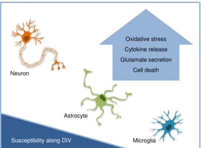
Fig. I. 10. Contribution of each cell-dependent injury to the general outcome of unconjugated bilirubin (UCB) toxicity. ... 24
Fig. II. 1. Schematic representation of the experimental model for chemotaxis assays. ... 31
Fig. II. 2. Representative steps of the chemotaxis assay. ... 32
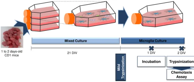
Fig. II. 3. Schematic representation of the experimental model for culture characterization. ... 33
Fig. III. 1. Free, unbound, unconjugated bilirubin (fUCB) do not induce chemotaxis of microglial cells in the Boyden chamber assay. ... 38
Fig. III. 2. Unconjugated bilirubin (UCB) exerts immunosuppressive effect on microglia migration. .... 39
Fig. III. 3. Immunosuppressive effects of unconjugated bilirubin (UCB) are maintained even in the presence of the chemotactic compounds ATP and S100B. ... 39
Fig. III. 4. Glycoursodeoxycholic acid (GUDCA) stimulates cell migration of unconjugated bilirubin (UCB)-treated microglia which is maintained in the presence of ATP and abolished in the presence of S100B. ... 40
Fig. III. 5. Microglia morphology evaluation point to 13 days in vitro (DIV) cultures as the most suitable for studies of reactivity ... 42
Fig. III. 6. Microglial cells at 13 in vitro (DIV) show the lowest levels of nuclear factor-kappaB (NF-κB) activation... 43
INDEX OF TABLES
Table II. 1. Solutions loaded to the bottom wells of the microchemotaxis chamber. ... 32 Table III. 1. Microglial cultures with 13 days in vitro (DIV) present the best representation of the phagocytic phenotypes variability. ... 44
ABBREVIATIONS
AD Alzheimer’s disease
Aβ Amyloid β
BBB Blood-brain barrier
BDNF Brain-derived neurotrophic factor
CNS Central nervous system
CR Complement receptor
CSF Colony stimulating factor
CY Cyanine
DIV Days in vitro
DMEM Dulbecco's Modified Eagle Medium
EDTA Ethylenediamine tetraacetic acid
eNOS Endothelial nitric oxide synthase
FBS Fetal bovine serum
FcR Fc receptor
GFAP Glial fibrillary acidic protein
GUDCA Glycoursodeoxycholic acid
HSA Human serum albumin
Iba1 Ionized calcium-binding adaptor molecule 1
IFN Interferon
IL Interleukin
LPS Lipopolysaccharide
Mac-1 Macrophage receptor 1
MAP-2 Microtubule associated protein-2
MHC Major histocompatibility complex
MMP Matrix metalloproteinase
NF-κB Nuclear factor-kappaB
NO Nitric oxide
PAMP Pathogen-associated molecular pattern
PBS Phosphate buffered saline
PD Parkinson’s disease
PKC Protein kinase C
PR Phosphatidylserine receptor PRR Pattern recognition receptor
PS Phosphatidylserine
PS1 Presenilin 1
PVP Polyvinylpyrrolidone
ROS Reactive oxygen species
ABBREVIATIONS Microglial clearance function: dependence on phenotypes
TAMs Tumor-associated macrophages
TGF Transforming growth factor
TH Helper T cell
TLR Toll-like receptor
TNF Tumor necrosis factor
TREM-2 Triggering receptor expressed on myeloid cells-2
I.
INTRODUCTION
1. Microglia: introduction and overview
The survival and proper function of neurons is ensured by the large number of glial cells (Streit, 2002). Among this group of cells, a unique population resembling neural environment-adapted macrophages comprises upwards 12% of the non-neuronal brain cells and is designed by microglia (Aloisi, 2001; Ladeby et al., 2005). These cells are ubiquitously distributed in non-overlapping territories throughout the central nervous system (CNS) including the spinal cord, but they vary in density, with the white matter generally containing fewer microglia than the grey matter (Soulet and Rivest, 2008; Ransohoff and Perry, 2009).
Since these cells derive from a mononuclear phagocyte lineage, they share several morphological features and functions with macrophages. Thus, microglia constitute the brain’s immune system contradicting the initial idea of CNS as an immune privileged site (Kreutzberg, 1996; Streit, 2002). For this reason, microglia play an essential role in both physiological and pathological conditions (Polazzi and Monti, 2010), as it will be further discussed.
1.1. Microglia phenotypic diversity
For a long time, microglia phenotypes were solely divided in a resting and an activated state. In healthy normal CNS, microglia would be in a resting state presenting small cell soma with rod-shaped nuclei and numerous branched processes. In pathological conditions, microglia would turn into an activated state undergoing several morphological alterations, like the acquisition of an amoeboid shape with processes retraction, upregulation of cell surface markers and production of a plethora of bioactive mediators (Kim and de Vellis, 2005; Hanisch and Kettenmann, 2007). Cells in the resting state have been for a long time considered to be quiescent and inactive. Also, it was described that conversion to an activated state occurred nonspecifically irrespective of the injury, that is, in a stereotyped manner with a predetermined program of functions to execute (Davis et al., 1994; Gehrmann et al., 1995).
Recent reports, however, demonstrate that although microglial cell bodies are relatively fixed, their fine processes present an elevated motility, higher than that of astrocytes, allowing them to constantly palpate and monitor their microenvironment (Davalos et al., 2005; Nimmerjahn et al., 2005). Formation of these processes occurs even in absence of any pathogenic stimulus suggesting that this formation may be associated with the integration of homeostatic signals throughout the CNS (Raivich, 2005; Carson et al., 2007). Thus, these cells are highly active in their presumed resting state and the term “surveillant” has been advanced to replace this traditional definition (Ransohoff and Cardona, 2010). Furthermore, the activation process is adaptive so that microglia response is specific for each stimulus and brain region, and depending on the circumstances, this response may have neuroprotective or neurotoxic outcomes. (Carson et al., 2007). For instance, expression of some surface molecules associated with immune function show brain regional variation (de Haas et al., 2008).
I. INTRODUCTION Microglial clearance function: dependence on phenotypes
Silva et al., 2010). In fact, microglial cells can detect microdamages in their neighborhood and play regenerative functions without undergoing a drastic transformation or initiating an inflammatory response. However, when stimuli are stronger or prolonged, microglial cells undergo more dramatic changes (Hanisch and Kettenmann, 2007).
Recently, it has been debated the concept of different states of activation of microglia accounting for the differing functional properties of these cells (David and Kroner, 2011). The idea emerged from research in non-CNS field in an attempt to perceive whether macrophages play a harmful or beneficial role after injury and which revealed important insights into macrophage polarization. Analogous to the T helper (TH) 1/ TH2 dichotomy of T cell polarization, macrophages can be polarized by the microenvironment to mount specific M1 or M2 functional programs (Porta et al., 2009). Classical (M1) activation, induced by interferon (IFN)-γ, is characterized by a robust pro-inflammatory response required for the elimination of extracellular pathogens. On the other hand, interleukin (IL)-4 triggers an alternative (M2) activation important for the immune response to parasites as well as for tissue repair (David and Kroner, 2011; Saijo and Glass, 2011). Since macrophages are innate immune cells, their primary function is to act as phagocytes in response to pathogens. Nevertheless, they are capable of calling adaptive immune cells, despite the limited ability to process and present antigen to T cells, so that both arms of immune response act in concert to restore the homeostasis (Town et al., 2005). Curiously, the populations of effector CD4 T cells intervening in the response are determined by macrophage polarization. For example, M1 cells release IL-12 that leads to activation of TH1 which, in turn, release IFN- γ that induce long-lasting classical activation (Martinez et al., 2009).
However, these simplified polarization states (M1 and M2) describe a complex process and are the extremes of a spectrum of functional states (Mantovani et al., 2004). In fact, subgroups of M2, like M2a, M2b and M2c, have been used to further define macrophage polarization, since this general designation encompasses cells with dramatic differences in their biochemistry and physiology (Gordon, 2003; Mantovani et al., 2007). Furthermore, it has also been proposed a distinct division of macrophage populations into three groups based on their homeostatic activities – host defense, wound healing and immune regulation (Mosser and Edwards, 2008).
The transposition of these concepts to microglia is quite simple in the case of classical activation and might also be applicable in the case of alternative activation. However, the definition of the steady-state in the case of microglia is more challenging and the determinants of this state might be very distinct from those imposed to macrophages. Moreover, it is not known if the deactivation of microglia results in a state functionally similar to the resting state. Notwithstanding, reports available are sufficient evidence to support the association between distinct phenotypes and pathology (Saijo and Glass, 2011)
Therefore, macrophages and microglia recognize signals from their microenvironment that induce specialized activation programs (Martinez et al., 2009) leading to divergent effects in response to CNS injury (Kigerl et al., 2009). Thus, depending on their differentiation status, microglia differently affects cellular function with either a deleterious or beneficial outcome in the repair (Miron et al., 2011).
Microglial clearance function: dependence on phenotypes I. INTRODUCTION
differences in activation states undergone by microglia to achieve appropriated effector responses for each challenge to CNS (Ransohoff and Cardona, 2010). The recent discovery of microglial involvement in neurogenesis, postlesional and “synaptic stripping,” underscores the existence of additional, functionally adapted microglial phenotypes (Graeber, 2010).
Fig. I. 1: Microglial phenotypic diversity.
A. Microglial cells show a great phenotypic heterogeneity and plasticity being capable of quickly adaption to
achieve an appropriated effector response for each challenge to CNS. B. Recently, it was proposed the concept of different states of activation, ranging from “classical” activation (or M1) to “alternative” activation (or M2), which represent the extremes of a spectrum of functional states. C. Other authors extended the classification into three groups based on their homeostatic activities. They are arranged according to the three primary colors, with red designating classically activated macrophages, yellow designating wound-healing macrophages and blue designating regulatory macrophages. Secondary colors, such as green, purple and orange, represent cells that share properties from two of those groups.
Functional and phenotypic heterogeneity of these cells imply that no simple expression signature exist for microglia (Graeber and Streit, 2010). Indeed, in uninjured CNS, macrophages and microglia in different regions of the brain show differences in morphology and surface markers. This allows “resting” microglia to be distinguished from activated macrophages/microglia by their low CD45 expression, but in the injured CNS, this is no longer possible (David and Kroner, 2011). Therefore, along with a new definition, it becomes essencial to seek for new means of identifying this complexity of microglial phenotypes.
M2 M1
Classically activated macrophages
Wound-healing macrophages
Regulatory macrophages
B
A
I. INTRODUCTION Microglial clearance function: dependence on phenotypes
1.2. Role of microglia in critical periods of brain development
Distribution of microglia in the CNS varies with the different stages of brain development and is accompanied by the acquisition of different functions (Cuadros and Navascues, 1998). The roles of microglia during early stages of brain development, namely neonatal/postnatal period, and in aging brain are critical for the proper functioning of this organ and determine, in part, the possible occurrence of long-term neurological disabilities. Therefore, the next two topics will be intended to review these issues.
1.2.1. Early brain development
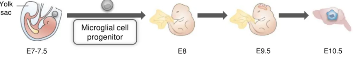
The origin of microglia has been at the centre of debate since their first documentation by del Rio-Hortega (1932) according to whom microglia were derived from the invasion of mesenchymal pial elements during embryonic development. Currently, it is generally accepted by most authors that these cells derive from progenitors, at the yolk sac, that arrive to the developing CNS through the bloodstream, ventricles and meninges, at an early embryonic stage (Imai et al., 1997; Chan et al., 2007; Bilbo and Schwarz, 2009) (Fig. I. 2). In contrast,other authors still believe that microglia have a neuroectodermal origin arguing that neuroectodermal matrix cells are able to differentiate into microglia locally (Fedoroff, 1995).
Fig. I. 2. Origin of microglial cells.
Microglial cells originate from primitive progenitors that arise in the yolk sac early during embriogenesis. They enter the embryo at the embryonic day (E) 8 and surround the neuroepithelium by E9.5. At E10.5, the earliest microglia are found in the brain. Adapted from Ransohoff, 2011 and Saijo, 2011.
There is a “developmental window” during which the microglial progenitors infiltrate into the CNS and that extends from embryonic day (E) 10 to E19 in rodents and from the latter half of the first trimester and throughout the early part of second trimester in humans (Rezaie and Male, 2002b; Rezaie and Male, 2002a). After entering the brain, these precursors originate microglial cells that still retain features of their progenitors presenting an amoeboid morphology and being highly motile, phagocytic and proliferative (Schlegelmilch et al., 2011). They then migrate by tangential and radial migration and proliferate as amoeboid microglia becoming distributed throughout the nervous parenchyma (Marín-Teva et al., 1998; Navascues et al., 2000). First appearance of these macrophage-like microglia occurs about E14 in rat brain and continues to increase in density reaching a peak within the first postnatal week, between postnatal day (P) 4 and P8, with slightly variability depending on brain region (Wu et al., 1992; Ling and Wong, 1993). During this time window, amoeboid microglia plays a central role in phagocyting cellular debris resulting from apoptosis of overproduced neurons (Kim and de Vellis, 2005; Schlegelmilch et al., 2011). Also, even survival and neuronal death itself can be regulated in part by microglia through the release of growth factors and
Yolk sac
E9.5 E10.5
E8 Microglial cell
Microglial clearance function: dependence on phenotypes I. INTRODUCTION
cytokines that not only influence neurons but all the other surrounding cells (Kreutzberg, 1996; Hanisch, 2002). Indeed, cytokines are involved in many important processes in which microglia are committed like neurogenesis, synaptogenesis and gliogenesis (Santambrogio et al., 2001; Nawa and Takei, 2006). Later in development, microglial cells also participate in neuritogenesis as well as in axonal growth and guidance of neurons (Streit, 2001) and it has also been suggested that they can promote vasculogenesis and angiogenesis (Pennell and Streit, 1997). Some of these processes extend from embryonic period throughout adolescence or even adulthood (Rice and Barone, 2000).
When microglia is mainly in an amoeboid/phagocytic state or present solely primitive ramification, they can respond promptly to infection or injury (Ling and Wong, 1993; Santos et al., 2008). Indeed, several studies report cases of neonatal pathological conditions in which microglia is involved, like hypoxic-ischemic injuries (Tahraoui et al., 2001; Kaur and Ling, 2009; Deng et al., 2010) and excitotoxic brain damage (Dommergues et al., 2003). In these cases, concomitant to the microglial activated phenotype, several surface markers are up-regulated, like major histocompatibility complex (MHC) class II and complement receptors (Greensmith and Navarrete, 1994; Ábrahám and Lázár, 2000). Also, inflammatory transcription machinery starts with nuclear factor-κappaB (NF-κB) activation (Nijboer et al., 2008), leading to the release of mediators and modulators of inflammation that contribute to aggravate the pathological condition (Lai and Todd, 2006). Although this is the general outcome, some factors can have neuroprotective role following trauma (Ellis et al., 2007; Nijboer et al., 2008), which will be further discussed.
Pathologies in the neonatal period can have significant repercussions later in life, since it has been demonstrated that they can result in a phenomenon called “glial priming”. In this case cells become sensitized by an insult such that subsequent responses to future challenges are exacerbated (León-Chávez et al., 2003; Perry et al., 2003) and can lead to cognitive and behavioral impairments (Perry et al., 2002; Nawa and Takei, 2006; Cooke and Abernethy, 2010). It is not without precedent that early development exposure to neurotoxic agents may determine latent effects on both morphological and behavioral endpoints which are manifest during the aging process (Barone et al., 1995). Furthermore, it should be noted that the first postnatal weeks are particularly critical due to maximal brain growth rate, implying that any alteration will impact on neurogenesis ability and brain function in later life (Georg Kuhn and Blomgren, 2011). At about P15 in rodents (and from late second to early third trimester in humans), microglia reach their final destination in CNS parenchyma and undergo differentiation, a last major process that follows proliferation and migration. This sequence of events often referred to as “developmental” plasticity culminates with amoeboid microglial cells lastly turning into fully mature, ramified microglia (Bilbo and Schwarz, 2009; Tambuyzer et al., 2009). These processes-bearing cells constitute the “resident” microglia of the adult brain.
1.2.2. Aging brain
I. INTRODUCTION Microglial clearance function: dependence on phenotypes
Microglia from aged brain shows amoeboid-like morphology, increased proliferation and antigen expression with up-regulation of MHC class II, as well as increase in inflammatory cytokines production (Perry et al., 1993; Cagnin et al., 2001). They also showed enhanced proliferation and resistance to a down-regulated phenotype, losing the sensibility to the anti-inflammatory cytokine transforming growth factor (TGF)-β1 (Rozovsky et al., 1998). However, the chronic use of non-steroidal anti-inflammatory drugs appears to be effective in treating Alzheimer’s disease (AD) by diminishing microglial activation (Mackenzie and Munoz, 1998). This is particularly true if nonsteroidal anti-inflammatory drugs are used at the earliest possible times as preventive instead as a recovering therapy (Varvel et al., 2009)
Reactive microglial profile can lead to an exaggerated release of pro-inflammatory cytokines in case of activation of innate immune system that may lead to behavioral and cognitive impairments (Perry et al., 2003; Cunningham et al., 2005). What triggers increased microglial activation is still unclear but oxidative stress, resulting from increased reactive oxygen species (ROS) appear to have a determinant role (Floyd and Hensley, 2002; Lu et al., 2004). In fact, gene-expression profile of aged human and mouse brains indicates an environment of inflammation and oxidative stress with reduced expression of genes related to synaptic function/transport, growth factors and trophic support (Lee et
al., 2000; Lu et al., 2004). These global changes paint a bleak picture of the aged brain where neurons encounter increased challenges and receive reduced support.
Despite morphological and phenotypic changes indicating microglia activation, it has been proposed that microglia may undergo senescence, becoming dystrophic (Streit et al., 2008). The morphological changes seen in this case are distinct from the ones that occur during microglia activation and include the loss of ramifications and even partial or complete fragmentation of cytoplasm, and nuclear condensation (Streit et al., 2004b). This degeneration in normal aging brain could explain the high incidence of microglial apoptosis in AD brain reported by Lassmann et al. (1995). Dystrophy of microglia may reduce the secretion of neurotrophic factors and downregulate phagocytosis. Indeed, older rats reveal less clearance of myelin after a toxin-induced demyelination lesion (Zhao et al., 2006). Furthermore, tau pathology was recently associated with dystrophic rather than activated microglia supporting the idea that it is the loss of microglial neuroprotection that contributes to the onset of sporadic AD (Streit et al., 2009). Once dystrophic microglia accumulates in the aging human brain it is assumed that microglia undergo progressive deterioration.
In conclusion, it is probably the joint action of increased levels of inflammatory mediators and diminished levels of neurotrophic functions that lead to neuronal loss and inefficient clearance of toxic aggregates in neurodegenerative diseases (Fig. I. 3).
2. Functional roles of microglia
Microglial clearance function: dependence on phenotypes I. INTRODUCTION
where they can perform protective or toxic functions that will determine the brain functionality in the future (Perry et al., 2010).
Fig. I. 3. Microglia in the aged brain.
During aging, microglial cells can become over-activated or senescent, resulting in increased pro-inflammatory response and decreased neuroprotective functions, respectively. Therefore, neurons encounter increased challenges and receive reduced support in the aged brain, accounting for neurodegeneration seen during age-related diseases.
The following sections will be dedicated to discuss the functional roles of microglia both in physiological and pathological brain.
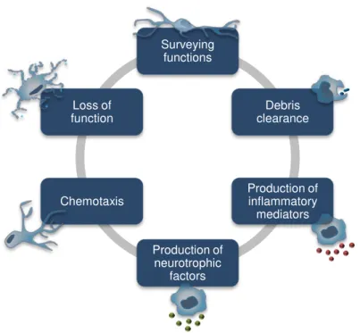
Fig. I. 4. Functional roles of microglia.
Resident microglia, when activated, adopt one of many diverse phenotypes and perform their functional roles during both healthy brain development and in response to pathological changes. Adapted from Silva, 2010.
Neurotrophic functions
Senescent Over-activated Cytotoxic
functions
Age-related neurodegeneration
Surveying functions
Debris clearance
Production of inflammatory
mediators
Production of neurotrophic
factors Chemotaxis
I. INTRODUCTION Microglial clearance function: dependence on phenotypes
2.1. Surveillance and repair
Microglia play a pivotal role in immune surveillance, host defense, and tissue repair, maintaining the homeostasis in the CNS (Liu et al., 2001). During CNS development there is an intense remodeling in the brain since neurons must be generated in right quantity and location, and neural connections have to be properly established. Therefore, formation of mature neural circuits requires the elimination of inappropriate synaptic connections, a process termed as pruning. Recently, Stevens et al. (2007) demonstrated that the proteins C1q and C3b, both involved in the classical complement cascade, are required for this process as deficiencies in one or both proteins lead to defects in synapse elimination. This finding points to members of the complement cascade as new immune system players with also non-immune functions. The model is based in an early model from Jennings (1994) in which unwanted synapses are tagged by C3b and then phagocytosed by the resident microglia which express C3 receptor.
Since microglia appeareed to be involved in synaptic pruning, it is suggested that they might also contribute to plasticity after lesions (Cullheim and Thams, 2007). Indeed, an increased number of microglial cells is present both during development of CNS and in neurological diseases, reinforcing the idea of microglia involvement in synaptic remodeling in postnatal brain, but also in adult brain during pathological scenarios. Blinzinger and Kreutzberg (1968) showed that microglial cells caused the displacement of synaptic boutons from injured neurons, a process termed “synaptic stripping”. Similar observations have been made where activated microglia migrate and strip synapses during inflammatory processes and appear to have neuroprotective consequences (Trapp et al., 2007). In fact, there are three ways of synapse modification or elimination that are supposed to be mediated by microglia: proteolytic modification of the perisynaptic environment, remodeling of dendritic spine morphology, and phagocytic engulfment of dendritic spines, as well as of axon terminals (Tremblay and Majewska, 2011).
Microglia have also been pointed to induce neuronal apoptosis with the subsequent engulfment of cellular debris as demonstrated in cerebellar slice cultures where microglia showed to induce Purkinje cell neurons to undergo apoptosis (Marin-Teva et al., 2004), and by a wide set of experiments evidencing that apoptotic cells are phagocytosed by microglia (Hume et al., 1983; Perry et al., 1985; Streit, 2001). This subject will be further discussed in section 2.2.3.
Microglial clearance function: dependence on phenotypes I. INTRODUCTION
mice expressing the wild-type PS1 or PS1 variants suggesting a role for these factors in mediating adult hippocampal neurogenesis (Choi et al., 2008).
Microglia also appear to exert functions that promote vascularization in the CNS. In an early study from Pennell and Streit (1997) it was observed that neural allografts were colonized by microglial cells before any blood vessels formation. Also, they continued to differentiate parallel to graft vascular development, and are often seen in close proximity to ingrowing vessels suggesting their participation in graft neovascularization. In fact, more recently, microglia were shown to differentiate from myeloid progenitors to facilitate vascularization in a model of ischemic retinopathy (Ritter et al., 2006). The existence of tumor-associated macrophages (TAMs) with pro-tumoral functions raised the possibility that microglia, as brain resident macrophages, could have a role in angiogenesis (Allavena et al., 2008). However, in this case, the outcome of this response might not be the one desired since TAMs produce factors that promote angiogenesis, remodel tissue and dampen the immune response to tumors (al-Sarireh and Eremin, 2000). Therefore, this particular case will be addressed later.
2.2. Role in innate and adaptive responses
Microglia reside in the healthy CNS as surveillant cells and are critically involved with both innate and adaptive immune system, regulating inflammation and cell damage in the brain (Chew et al., 2006). They respond rapidly to changes in the CNS microenvironment and, depending on the condition, their response may have neuroprotective or neurotoxic outcomes (Ladeby et al., 2005), as already mentioned.
Activation of microglia involves several features including cell morphological changes, upregulation of several cell surface markers, production of inflammatory mediators, as well as the ability to present antigens and to perform phagocytosis (Hanisch and Kettenmann, 2007). These features do not define one single microglial phenotype per every challenge to CNS, as aforementioned. Indeed, Town et al. (2005) consider that exists a continuum of microglial activation, with phagocytic response (innate activation) at one end and antigen presenting cell function (adaptive activation) at the other. Therefore, the nature and duration of stimuli influences the pathways of gene expression and the phenotypical changes that will occur afterwards, reinforcing the idea of plasticity in microglial response (Ransohoff and Perry, 2009; Parkhurst and Gan, 2010).
2.2.1. Signals triggering activation
I. INTRODUCTION Microglial clearance function: dependence on phenotypes
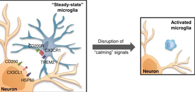
deregulates microglia responses resulting in neuronal death in vivo,as observed in several models of CNS insult (Cardona et al., 2006; Ré and Przedborski, 2006). Interestingly, this function depend on cell-to-cell contact but this membrane-tethered chemokine can be cleaved and act as a soluble molecule with chemoattractive activity (Chapman et al., 2000), an issue to be discussed in the next section. Interaction of the myeloid restricted molecule CD200R with its widely expressed ligand CD200 is also determinant to block microglia activation (Taylor et al., 2011). Another example of an inhibitory signal derives from endogenous ligands of neurons that act on triggering receptor expression by myeloid cells-2 (TREM2), promoting phagocytosis and retarding inflammation (Hsieh et al., 2009). When neurons are damaged, the communication through these “calming signals” is disrupted leading to microglia activation with the consequent increase of inflammatory response and neurotoxicity (Cardona et al., 2006; Ré and Przedborski, 2006).
Fig. I. 5. Steady-state microglia and homeostasis in the central nervous system (CNS).
Under steady-state conditions, microglia exhibit an extensively ramified morphology and a resting phenotype. This phenotype is maintained in part through neuron-derived signals, including CX3CL1 and CD200, which act through corresponding receptors expressed by microglia. Also, functional triggering receptor expressed by myeloid cells-2 (TREM2) is essential to prevent excessive innate and adaptive immune responses. Disruption of these calming signals leads to microglia activation with a switch to a characteristic amoeboid morphology.
Microglia can also detect some factors that can lead to their activation. For example, serum constituents crossing the disrupted blood-brain barrier (BBB) (Ransohoff and Perry, 2009), altered synaptic activity leading to variations in neurotransmitters availability such as glutamate (Taylor et al., 2003) and ATP (Davalos et al., 2005), thus resulting in microglia activation. In the case of purine ATP that is released by damaged neurons, it acts on microglia to mediate processes extension (Gyoneva
et al., 2009) and the function of microglia as phagocytes (Koizumi et al., 2007). Plasma fibrinogen can also bind to an engaged macrophage receptor 1 (Mac-1) /complement receptor (CR) 3 on activated microglia leading to increased phagocytic activity. Several cytokines and colony-stimulating factors (CSFs) also induce proliferation or phenotypic and morphological changes consistent with microglial activation (Kreutzberg, 1996).
Disruption of
“calming” signals
CX3CL1 HSP60 CD200
CX3CR1
TREM2 CD200R
Neuron
“Steady-state” microglia
Neuron
Microglial clearance function: dependence on phenotypes I. INTRODUCTION
In addition to endogenous signals, non-self components like bacterial and viral molecular patterns, recognized by toll-like receptors (TLRs) expressed on microglia, also lead to activation of these cells. Lipopolysaccharide (LPS), an Gram-negative bacterial endotoxin acting on TLR4, is widely used as an experimental activator since by changing microglia morphology and inducing the production of harmful factors. Indeed, LPS leads to microglial production of nitric oxide (NO), tumor necrosis factor (TNF)-α and IL-1β through the mediation of conventional protein kinase C ((Nakajima et al., 2003).
2.2.2. Motility
Microglial cells are able to sense extracellular directional cues and to respond with asymmetric changes in cell morphology and motility.
In the healthy brain, “resting” microglia display a baseline motility characterized by the extension and retraction of their cellular processes without cell body movement (Davalos et al., 2005; Nimmerjahn et al., 2005) but, after CNS injury, they can migrate to rapidly populate the site of injury (Morgese et al., 1983). This movement of microglial cells leading to their accumulation at the site of lesion (a process called homing) is essential for the cellular immune responses of microglia and is triggered by “find-me” signals released by the damaged tissue (Azuma et al., 2001; Kurpius et al., 2007). Movement of microglia can be the result of a random, non-vectorial motility (chemokinesis), or a directed migration that depends from a chemical gradient to organize the movement (chemotaxis), or both (Devreotes and Janetopoulos, 2003; Miller and Stella, 2009).
I. INTRODUCTION Microglial clearance function: dependence on phenotypes
Microglial cells also migrate towards neurotrophic factors such as the epidermal growth factor (Nolte et al., 1997) and nerve growth factor (De Simone et al., 2007), corroborating the role of microglia in brain development. Pathological protein aggregates, such as amyloid-β (Aβ), have also been shown to induce microglial migration (Rogers and Lue; Gyoneva et al., 2009).
Purines that are released from injured tissue in higher levels into the extracellular space by neurotransmission, lead to microglia chemotaxis (Parkhurst and Gan, 2010) (Fig. I. 6). Indeed, the baseline motility of microglial processes in the intact brain is modulated by some of the ATP signaling mechanisms mediating injury-induced microglial responses (Davalos et al., 2005). Until now, process extension and migration is known to involve chloride channels (Hines et al., 2009), ATP G protein-coupled purinergic receptors (P2YR) (Honda et al., 2001; Haynes et al., 2006), outward potassium currents (Wu et al., 2007), astrocytic connexin hemichannels (Suadicani et al., 2006), integrin β1 (Ohsawa et al., 2010), signaling through phosphoinositide 3-kinase, Akt (Irino et al., 2008; Franke et
al., 2009) and non-transcriptional activation of matrix metalloproteinase-9 (Choi et al., 2010). In fact, microglial cells from mice lacking P2Y12R receptor are unable to polarize, migrate or extend processes towards nucleotides, either in vitro or in vivo (Haynes et al., 2006). Besides P2YR, ATP acts on ionotropic P2X receptors (P2XR) and P2X4R which are also involved in microglial chemotaxis as demonstrated by Ohsawa and his colleagues (2007). Moreover, the P2X7R mediate ATP release,
Microglial clearance function: dependence on phenotypes I. INTRODUCTION
Another curiousl aspect of this purinergic signaling is its involvement in the retraction of microglia processes. Indeed, a switch of the chemotactic response to ATP occurs in activated microglial cells, such as the one observed in LPS-pretreated microglia (Gyoneva et al., 2009). This chemorepulsion away from ATP is a consequence of upregulation of a G protein-coupled adenosine receptor (AR), A2AR, which is activated by adenosine resulting from rapid ATP breakdown by nucleotidases (Orr et al., 2009). Also, simultaneously, there is the downregulation of P2Y12R as a result of TLR activation by LPS (Gyoneva et al., 2009). Therefore, microglia extend their processes and migrate towards the local of lesion where they envelop the area and adopt an amoeboid morphology as the one seen during brain neuroinflammation and neurodegeneration.
Fig. I. 6. Purinergic signaling involved in microglia motility.
Presynaptic neuron release ATP as a cotransmitter, by exocytosis. The released ATP acts postsynaptically on P2X and P2Y receptors activated by ADP, UTP and UDP, as well as ATP. ATP is also released from astrocytes (and probably from microglia, as well) to participate in glial–neuron interactions. Both P2X and P2Y receptor subtypes are expressed by astrocytes. Activation of P2Y receptors on astrocytes triggers a calcium (Ca2+) wave inducing the release of ATP through connexin hemichannels. P2X4 and P2Y12 receptors expressed on resting microglia mediate migration, whereas P2Y6 receptors that are expressed on the activated amoeboid microglia mediate phagocytosis of debris at the site of damage. Also, activated microglia express A2A receptors that are activated by adenosine (ADO) resulting from ATP breakdown by ectonucleotidases. Activation of such receptors induces chemorepulsion of microglial cells. Not depicted is the activation of potassium and chloride channels as well as signaling through phosphoinositide 3-kinase and Akt resulting from activation of P2Y12 receptors.
Another compound with chemoattractive function that was recently described is S100B. This protein is abundantly expressed in astrocytes but also in microglia and is released during the course of acute and chronic brain disorder (Ellis et al., 2007). Once released, S100B exerts autocrine and
ADO P2X4 P2Y12 “Resting” microglia Activated microglia Astrocyte Neuron Postsynaptic terminal P2X P2Y Connexin hemichannel PROCESS RETRACTION/ CHEMOREPULSION PHAGOCYTOSIS Neuron Presynaptic terminal ? UDP/UTP A2A P2Y6 ATP P2Y P2X Vesicles ATP PROCESS EXTENSION/ CHEMOATTRACTION
I. INTRODUCTION Microglial clearance function: dependence on phenotypes
paracrine effects mostly by engaging RAGE (receptor for advanced glycation end products) on responsive cells. In low concentrations this protein might exert trophic functions, while in high concentrations, like those shown to be present in the extracellular milieu in case of brain damage, has inflammatory effects leading to activation of microglia, enhancing migration and secretion of pro-inflammatory mediators (Bianchi et al., 2011). For these reasons, S100B has been suggested to play a role in the pathophysiology of neurodegenerative and inflammatory brain diseases (Sorci et al., 2010).
The signaling pathways involved in migration are diverse, as described above, and control different types of primary microglial cells movement (Miller and Stella, 2009).
2.2.3. Phagocytosis
Cells can take up macromolecules and particles from the surrounding medium in a process termed endocytosis. This is essential for ingestion and uptake of extracellular nutrients, for antigen presentation, and for removal of microbial pathogens. During endocytosis cells absorb the material or organisms from the extracellular space by engulfing them with their plasma membrane which then buds off inside the cell to form a vesicle containing the ingested material. Three principal mechanisms of endocytosis have been described. One of the best-characterized endocytic processes is receptor-mediated endocytosis via clathrin-coated pits. In this case cells internalize molecules by the inward budding of plasma membrane vesicles containing proteins with receptor sites specific for the compounds. The term pinocytosis refers to the ingestion of dissolved material or single molecules from the extracellularspace. During this process the cytoplasmic membrane invaginates and pinches off small pinocytic vesicles which are then transferred to the cytosol. Phagocytosis isthe third form of endocytosis involving the vesicular internalization of solid particles, such as microbial pathogens or apoptotic cell debris. Binding of the particle to receptors on the surface of the phagocytic cells stimulates the extension of pseudopodia, which eventually surround the particle, and fuse to form a large intracellular vesicle called phagossome. Once entered into the cell, the phagosome fuses with the lysosome, producing phagolysosomes in which the ingested material is digested (Silverstein et al., 1977; Mukherjee et al., 1997) .
Phagocytosis is a major function of activated microglia. This function is critical for the uptake and degradation of infectious agents and senescent cells, and it participates in development, tissue remodeling, immune response, and inflammation (Chew et al., 2006). In general, microglial phagocytosis can be divided into two distinct responses: phagocytosis of pathogens and stimulation of TLRs inducing a pro-inflammatory cascade (Fig. I. 7A) and clearance of apoptotic cell membranes and recognition of phosphatidylserine (PS) residues inducing an anti-inflammatory response (Fig. I. 7B).
Microglial clearance function: dependence on phenotypes I. INTRODUCTION
(Janeway, 1992; Aderem and Underhill, 1999). The major PRRs include the Fc receptors (FcRs) and the complement receptors (CRs) which recognize immunoglobulins and complement proteins coating the particles, acting as opsonins. Besides these, other receptors are involved in particle uptake, such as lectins, like mannose receptor that recognize mannans, other non-complement-receptor integrins and scavenger receptors (SR) that recognize surface components on bacteria including LPS(Aderem and Underhill, 1999; Underhill and Ozinsky, 2002). Multiple receptors are simultaneously engaged to mediate internalization, activate microbial killing, and induce the production of inflammatory cytokines and chemokines. Collectively, these cellular processes constitute the phagocyte response. Certain phagocytic receptors such as FcRs trigger inflammatory responses directly, whereas others, such as complement receptors, often do not stimulate inflammatory responses (Ravetch and Clynes, 1998). In many cases, these responses are regulated by additional receptors (that are not themselves phagocytic), such as TLRs, which are widely expressed on microglia. During phagocytosis, TLRs are recruited to the phagosomes, although recruitment does not require receptors activation (Underhill and Ozinsky, 2002). Nevertheless, they can sample the contents to determine the nature of the pathogen and participate in the formulation of an inflammatory response appropriate for defense (Aderem and Ulevitch, 2000). Also, activation of TLR signaling pathway by bacteria regulate internalization and phagosome maturation (Blander and Medzhitov, 2004). Concomitantly, TLRs activate NF-κB (Kawai and Akira, 2007) which can transactivate promoters of genes involved in immune and inflammatory responses (Baeuerle, 1991). Indeed, TLR4 can mediate LPS-induced macrophage activations of IL-1β and IL-6 gene expression, chemotaxis, phagocytosis, and oxidative ability (Wu et al., 2009).
Microglial cells also play an important role in the recognition and clearance of apoptotic cells. Removal of apoptotic cells usually involves three central elements. First, the attraction of phagocytes via soluble "find-me" signals released signals by damaged cells, an issue already discussed in previous section. Second, recognition and phagocytosis via cell surface-presenting "eat-me" signals and finally, suppression or initiation of inflammatory responses depending on additional innate immune stimuli (Napoli and Neumann, 2009).
The best studied “eat-me” signal is PS, which translocates from the inner to the outer leaflet of plasma membrane at the early stage of apoptosis. Interestingly, it was recently demonstrated that viable cells can also express PS without leading to their phagocytosis indicating that the exposure of these residues alone is not sufficient to be recognized by microglia as an “eat-me” signal (Segawa et al., 2011). The exposed PS on apoptotic cell is recognized by several phagocyte receptors including a
presumptive PS receptor (PSR) (Savill et al., 1993), even though experimental demonstration of such
a receptor has been quite controversial (Williamson and Schlegel, 2002). Recently, several groups have identified receptors that both directly recognize PS and induce phagocytosis of apoptotic cells. These receptors include the brain-specific angiogenesis factor 1 (Park et al., 2007a), the T-cell
immunoglobulin domain and mucin domain 4 (Miyanishi et al., 2007; DeKruyff et al., 2010; Freeman et al., 2010), and stabilin-2 (Park et al., 2007b).
I. INTRODUCTION Microglial clearance function: dependence on phenotypes
receptor on the phagocyte, thus acting as a collectin or opsonin (Fuller and Van Eldik, 2008). The arrest-specific gene 6 helps phagocytic cells in recognizing surface PS expression and facilitates the clearance of PS-expressing cells (Ishimoto et al., 2000). SRs, such as CD36, appear to recognize membranes of apoptotic cells which are negatively charged (Husemann et al., 2002) and CRs expressed on microglia recognize the complement protein C1q, which is able to bind to PS and act as a bridging molecule in apoptotic cell recognition during early stages of apoptosis (Paidassi et al., 2008). Indeed, both receptors mediate myelin phagocytosis by microglia even though CR3 has a dominant role (Reichert and Rotshenker, 2003; Rotshenker, 2003). TREM2 interacts with ligands on apoptotic neurons, stimulating their removal and counter-regulating pro-inflammatory signals to allow repair (Neumann and Takahashi, 2007; Hsieh et al., 2009).
Microglial clearance function: dependence on phenotypes I. INTRODUCTION
Fig. I. 7. Phagocytic receptors of microglia.
Microglia recognize specific structural patterns (PAMPs) of most microbial pathogens via their TLRs, complement receptors (CRs), Fc Receptors (FcRs) or scavanger receptors (SRs), leading to a pro-inflammatory response. B. Microglia recognize apoptotic cells through phosphatidylserine residues (PS) expressed on the membrane of the death cells. These residues are recognized by PS receptors, like Tim4, which are supported via additional phagocytic receptors including TREM2. Phagocytosis of apoptotic cells induces the release of anti-inflammatory cytokines Adapted from Neumann, 2009 and Napoli, 2009.
In conclusion, phagocytosis is a cell response with important functions in the course of an immune response, but also during tissue remodeling and wound healing. However, it is not a cell response that occurs as an isolated event, and a phagocytic stimulus triggers, besides associated cell responses of destructive nature, an immuno-modulatory cell response as well.
2.2.4. Neuroprotection vs. neurodegeneration: production of mediators
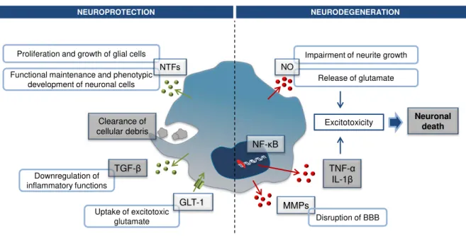
Microglial cells are the source of an array of mediators that may either have a neurotoxic or neuprotective effect (Fig. I. 8). During brain development and in physiological conditions, microglia contributes to growth, functional maintenance and phenotypic development of neuronal cells, as well as to proliferation and growth of glial cells by producing neurotrophic factors, such as the glial cell line-derived neurotrophic factor, basic fibroblast growth factor, brain-line-derived neurotrophic factor (BDNF), nerve growth factor and neurotrophins (Lindvall et al., 1994; Nakajima et al., 2001a).
I. INTRODUCTION Microglial clearance function: dependence on phenotypes
Activation of microglia upon CNS injury has been considered as a detrimental event. This idea came from considering the inflammatory and cytotoxic phenotype acquired by LPS-stimulated microglia in cell-cultures as typical of activation (Schwartz et al., 2006; Hanisch and Kettenmann, 2007). However, recent studies suggest that under pathological conditions, microglia exert neuroprotective functions through the production of neurotrophic molecules and by clearance of cell debris (Kitamura et al., 2009). In fact, microglial activation by an acute CNS injury intends to ameliorate primary tissue damage and promote repair. Thus, release of such mediators is a first step in creating an environment propitious to regeneration which involves the recruitment of phagocytic and neuronal precursor cells to the site of injury (Streit, 2002; Neumann et al., 2009).
There is accumulating evidence suggesting a role for these mediators in the regeneration of brain tissue after injury. These include cytokines in a soluble form for auto- and paracrine signaling or membrane-associated for cell-to-cell interaction, and which participate in a multitude of biological
processes by serving cellular communication. Indeed,
s
everal studies show that microglia protect neurons in damaged brain by secreting anti-inflammatory cytokines and growth factors like IL-10, TGF-β and BDNF (Streit, 2005). TGF-β appears to act as an autocrine mediator by regulating negatively inflammatory and immunoregulatory functions of activated microglia, such as suppresion of cytokine production and ROS formation (Suzumura et al., 1993). Also, pro-inflammatory cytokines, such as IL-1β, il-6 and TNF- α, can also be released to play a role in neuroprotection (Imai et al., 2006). In fact, TNF-α released by activated microglia is suggested to induce expression of potent angiogenic factors that promote retinal neovascularization during post-ischemic inflammation (Yoshidaet al., 2004). Also, injection of exogenous microglia itself protected CA1 pyramidal neurons against ischemia-induced neuronal degeneration possibly through a neurotrophin-dependent mechanism (Hayashi et al., 2006; Imai et al., 2006).
Neuroprotection by microglia also involves their ability to uptake excitotoxic glutamate by expressingglutamatetransporter 1 (Nakajima et al., 2001b). Interestingly activation of this transporter by glutamate leads to production of neurotrophic factors by microglia via PKC pathway (Liang et al., 2010).
Microglial clearance function: dependence on phenotypes I. INTRODUCTION
treatment, (Meda et al., 1995), studies using organotypic hippocampal slice cultures demonstrated that they act neuroprotectively when pre-activated with IL-4 (Butovsky et al., 2006).
NO is the mainly free radical produced by murine microglia upon activation by several stimulants like LPS (Tambuyzer et al., 2009). In co-cultures with neurons, microglia production of ROS lead to the reduction of neurite growth during brain development (Thery et al., 1991). Also, NO induces glutamate release from neurons that leads to NMDA receptors activation and consequent excitotoxicity (Brown, 2010). Years of research using LPS model of microglia activation, have shown that LPS acts on the specific microglial receptors TLR4 and Mac-1 triggering a signaling pathway that results in pro-inflammatory gene expression mediated by NF-κB activation and neuronal death (Loane and Byrnes, 2010).
Microglia are also a source of matrix MMPs, namely MMP-2 and MMP-9 which expression can be induced by LPS stimulation. These enzymes have been shown to degrade components of the basal lamina, leading to the disruption of the BBB and thus, contributing to the neuroinflammatory response in many neurological diseases (Rosenberg, 2002).
Fig. I. 8. Examples of mediators produced in neuroprotection and neurodegeneration.
Microglia exert neuroprotective functions through the production of neurotrophic factors (NTFs) and by the clearance of cell debris, during brain development and under pathological conditions,. Also, they express a glutamate transporter (GLT-1) responsible for the uptake of excitotoxic glutamate. In certain conditions, activation of microglia can exert neurotoxic functions by producing nitric oxide (NO) that intertferes with neurite growth and contributes to the release of glutamate. Also, pro-inflammatory gene expression by nuclear factor (NF)-κB result in pro-inflammatory cytokines production, such as tumor necrosis factor (TNF)-α and interleukin (IL)-1β. Release of these mediators contributes to excitotoxicity resulting in neuronal death. In addition, metalloproteinases (MMPs) are released leading to the disruption of the blood-brain barrier.
Recalling the issue of the different states of activation of microglia and their corresponding functions, we can, in a very simplistic way, define these cells as anti-inflammatory M2 cells or pro-inflammatory M1 cells. However, as already mentioned, microglia response to injury is complex and multifaceted. Nevertheless, it should be always kept in mind that microglia primary purpose is to limit
Impairment of neurite growth
Release of glutamate Functional maintenance and phenotypic
development of neuronal cells
Downregulation of inflammatory functions
Uptake of excitotoxic glutamate Proliferation and growth of glial cells
NTFs TGF-β GLT-1 Clearance of cellular debris NO TNF-α IL-1β
NF-κB
MMPs
Neuronal death
Excitotoxicity
Disruption of BBB