Declaração de autoria de trabalho
Declaro ser a autora deste trabalho, que é original e inédito. Autores e trabalhos consultados estão devidamente citados no texto e constam da listagem de referências incluída.
________________________________________________________ (Francisca Inês Gomes Novais Ribeiro)
© Francisca Ribeiro
A Universidade do Algarve reserva para si o direito, em conformidade com o disposto no Código do Direito de Autor e dos Direitos Conexos, de arquivar, reproduzir e publicar a obra, independentemente do meio utilizado, bem como de a divulgar através de repositórios científicos e de admitir a sua cópia e distribuição para fins meramente educacionais ou de investigação e não comerciais, conquanto seja dado o devido crédito ao autor e editor respetivos.
Polystyrene Microplastics Accumulation
i
AGRADECIMENTOS
Em primeiro lugar gostaria de agradecer à Professora Maria João Bebianno por me ter dado a oportunidade de trabalhar num tema à minha escolha, pela experiência e conhecimentos transmitidos, e acima de tudo, por todo o apoio, disponibilidade e orientação ao longo deste trabalho.
Só consegui realizar este projeto porque tive a sorte de ter pessoas que me acolheram e que tiveram a disponibilidade e interesse em transmitir conhecimento. À Nélia Mestre, à Tainá Fonseca, à Cátia Cardoso, ao Thiago Rocha e à Manon Auguste um obrigada sincero. Mais do que colegas de trabalho, tornaram-se verdadeiros amigos durante este ano. Agradeço ainda às minhas duas companheiras de laboratório Maria Fonseca e Beatriz Pereira, pela companhia durante as longas horas passadas no laboratório.
Queria agradecer também ao Paulo Pedro pela ajuda prestada na recolha das lambujinhas no campo.
À professora Margarida Ribau Teixeira e à Vânia Sousa pela ajuda prestada na caracterização dos microplásticos.
À professora Laura Ilharco e à professora Ana Rosa Garcia por me terem recebido tão bem no Instituto Superior Técnico de Lisboa e por me terem introduzido a uma nova técnica laboratorial (DRIFT).
Ao João Quintela pelo auxílio dado durante a parte experimental e ao Paulo Santana por me ter permitido a utilização de equipamentos que foram fundamentais para a concretização deste trabalho.
Aos meus pais pela força e encorajamento ao longo da minha vida académica. Sem o seu apoio, a todos os níveis, não teria chegado até aqui.
À Marcia Barros e ao José Pedro Costa por terem feito a capa desta tese. Ficou linda!
E finalmente, à minha família emprestada durante a minha estadia em Faro – Mariana Santos e Teófilo Morim – pelo apoio e compreensão em todos os momentos, principalmente quando as coisas corriam menos bem.
ii
ABSTRACT
Nowadays there is an increasing resilience of plastics as an everyday item. With the rapid increase in their production and spread, plastic debris are accumulating in the marine environment where they are fragmented into smaller pieces. One of the most produced polymer, and accordingly, more common in the marine environment is the polystyrene (PS). Ranges of organisms, especially invertebrates, are vulnerable to the exposure of microparticles. However, the impacts of microplastics (< 5mm) in the marine systems are poorly understood. The aim of this study was to assess the ecotoxicity of PS microplastics in different tissues of the peppery furrow shell Scrobicularia plana and select the most appropriate biomarkers to evaluate microplastics effects.
Clams were exposed to 1 mg L-1 of PS microplastics (20 µm) for 14 days, followed
by a 7 days depuration. Microplastics accumulation in gills and digestive gland was analysed through Diffuse Reflectance Infrared Fourier Transform Spectroscopy (DRIFT) and their effects by a battery of biomarkers of oxidative stress (superoxide dismutase, catalase, glutathione peroxidases and glutathione-S-transferases), genotoxicity (comet assay to evaluate DNA damage), neurotoxicity (acetylcholinesterase activity) and oxidative damage (lipid peroxidation).
Results indicate that microplastics were accumulated in both organs, but more significant in the gills and were not completely eliminated after 7 days of depurarion. Microplastics accumulation induced an oxidative stress response in both tissues. The overall results on oxidative stress biomarkers indicated that short-term exposure to PS microplastics induce major perturbations, as revealed by the effects on the total antioxidant capacity, DNA damage, neurotoxicity and thus oxidative damage.
The results highlighted the potential source of PS toxicity for human health and the marine environment and that S.plana is a significant target of PS microplastics ecotoxicity and can be a suitable biomonitor for assess their environmental risk.
Key words: ecotoxicology, accumulation, neurotoxicity, oxidative stress, genotoxicity, Scrobicularia plana
iii
RESUMO
Hoje em dia há uma resiliência crescente dos plásticos como um item do dia-a-dia para fins comerciais, industriais e terapêuticos. No entanto, a sua produção, o rápido crescimento e distribuição tem dado origem a sérias implicações ambientais. O consumo de plásticos em muitos países europeus indica que as resinas plásticas mais utilizadas desde 2007 são polietileno de baixa densidade (PEBD) e polietileno de alta densidade (HDPE), polipropileno (PP), cloreto de polivinilo (PVC), polietileno tereftalato (PET) e poliestireno (PS). O poliestireno (PS) é um dos plásticos mais utilizados em todo o mundo e tem sido detetado nos oceanos sob a forma de micro e nano partículas. Tem-se verificado que o PS tem um impacto ambiental considerável, nomeadamente em espécies marinhas. Recentemente, foram identificadas no ambiente marinho partículas microscópicas omnipresentes - os microplásticos - definidos como partículas com menos de 5 mm de diâmetro, de acordo com a National Oceanic and Atmospheric Administration dos Estados Unidos da América. São considerados um poluente marinho emergente e, até à data, têm sido detetados em muitos habitats e numa variedade de espécies marinhas e de água doce. Assim, é importante entender a sua distribuição no ambiente marinho e as implicações sobre os habitats, biodiversidade e bem-estar das espécies marinhas.
Os efeitos biológicos dos microplásticos nos organismos dependem do seu tamanho sendo que, quanto menor, maior será a acumulação e o efeito a nível celular. Apesar da preocupação relacionada com a ingestão, os efeitos dos microplásticos em populações marinhas e as suas implicações para a cadeia alimentar ainda não são bem conhecidos. Os invertebrados marinhos são particularmente suscetíveis aos microplásticos, por causa do tamanho e modo de alimentação. Uma vez que o modo de ação e o risco biológico dos microplásticos ainda não são muito claros, esta dissertação avaliou a acumulação e os efeitos dos microplásticos de poliestireno (20 µm) na lambujinha Scrobicularia plana, de forma a avaliar o potencial risco ecotoxicológico para os diferentes níveis de organização biológica e selecionar o biomarcador mais apropriado para determinar os efeitos dos microplásticos.
Relativamente à parte experimental, após a recolha, os animais tiveram um periodo de aclimatação de 7 dias. Seguidamente foram expostos a uma concentração de PS microplásticos (1 mg L-1) durante 14 dias, juntamente com um grupo de controlo, ao
qual se seguiu um período de depuração de 7 dias. Os animais foram recolhidos em diferentes dias de exposição, nomeadamente nos dias 0, 3, 7, 14 e 21.
iv
Inicialmente, as características dos microplásticos e o seu comportamento na água do mar foram analisados em termos de forma, tamanho, carga superficial (potencial zeta), agregação, turbidez e taxa de sedimentação. Seguidamente, avaliou-se a acumulação dos microplásticos nas brânquias e na glândula digestiva através da observação ao microscópio ótico das partículas presentes na hemolinfa, e pela técnica de espectroscopia por refletância difusa no infravermelho com transformação de Fourier (DRIFT).
Para o estudo da toxicidade dos microplásticos de PS uma bateria de biomarcadores foi analisada nas brâquias e na glândula digestiva incluindo: stress oxidativo (superóxido dismutase - SOD, catalase - CAT, glutationa peroxidases - GPx e glutationa-S-transferases - GST), genotoxicidade (danos no ADN), neurotoxicidade (actividade da enzima acetilcolinesterase), e dano oxidativo (peroxidação lipídica).
Os microplásticos usados neste estudo foram micropartículas esferóides com um tamanho de 20 ± 0.02 µm e densidade de 1.05 g cm-3. Em água do mar, os microplásticos de PS tendem a formar pequenos agregados com uma carga superficial negativa (potencial zeta = -12.4 ± 2.36 mV). A taxa de sedimentação rápida e lenta dos microplásticos na água do mar foi de 1.04 x 10⁻¹ e 1.16 x 10⁻³ h-1, respetivamente, confirmando a sua tendência para sedimentar nos tanques de exposição, após as primeiras duas horas.
No que diz respeito à acumulação dos microplásticos de PS nos tecidos, as brânquias apresentaram um padrão de acumulação crescente ao longo do tempo de exposição, com uma possível recuperação no final do período de depuração, através da eliminação dos microplásticos de PS quase na sua totalidade. Em relação à glândula digestiva, a acumulação de microplásticos de PS é evidente, no entanto, não apresentou o mesmo padrão de aumento observado nas brânquias. Os resultados indicaram que a acumulação foi mais eficiente nas brânquias do que na glândula digestiva.
A toxicidade dos microplásticos de PS nas lambujinhas é dependente do tecido e do tempo de exposição e envolve mudanças na atividade das enzimas antioxidantes, stress oxidativo, neurotoxicidade e danos no ADN.
As brânquias são o orgão que responde mais ativamente ao stress oxidativo induzido pelos microplásticos de PS, com efeitos associados ao aumento da atividade das enzimas antioxidantes (SOD, CAT, GPx) e de biotransformação (GST). Na glândula digestiva verificou-se um aumento da atividade da SOD, CAT e GPx. Comparando as atividades das enzimas antioxidantes e de biotransformação dos dois órgãos (brânquias e glândula digestiva), a atividade da CAT foi a única que aumentou na glândula digestiva
v
em relação às brânquias. Após o período de depuração verificou-se um aumento da atividade da SOD e GPx nas brânquias. Na glandula digestiva ocorreu um aumento da atividade da CAT e uma diminuição da atividade da GST.
Verificou-se ainda um efeito genotóxico e neurotóxico causado pelos microplásticos de PS. O efeito genotóxico traduziu-se pelo aumento da percentagem de ADN presente na cauda do cometa (DNA Tail) e no comprimento da cauda do cometa e pela proporção de ADN presente na cauda (Olive Tail Moment). Também no período de depuração se verificou um aumento para estes dois parâmetros. O efeito neurotóxico dos microplasticos de PS é suportado pela diminuição da atividade da acetilcolinesterase após o primeiro dia de exposição.
De uma forma geral, o dano oxidativo foi maior na glândula digestiva do que nas brânquias. Nas brânquias o dano foi menor após o ínicio da exposição aos microplásticos. Na glândula digestiva verificou-se um aumento progressivo até ao 7º dia. Após o período de depuração, apenas se verificaram diferenças significativas na glândula digestiva, com uma diminuição do nível de LPO em relação ao ultimo dia de exposição (dia 14).
Analisando os resultados no seu conjunto, as brânquias aparentam ser um órgão essencial na proteção de S. plana contra o efeito dos microplásticos de PS, uma vez que a resposta das enzimas antioxidantes e de biotransformação foi mais notória neste orgão do que na glândula digestiva, traduzindo-se numa maior toxicidade. Estes resultados indicam que, possivelmente, S. plana lida com a produção de espécies reativas de oxigénio (ROS) através da indução das defesas antioxidantes, o que, por conseguinte, limita o ataque de ROS nas membranas celulares, impedindo que haja peroxidação lipídica nas brânquias.
O período de depuração não parece ser suficiente para a eliminação dos microplásticos de PS. Durante o período de depuração, as brânquias de S. plana aparentam possuir baixa capacidade de eliminação de PS, sendo o principal órgão de contacto com os microplásticos, enquanto que a glândula digestiva parece eliminar mais facilmente as micropartículas.
Palavras-chave: ecotoxicologia, acumulação, neurotoxicidade, stress oxidativo, gentoxicidade, Scrobicularia plana
vi INDEX AGRADECIMENTOS ... i ABSTRACT ... ii RESUMO ... iii INDEX ... vi FIGURE INDEX ... ix
TABLE INDEX ... xii
LIST OF ABBREVIATIONS ... xiii
CHAPTER 1. INTRODUCTION ... 1
1.1. Plastics production ... 1
1.1. Origin and the presence of plastics in the marine environment... 3
1.2. The impact of plastics in marine organisms ... 6
1.3. Microplastics as contaminants to marine organisms... 12
1.4. Biomarkers... 13 1.5. Oxidative stress ... 14 1.5.1. Antioxidant enzymes ... 15 1.6. DNA damage ... 17 1.7. Neurotoxicity ... 17 1.8. Lipid peroxidation ... 18
1.9. Scrobicularia plana characterization ... 19
1.9.1. S. plana as a bioindicator of environmental contamination ... 21
1.10. Objectives ... 22
CHAPTER 2. MATERIALS AND METHODS ... 23
2.1. Microplastics characterization ... 23
vii
2.3. Laboratory assay ... 24
2.4. Condition index... 25
2.5. Tissue ppreparation for microplastics accumulation ... 25
2.5.1. Diffuse Reflectance Infrared Fourier Transform Spectroscopy (DRIFT) 25 2.6. Tissue preparation for biomarker analysis ... 26
2.7. Total protein concentrations ... 26
2.7.1. Superoxide dismutase (SOD) ... 27
2.7.2. Catalase (CAT) ... 27
2.7.3. Glutathione peroxidase (GPx) ... 27
2.7.4. Glutathione-S-transferase (GST) ... 28
2.8. Oxidative damage ... 28
2.9. Acetylcholinesterase (AChE) activity ... 28
2.10. Genotoxicity ... 29 2.10.1. Cell viability ... 29 2.10.2. Comet assay ... 29 2.11. Statistical analysis ... 31 CHAPTER 3. RESULTS ... 31 3.1. PS microplastics characterization ... 31 3.2. Sedimentation rate... 33 3.3. Condition index... 35 3.4. Microplastics accumulation ... 35
3.4.1. Diffuse Reflectance Infrared Fourier Transform Spectroscopy (DRIFT) 36 3.5. Enzymatic activity... 40
3.6. Comet assay ... 43
3.7. AChE activity ... 45
3.8. Oxidative damage ... 45
viii
CHAPTER 4. DISCUSSION ... 49
5.1. Conclusions... 56
5.2. Future perspectives... 57
ix
FIGURE INDEX
CHAPTER 1. INTRODUCTION
Figure 1.1. Worldwide plastic production between 1950 and 2014 (Source: World Economic Forum (2016), adapted from Plastics Europe (2015))...2 Figure 1.2. Generalized scheme for oxifative damage to macromolecules of living organisms (adapted from: Lackner (1998))...15 Figure 1.3. Representative scheme of the action of antioxidant enzymes (SOD, CAT; GPx and GR) and GST ...16 Figure 1.4. Specimens of S. plana A – interior of the shell B – outer surface of the shell...20 Figure 1.5. Internal appearance of S. plana...20 Figure 1.6. World distribution of Scrobicularia plana (Source: Discover life. http://www.discoverlife.org)...21
CHAPTER 2. MATERIALS AND METHODS
Figure 2.1. Representative scheme of the experiment...24 Figure 2.2. Diagram of typical comet showing distribution of DNA in head and tail...30
CHAPTER 3. RESULTS
Figure 3.1. PS microplastics in aqueous solution observed under the OM (magnification: x100). One ocular unit (o.u.) corresponds to 20 µm ...32 Figure 3.2. PS microplastics in natural seawater (100 mg L-1) observed under the OM (magnification: x100). One ocular unit (o.u.) corresponds to 20 µm...33 Figure 3.3. Turbidity of PS microparticles for 24 hours in MQ water and in sea water. C/C0 is the normalised microparticle turbidity where C is the turbidity at time t and C0 the
initial turbidity at time 0………...……..….34 Figure 3.4. Fast SR (A) (over the first 2 hours) and slow SR (B) (over the remaining 22 hours) of PS microplastics suspended in MQ water and sea water...34 Figure 3.5. PS microplastics in the hemolymph, observed under the OM (magnification 40x). The presence of PS is highlighted by the dark circles...35 Figure 3.6. A. DRIFT spectrum of digestive gland from negative control (Dg-T0), positive controls (Dg – Ctr- p1 and p2), a digestive gland from a clam exposed to
x
microplastics (Dg-T7) and the PS used in the assay (PolyStyr). B. Expansion of the region 900-450 cm-1 of DRIFT spectrum from figure A. The presence of PS is highlighted by the blue circle...37 Figure 3.7. Expansion of the regions 3800-2500 cm-1 and 1000-450 cm-1 of DRIFT spectra from a positive control (Gi-Ctr-p) and negative controls (Gi-g3, 4, 5 and 6-T0), in gills of S. plana. The presence of PS is highlighted by the blue circle...37 Figure 3.8. Expansion of regions 1000-450 cm-1 of DRIFT spectra from in gills of S. plana from day 0 and day 3 of the experiment (A), day 0 and day 7 (B) day 0 and 14 (C) and day 14 and 21 (D). The presence of PS is highlighted by the blue circle...38 Figure 3.9. . Expansion of regions 1000-450 cm-1 of DRIFT spectra from gills of S. plana from day 0 and day 21 of the experiment . The presence of PS is highlighted by the blue circle...39 Figure 3.10. Expansion of regions 1000-450 cm-1 of DRIFT spectra ofdigestive gland of S. plana from day 0 and day 3 of the experiment (A), day 0 and day 7 (B), day 0 and 14 (C) and days 14 and 21 (D)...40 Figure 3.11. SOD, CAT, GPx and GST activities (mean ± SD) in the gills (A, C, E and G) and digestive gland (B, D, F and H) of S. plana for control (CTR) and PS microplastics (MICR 1 mg L-1) during exposure and depuration. Statistical differences between
treatments at each time of exposure are indicated with #, small letters indicate statistical differences between controls and capital letters among MICR treatment (p < 0.05)...41 Figure 3.12. DNA damage (average ± SEM) in the haemocytes of S.plana expressed as tail DNA % (A), OTM (a.u.) (B) and Tail length (µm) (C) for control (CTR) and PS microplastics (MICR). Statistical differences between treatments at each time of exposure are indicated with #, small letters indicate statistical differences between controls and capital letters among MICR treatment (p < 0.05)...44 Figure 3.13. . AChE activity in the gills of S. plana (average ± SD) for control (CTR), and microplastics (MICR). Statistical differences between treatments at each time of exposure are indicated with #, small letters indicate statistical differences between
controls and capital letters among MICR treatment (p <
0.05)...45 Figure 3.14. Figure 3.14. LPO (mean ± SD) in gills (A) and digestive gland (B) of S. plana for control (CTR) and microplastics (MICR). Statistical differences between
xi
treatments at each time of exposure are indicated with #, small letters indicate statistical differences between controls and capital letters among MICR treatment (p < 0.05)...46 Figure 3.15. Principal component analysis (PCA) of a battery of biomarkers in the gills (A) and digestive gland (B) of S. plana from exposed clams...48
xii
TABLE INDEX
CHAPTER 1. INTRODUCTION
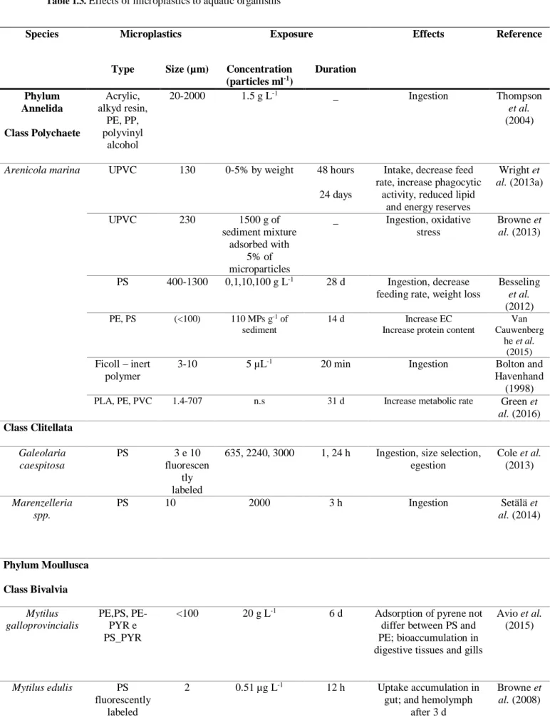
Table 1.1. Plastic resins and their applications (adapted from: Pinto (2012)) ... 2 Table 1.2. Evidence of microplastics ingestion in marine organisms ... 7 Table 1.3. Effects of microplastics to aquatic organisms... 8 Table 1.4. Scientific classification of Scrobicularia plana (Source: WoRMS
xiii
LIST OF ABBREVIATIONS
ACh Acetylcholine
AChE Acetylcholinesterase
BHT Butylated hydroxytoluene
BSA Bovine serum albumin
CAT Catalase
CDNB 1-Chloro-2,4-dinitrobenzene
ChE Cholinesterase
CI Condition Index
CTR Control treatment
DAPI 4',6-Diamidino-2-Phenylindole Dihydrochloride
DDT Dichlorodiphenyltrichloroethane
DLS Dynamic Light Scattering
DNA Deoxyribonucleic acid
DRIFT Diffuse Reflectance Infrared Fourier Transform Spectroscopy
DTT Dithiothreitol
EDTA Ethylenediaminetetraacetic acid
ELS Electrophoretic Light Scattering
GPx Glutathione peroxidases GR Glutathione reductase GSH Glutathione GSSG Oxidized glutathione GST Glutathione S-Transferases H2O2 Hydrogen peroxide
HDPE High density polyethylene
KBr Potassium bromide
LMA Low melting point agarose
LPDE Low density polyethylene
LPO Lipid peroxidation
MCT Mercury cadmium telluride
MDA Malondialdehyde
MICR Microplastics treatment
xiv
NaCl Sodium chloride
NADPH Nicotinamide adenine dinucleotide phosphate
NMA Normal melting point agarose
NPs Nanoparticles
OM Optical microscope
OTM Olive Tail Moment
PAH Polycyclic aromatic hydrocarbons
PBDEs Polybrominated diphenyl ethers
PBTs Polychlorinated biphenyls
PCA Principal Component Analysis
PET Polyethylene terephthalate
POPs Persistent Organic Pollutants
PP Polypropylene
PS Polystyrene
PSW Plastic Solid Waste
PVC Polyvinyl chloride
RNS Reactive nitrogen species
ROS Reactive oxygen species
SEM Standard error of the mean
SOD Superoxide dismutase
SR Sedimentation rate
STD Standard deviation
ζ-potential Zeta potential
1
CHAPTER 1. INTRODUCTION
1.1. Plastics production
The term “plastic” defines a sub-category of a larger class of materials called polymers. Polymers are very large molecules that have long chain molecular architecture and, therefore, high average molecular weight. They may consist of homopolymers, which are repeating identical units or different sub-units in various possible sequences - copolymers. These polymers, which can be shaped by heat, are generally referred to as “plastic” materials. These include both virgin plastic resin pellets (easily transported prior to manufacture of plastic objects) as well as resins mixed with numerous additives to enhance the performance of the material (Kershaw, 2015).
Nowadays there is an increasing resilience on plastics as an everyday item, however, their rapid growth production and distribution has serious environmental implications (Lusher, 2015). Plastics are used in everyday life and in several items: in cars, electronic equipment, furniture, footwear, construction, food packages, among others (Pinto, 2012). The largest plastics producers are the sectors of packaging (39%) and construction (20.6%), followed by transportation, agriculture, household and electronics (Pinto, 2012).
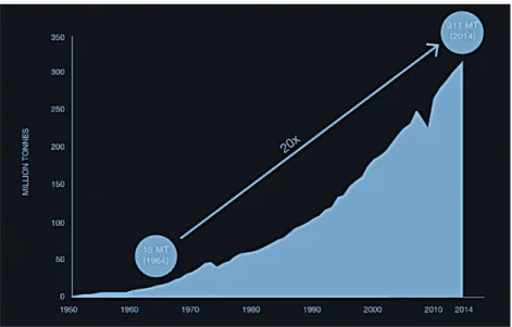
Its high durability and resistance to degradation, coupled with high consumption and low recycled volumes, contributed to the continuous increase of plastics in the environment in recent decades (Keane, 2007). Thus, there is an accumulation of plastic material and a growing need for the production of new ones (Pinto, 2012). In modern society, plastics have reached a critical status for medical, commercial and industrial applications. The annual production of plastics increased considerably from 1.5 million tons in 1950 (decade where the commercial development of polyolefins, polypropylene and polyethylene - started), to approximately 311 million tons in 2014 (PlasticsEurope, 2015; Wright et al., 2013b), representing an increase of 9% per year, approximately. Figure 1.1 shows the evolution of world plastic production during that period.
The world's greatest producer of plastics in 2014 was China, with a production of 67.6 million tons, followed by the European Union and North America, with 52 and 49.4 million tons, respectively (PlasticsEurope, 2015). Of the total production, about 60% was transformed into plastic solid waste (PSW) (Al-Salem et al., 2010).
2
The consumption of plastics in many European countries indicates that the plastic resins most used since 2007 are low density polyethylene (LDPE) and high density polyethylene (HDPE), polypropylene (PP), polyvinyl chloride (PVC), polyethylene terephthalate (PET) and polystyrene (PS). Table 1.1 features the different plastic resins and their applications.
Table 1.1. Plastic resins and their applications (adapted from: Pinto (2012))
Types of resins Characteristics Applications
Low density polyethylene (LDPE) Low electrical and thermal conductivity; resistant to chemical action. Many features and mechanical properties are maintained below 60 ° C.
Electronic material, agriculture and construction.
High density polyethylene (HDPE) High density. Opaque material. Easy to be processed, tougher and with better mechanical properties than LDPE. Resistance to chemicals, but not to strong oxidizing agents.
Packages, electronic material.
Polypropylene (PP) Homopolymer: electrical and mechanical strength; resistant to high temperatures.
Copolymer: transparent, more flexible and resilient than the homopolymer.
Car industry, packages, toys and electronic material.
Polyvinyl chloride (PVC) Resistant to high temperatures; flexible with much elasticity; resistance to many chemicals; good mechanical strength. High resilience to low temperatures. Easy to be sterilized.
Construction, packages, industrial processes, toys, footwear.
Figure 1.1. Worldwide plastic production between 1950 and 2014 (source: World
3 Polystyrene (PS) Crystal: electrical insulating of high
molecular weight and low water absorption. Bright, transparent and sensitive to light. Good thermal stability.
Expanded: high mechanical strength; looses properties at temperatures ≥ 88 °C. Resistant to acids, bases and salts. Flammable. Low adsorption of water. High and medium impact: sensitive to UV radiation. Translucent or opaque. Rigid and impact-resistant; not resistant to high temperatures. Thermally stable.
Packages and electronic material.
Polyethylene terephthalate (PET) Good mechanical strength and lower impact resistance. Impermeable to gases. Water adsorption capacity.
Packages and electronic material.
This production volume (Figure 1.1), coupled with their high durability and low weight, leads to the widespread and accumulation of discarded plastic in landfills and, as litter, in terrestrial and aquatic habitats worldwide (Derraik, 2002; Moore, 2008; Thompson et al., 2004).
1.1. Origin and the presence of plastics in the marine environment
The presence of plastics in the oceans was first reported in 1970, however it drew minimal interest of the scientific community (Andrady, 2011; Fowler, 1987). In the following decades, with the ecological effects of plastics, the subject began to gain scientific notoriety (Fowler, 1987).
The literature indicates the predominance of plastics amongst the marine litter (Gregory & Ryan, 1997). It is not possible to obtain reliable estimates of the amount of plastic debris reaching the marine environment, but the amounts are, however, quite substantial (Derraik, 2002). The production trend or the volume of particular polymer types does not correspond to the pattern of plastic litter observed. In fact, the variety of resin types produced reflects the composition of plastic debris recovered from the marine environment, but there are many social, economic, technical and environmental factors that influence the distribution and composition of plastic litter (Kershaw, 2015).
The major sources of plastic debris in the sea are fishing fleet (Cawthorn, 1989), recreational fishing and boats (UNESCO, 1994). Plastic materials also end up in the marine environment due to marine recreational activities (Pruter, 1987; Wilber, 1987).
4
Plastic also reaches the sea, as litter, carried by rivers and municipal drainage systems (Williams & Simmons, 1997). The major inputs of plastic litter from land sources occur in densely populated or industrialized areas (Derraik, 2002). It is estimated that with the migration of population to coastal areas, the influx of plastic waste in the ocean increased (Andrady, 2011) and that about 10% of the produced plastics enters the sea (Thompson et al., 2004).
In the sea, these versatile and non-biodegradable polymers are found in the form of larger items (macroplastics), including hulls of boats and fishing nets many meters long, and tiny fragments, micrometres in length (microparticles), and potentially, also at the nano-scale level (Browne et al., 2008; Canesi et al., 2015).
Particles of macroplastic (>1 mm) can be transported thousands of kilometres and contaminate relatively distant locations (Browne et al., 2010). Plastic debris accumulate along strandlines (Thornton & Jackson, 1998), in the open ocean (Shaw & Day, 1994), and on the seafloor (Galgani et al., 2000). Many data suggest that physical factors determine the spatial distribution of plastic, such as wind (Williams & Tudor, 2001), wave-action (Thornton & Jackson, 1998), and density of plastic (Thiel et al., 2003). This last factor will determine whether they float or sink and the role in influencing spatial patterns of accumulation. The polystyrene is the third densest resin among the more common plastics found in the marine environment, with a specific gravity of about 1.04 – 1.09 g cm-3 (Andrady, 2011).
Most plastics are resistant to biodegradation, but will break down gradually through mechanical action, since the mechanical integrity of plastic depends on its molecular weight and, therefore, any significant degree of degradation inevitably weakens the material (Thompson et al., 2004). When exposed to UV-B radiation, to the oxidative properties of the atmosphere and to the hydrolytic properties of seawater, these plastics brittle and break into smaller pieces, until they become microplastics.
Microplastics are defined as particles that are less than 5 mm in diameter, according to the National Oceanic and Atmospheric Administration of the United States of America (NOAA, 2015). Its presence in the ocean comes from a variety of sources and are distributed according to the currents (Lusher, 2015). Their origin can be natural (such as silk or cotton) or synthetic, such as polystyrene (PS) or polyethylene (PE) (Pinto, 2012).
The distinction between primary and secondary microplastics is based on whether the particles were originally manufactured to be that size (primary) or whether they have
5
resulted from the breakdown of larger items (secondary) (Kershaw, 2015). The primary source of microplastics may include polyethylene (PE), polypropylene (PP) and polystyrene (PS) from cleaning products or cosmetics (Fendall & Sewell, 2009), or from industries or industrial effluents (Lusher, 2015).
The secondary source is the degradation of plastics under marine conditions, that dramatically reduces the molecular weight of the polymers (Andrady, 2011). Ideally, these particles may also undergo further degradation by microbial action, releasing carbon (Andrady, 2011). The digestion is complete when the carbon present in plastics is converted into CO2, water and biomass (Andrady, 1994). To date, there is no information
on the complete mineralization of plastic in the marine environment because of the temperature and oxygen concentration (Andrady, 2011).
An exhaustive quantitative description of the relative abundance of microplastic compared to macroplastic debris remains to be accomplished. Although microplastics greatly exceed large plastic items in marine systems, they are still only a small proportion of the total mass of plastics in the ocean. This means that even if we were able to stop the discharge of macroplastics into the sea, the on-going degradation of the larger items already in the sea and on the beaches would result in a sustained increase in microplastics for many years to come (Browne et al., 2010).
Microplastics have become a growing issue in such a way that the Marine Strategy Framework Directive (MSFD Nº 2008/56/EC) highlights microplastics and its associated chemicals as one of the major policy descriptors to determine the impact on the marine environment (Zarfl et al., 2011).
In general, microplastics have been documented in most habitats in the open ocean, seas and beaches, in surface waters, in the water column and in the deep ocean (Lusher, 2015), and recently, in freshwater systems (Eerkes-Medrano et al., 2015). In Portugal, 62% of microplastics were identified in the North Atlantic by trawling, with a density of 580 000 particles per km2. 61% of the water samples collected in the Portuguese coast contained microplastics, and the concentrations were higher in the Costa Vicentina and Lisbon (0.036 and 0.033 particles m-3, respectively) than in the Algarve and Aveiro (0.014 and 0.002 particles per m3, respectively) (Lusher, 2015).
Manufactured nanoparticles are the latest trend of nanotechnology. They are used in a variety of applications including cosmetics, electronics, molecular biology, medicine, between others (Ward & Kach, 2009). Plastic nanoparticles - nanoplastics – are commonly defined as particles of plastic which are less than 100 nm (Koelmans, 2015).
6
These particles derived from post-consumer waste as well as from meso and microplastics after suffering degradation. However, the question remains about the origin of nanoparticles from the plastics breakdown (Andrady, 2011).
1.2. The impact of plastics in marine organisms
The occurrence of plastic in the ocean and the potential impact to marine organisms are of the growing concern (Canesi et al., 2015). The fact of microplastics have such a small size, actively contributes to its bioavailability and accumulation in organisms of lower trophic classes. Many of them, as they have little selectivity, catch everything that has an appropriate size and may easily be ingested by marine invertebrates, which are the basis of most food chains (Thompson et al., 2004). Therefore, ingestion is the main interaction between marine organisms and microplastics (Lusher, 2015), probably due to confusion with the prey (Andrady, 2011; Moore, 2008). As the particles interact with plankton and sediments, both organisms that feed on suspended particles, and the ones that feed on the bottom, are at risk of, accidentally or selectively, ingest plastic (Lusher, 2015). Plastics were ingested by marine mammals (Laist, 1997), cetaceans (Clapham et al., 1999), birds (Mallory, 2008), sea turtles (Mascarenhas et al., 2004), zooplankton (Cole et al., 2013), larvae and adult fish (Browne et al., 2013; Lusher, 2015; Rochman et al., 2014).
However, the selectivity is related to the size of the affected organisms and the particles they find (Lusher, 2015). Particles with less than 20 µm are likely to be ingested and egested (Lee et al., 2013) by small organisms (Thompson et al., 2004; Wright et al., 2013b). Microplastics with size between 1 and 5 mm can compromise the nutrition and digestion (Codina-García et al., 2013). The ingestion of plastics with a greater size, can cause serious external and internal injuries, ulcers, digestive tract blockage, false sense of fullness, loss of feeding capacity, impairment, inability to avoid predators or death (Gall & Thompson, 2015).
Microplastic ingestion was also documented for a wide range of marine vertebrates and invertebrates. Interactions were recorded from field of wild populations (Table 1.2) and during controlled laboratory studies (Table 1.3), both indicating microplastic ingestion. From ecotoxicological studies involving microplastics and their interactions and effects on aquatic organisms in seawater species, fish are the main group
7
studied, followed by Malacostraca, Bivalvia, Polychaeta, Mammalia and Echinoidea (Lusher, 2015).
Table 1.2. Evidence of microplastics ingestion in marine organisms
Whilst it is apparent that microplastics have become widespread and ubiquitous, the information concerning the accumulation, mode of action and biological impacts of this emerging pollutant in marine organisms is still scarce (Wright et al. (2013b). Table 1.3 summarizes the information about the ecotoxicological effects of microplastics in several aquatic organisms.
Species Microplastics
(%)
Mean (± SD) number of particles/ individual
Type and size (µm)
Reference
Phylum Arthropoda
Gammarus pulex _ _ Acrilic
29.5 ± 26
Imhof et al. (2013)
Notodromas monacha _ _ Acrilic
29.5 ± 26
Imhof et al. (2013) Phyllum Annelida
Lumbriculus variegatus _ _ Acrilic
29.5 ± 26
Imhof et al. (2013) Phylum Mollusca
Mytilus edulis _ 3.7 per 10 g tissue Fibres
300-1000
De Witte et al. (2014)
Mytilus edulis _ 0.36 (± 0.07) g-1 5 - 25 Van Cauwenberghe
and Janssen (2014)
Cassostrea gigas _ 0.47 (± 0.16) g-1 5 - 25 Van Cauwenberghe
and Janssen (2014) Phylum Crustacea
Lepas spp. 33.5 1-30 1.41 Goldstein and
Goodwin (2013)
Nephrops norvegicus 83 _ _ Murray and Cowie
(2011)
Crangon crangon _ 11.5 fibres per 10 g shrimp 300-1000 Devriese et al. (2015)
Phylum Chaetognatha
Parasagitta elegans 100 _ 0.1-3 PS Carpenter and Smith
(1972)
8
Table 1.3. Effects of microplastics to aquatic organisms
Species Microplastics Exposure Effects Reference
Type Size (µm) Concentration
(particles ml-1) Duration Phylum Annelida Class Polychaete Acrylic, alkyd resin, PE, PP, polyvinyl alcohol 20-2000 1.5 g L-1 _ Ingestion Thompson et al. (2004)
Arenicola marina UPVC 130 0-5% by weight 48 hours
24 days
Intake, decrease feed rate, increase phagocytic
activity, reduced lipid and energy reserves
Wright et al. (2013a) UPVC 230 1500 g of sediment mixture adsorbed with 5% of microparticles _ Ingestion, oxidative stress Browne et al. (2013) PS 400-1300 0,1,10,100 g L-1 28 d Ingestion, decrease feeding rate, weight loss
Besseling et al. (2012) PE, PS (<100) 110 MPs g-1 of sediment 14 d Increase EC
Increase protein content
Van Cauwenberg he et al. (2015) Ficoll – inert polymer
3-10 5 µL-1 20 min Ingestion Bolton and
Havenhand (1998)
PLA, PE, PVC 1.4-707 n.s 31 d Increase metabolic rate Green et al. (2016) Class Clitellata Galeolaria caespitosa PS 3 e 10 fluorescen tly labeled
635, 2240, 3000 1, 24 h Ingestion, size selection, egestion Cole et al. (2013) Marenzelleria spp. PS 10 2000 3 h Ingestion Setälä et al. (2014) Phylum Moullusca Class Bivalvia Mytilus galloprovincialis PE,PS, PE-PYR e PS_PYR
<100 20 g L-1 6 d Adsorption of pyrene not differ between PS and PE; bioaccumulation in digestive tissues and gills
Avio et al. (2015) Mytilus edulis PS fluorescently labeled 2 0.51 µg L-1 12 h Uptake accumulation in gut; and hemolymph
after 3 d
Browne et al. (2008)
9 4-16 3 and 9.6 3 d PS 10 50 14 d Greater accumulation of smaller particles; no significant effects on metabolism Van Cauwenber ghe et al. (2015) 30 50 90 10 110 ml-1 (Total concentration) HDPE 0-80 2.5 g L-1 3, 6, 12, 24, 48 and 96 h
Uptake; retention in gut and transfer into the
lymphatic system; immune response von Moos et al. (2012) Köhler (2010) PS Microspheres fluorescently labeled 0.5 50 μg per 400 ml seawater
1 h Uptake; trophic transfer to Carcinus maenas
Farrell and Nelson (2013)
PS 10 2 × 10⁴ 30 min Intake Ward and
Targett (1989) PS 10, 30 3.10 × 10⁵ _ Intake Claessens et al. (2013) PS 30 nm 0.1-0.3 g L-1 8 h Excretion in pseudofaeces; decrease of feeding rate Besseling et al. (2012)
Mytilus trossulus PS 10 1000 _ Intake Ward et al.
(2003) Crassostrea
virginica
PS 10 1000 45 min Intake and egestion
Ward and Kach (2009)
Crassostrea gigas PS 2, 6 µm 0.023 mg L−1 2 month Decreases in oocyte
number, diameter, and sperm velocity; decrease
of larval development; endocrine disruption Sussarellu et al. (2016) Placopecten magellanicus PS 15, 10, 16, 18, 20
1.05 1 h Intake, retention and egestion Brillant and MacDonal d (2002) Phylum Echinodermata Class Holothuridea Apostichopus californicus
PS 10, 20 µm 2.4 µl-1 - Intake, retention Hart
10 Thyonella gemmata PVC, nylon, resin pellets 0.25-15 mm 10 g PVC, 60 g resin 20 - 25 h Selective intake Graham and Thompson (2009) Holothuria grisea, Holothuria floridana, Cucumaria frondosa 2 g nylon line added to 600 ml of silica sand Class Echinoidea Tripneustes gratilla PE fluorescently labeled
32-35 1,10,100,300 5 d Intake and egestion Kaposi et al. (2014) Dendraster
excentricus
PS 10, 20 2.4 µL-1 _ Intake and egestion Hart
(1991) Strongylocentrotu
s sp.
PS 10, 20 2.4 µL-1 _ Intake and egestion Hart
(1991) Lytechinus variegatus PE n.s. 250 ml of MPs L-1 24 h Anomalous larvae development Nobre et al. (2015) Paracentrotus lividus PS 0.04-0.05 2.61-50 µg ml-1 48 h Increase of MPs accumulation Increase Abcb1 gene
Increase cas8 gene
Della Torre et al. (2014) Class Ophiuroidea Ophiopholis aculeata
PS 10, 20 2.4 µL-1 _ Intake and egestion Hart
(1991) Class Asteriodea
Dermasterias imbricata
PS 10, 20 2.4 µL-1 _ Intake and egestion Hart
(1991) Phylum Arthropoda Subphylum Crustacea Class Maxillopoda Semibalanus balanoides Synthetic polymers 20-2000 1 g L-1 _ Intake Thompson et al. (2004) Subclass Copepoda Tigriopus japonicus PS 0.05 9.1 x 10¹¹ 24 h Intake, egestion, mortality, reduced fertility rate Lee et al. (2013) 0.5 9.1 x 10⁸
Acartia tonsa Plastic particles
6 5.25 x 10⁵ 15 min Intake, particle size selection
Wilson (1973) 10-70 3000-4000
Acartia spp. PS 10 2000 3 h Intake Setälä et
al. (2014) Eurytemora
affinis
PS 10 1000, 2000,
10000
3 h Intake and egestion Setälä et al. (2014)
11 Limnocalanus macrurus PS 10 1000, 2000, 10000 3 h Intake Setälä et al. (2014) Temora longicornis PS fluorescently labeled 20 100 12 h Ingestion 10.7± 2.5
particles per organism
Cole et al. (2014) Calanus
helgolandicus
PS 20 75 24 h Intake and egestion Cole et al.
(2015) Class Branchipoda
Daphnia magna Acrilic 29.5 ± 26 µm _ _ Intake Imhof et al. (2013) Class Malacostraca Orchestia gammarellus Acrylic, alkyd resin, PE, PP, polyvinyl alcohol 20-2000 1 g per organism (n=150) _ Intake Thompson et al. (2004)
Talitrus saltator PE 10-45 10% per dry
weight food
24 h Intake, egestion after 2 h Ugolini et al. (2013) Allorchestes
compressa
LDPE 11-700 0.1 g/L 72 h Intake, egestion after 36 h
Chua et al. (2014)
Neomysis integer PS 10 2000 3 h Intake Setälä et
al. (2014)
Mysis relicta PS 10 2000 3 h Intake and egestion Setälä et
al. (2014)
Carcinus maenas PS
fluorescently labeled
8 – 10 4 x 10⁴ 24 h, 21 d Uptake by gills and mouth, retention and
excretion Watts et al. (2014) PP fibres 500 0, 0.6, 1.2, 2.0 mg added to 2 g of food
4 weeks Decrease intake of food over time; growth rate reduction; microfibers accumulation in digestive
tract - changing of microfibers properties by
passing the digestive tract Watts et al. (2015) Nephrops norvegicus PP microfibers 5 mm 10 fibres per 1 cm3 of organism _ Intake Murray and Cowie (2011) Class Branchiopoda Bosmina coregoni PS 10 2000, 10000 3 h Intake Setälä et al. (2014) Phylum Chordata Pomatoschitus microps 1-5 18.4, 184 µg L-1 96 h Intake, modulation of bioavailability and biotransformation of pyrene, decrease of energy, inhibition of AChE activity Oliveira et al. (2013)
12
1.3. Microplastics as contaminants to marine organisms
In addition to the direct physical impact caused by the intake of microplastics, they can be a vehicle for accumulation and transfer of other pollutants (Browne et al., 2013). These chemicals are divided into two groups: the first comprises the additives, monomers and by-products of the molecules component of the plastics, and the second,
PE 1-5 18.4, 184 µg L-1 96 h Intake, modulation of bioavailability and biotransformation of pyrene, decrease of energy, inhibition of AChE activity Oliveira et al. (2013) 420-500 30 particles per 300 ml water
96 h Intake (confusion with food– Artemia nauplii)
de Sá et al. (2015)
PE 1-5 0.184 mg L-1 96 h Inhibition of AChE activity
Luís et al. (2015)
Gadus morhua Plastic
particles
2, 5 mm _ _ Intake, egestion, particles of 5 mm for a long period. Emptying of plastics improved by giving additional meals.
Dos Santos and Jobling (1992) Sfellifer brasiliens
Nylon _ _ _ Intake (higher in adults
than larvae) ; presence of biofilms in microplastics Dantas et al. (2012) Cathorops spixii, Cathorops agasizii, Sciades herzbergii Nylon _ _ _ Intake by 17 to 33% of total organisms. Selective intake according to size Possatto et al. (2011) Eugerres brasilianus, Eucinostomus melanopterus, diapterus rhombeus Nylon 1-5 mm _ _ Intake by 4.9 to 33.4 % of total organisms. Selective intake according to size Ramos et al. (2012)
Gobio gobio Fibers and
plastic pellets
_ _ _ Intake by 12 of total
organisms. 7 in 11 fishes had microplastics in the
system.
Dris et al. (2015)
Oryzias latipes PE 3 mm 10% particles per
weight
2 months chronic exposure
Altered gene expression, decreased choriogenin regulation in males and decreased vitellogenin and choriogenin in females Rochman et al. (2014)
LDPE 3 mm 10% particles per weight
2 months chronic exposure
Toxicity in the liver, pathologies, hepatic stress Rochman et al. (2013) Dicentrarchus labrax PE 10-45 10-100 MPs mg-1 of diet 36 d Increase mortality Increase CYP P450 Mazurais et al. (2015)
PS (Polystyrene) PE (Polyethylene) PP (Polypropylene) LDPE (Low Density Polyethylene) HDPE (High-density Polyethylene) UPVC (Polyvinyl chloride) PE-PYR (Pyrene Treated Polyethylene) PS_PYR (Pyrene Treated Polystyrene)
13
hydrophobic chemicals that are adsorbed from surrounding seawater, due to affinity of those chemicals for the hydrophobic surface of the plastics (Teuten et al., 2009). This includes persistent organic pollutants (POPs) and bioccumulative and toxic substances (Browne et al., 2013; Engler, 2012), including polychlorinated biphenyls (PBTs), polybrominated diphenyl ethers (PBDEs), dichlorodiphenyltrichloroethane (DDT), polycyclic aromatic hydrocarbons (PAHs) and the other petroleum hydrocarbons (Chua et al., 2014; Mato et al., 2001; Rios et al., 2007; Teuten et al., 2009). Marine microplastics can also be covered with biofilm communities that act like a reservoir for POPs. In this case, there is a great probability of transfer of these chemicals along with the microplastics to marine organisms (Eerkes-Medrano et al., 2015), especially to lower trophic levels (Arnot & Gobas, 2004).
With the increasing microplastic contamination in marine ecosystems, large concentrations of microplastics and additives can harm ecophysiological functions performed by organisms (Browne et al., 2013). Ingestion of contaminated microplastics and bioaccumulation of sorbed chemicals have been documented in several organisms. In fish (Rochman et al., 2013) where, when exposed to a mixture of polyethylene with chemical pollutants sorbed from the marine environment, they bioaccumulate and suffer liver toxicity and pathology. Lugworms were also affected (Browne et al., 2013), by the uptake of Triclosan from PVC, which reduced the ability of worms to engineer sediments and cause mortality. The marine amphipod Allorchestes compressa assimilated PBDEs, derived from microplastics, into the tissues (Chua et al., 2014). Avio et al. (2015) observed PAH accumulation in digestive tissues, hemolymph and gills of M. galloprovincialis.
If marine organisms ingest microparticles, it is possible that microplastics-associated POPs and other additive chemicals are delivered to different tissue types and locations (Zettler et al., 2013) although, until to date, this was not totally clarified (Browne et al., 2013; Chua et al., 2014).
1.4. Biomarkers
In bivalves, the microplastics uptake relates directly to their feeding strategy, since they are filter feeders. The microparticles are inert and, after ingestion, pass through the cell membrane and are incorporated into tissues, particularly in the gut cavity (Wright et al., 2013b). After passing to the circulatory system, microplastics are transferred to other
14
organs, via hemolymph, where they can be retained for several weeks, accumulate in several organs and cause adverse effects (Browne et al., 2008). Exposure experiments demonstrated significant biological effects including weight loss, reduced feeding activity, increased phagocytic activity, transference to the lysosomal system and inhibition of AChE activity (Lusher, 2015). Microplastics accumulation can also be transferred to higher trophic levels (Wright et al., 2013b).
To clarify any effects of exposure to microplastics, a set of biomarkers are employed: the analysis of DNA damage, the activities of antioxidant enzymes that enable to maintain the cellular integrity (SOD, CAT, GPx), and glutathione S-transferases (GST) participating in biotransformation and protection against oxidative stress, the activity of the enzyme acetylcholinesterase (AChE) involved in neuro and neuromuscular transmission, and the levels of lipid peroxidation (LPO) as indicative of oxidative damage in lipids.
1.5. Oxidative stress
Most of the oxygen consumed by the animals is reduced to water along with oxidation of food intake and energy production. It is estimated that about 1 to 3% of oxygen consumed by animals is converted into ROS. These compounds are continuously produced due to the presence of xenobiotics/contaminants, mainly as "unwanted bi-products" of the biotransformation from endogenous processes, which include self-oxidation of heme proteins and electrons nuclear transport (de Zwart et al., 1999; Livingstone, 2001).
Even without pollution and stress factors, the cellular metabolism of aerobic organisms involves the production of reactive oxygen species (ROS), that include the anion superoxide (O2●-), hydrogen peroxide (H2O2), hydroxyl radical (●OH), the peroxyl
radical, the hidroperoxil radical, hypochlorous acid and peroxynitrite (considered by some as a reactive nitrogen species – RNS) (Halliwell & Gutteridge, 1999; Kappus, 1987; Valavanidis et al., 2006). These ROS are essential for the physiological control of cell functions in biological systems (Halliwell & Gutteridge, 1999).
Organisms have developed non-enzymatic and enzymatic antioxidant defence mechanisms to prevent and eliminate the effects of ROS, and repair of oxidized component systems. Aquatic organisms in the presence of a stressor agent can induce the
15
production of ROS, which can cause oxidative damage, indicative of toxicity (Livingstone, 2001).
The impact of plastic ingestion (stressor agent) in marine organisms include oxidative stress, and thus, oxidative damage (Table 1.3) (Browne et al., 2013; Galloway & Lewis, 2016; Luís et al., 2015; Oliveira et al., 2012; Oliveira et al., 2013).
When there is an imbalance between the production of ROS and the detoxification of biological systems, oxidative stress occurs (Valavanidis et al., 2006).
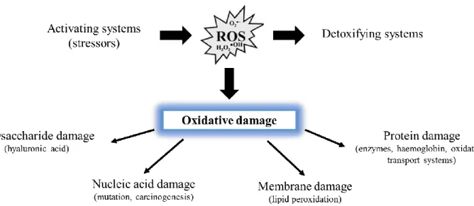
Due to the high reactivity of ROS, most components of cellular structures and function can be targets of oxidative damage (Kappus, 1987). ROS production associated with exposure to contaminants can inhibit the activity of antioxidant defences, leading to oxidation of essential cellular components such as proteins, DNA, carbohydrates and lipids, in the tissues of exposed organisms (Halliwell & Gutteridge, 1999; Shi et al., 2005) (Figure 1.2).
1.5.1. Antioxidant enzymes
The production of ROS needs to be balanced through enzymatic and non-enzymatic antioxidant defences. In aerobic organisms, intracellular antioxidant enzymes are responsible for the neutralization of ROS (Fenech & Ferguson, 2001).
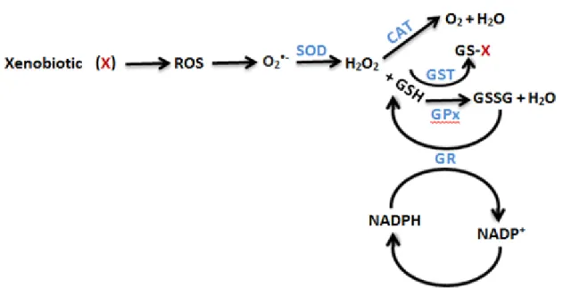
The use of antioxidant enzymes as biomarkers have been widely applied in toxicity studies with aquatic organisms (e.g. Bebianno et al., 2014, Silva et al., 2012) to evaluate the effects caused by a contaminant. In figure 1.3 there is an explanatory diagram
16
of the mechanism of action of each enzyme, and how they act in a coordinated fashion to be effective in the ROS removal.
Superoxide dismutase (SOD) catalyses the dismutation of the radical superoxide (a major reactive oxygen species) to hydrogen peroxide. This enzyme occurs in the cytoplasm and mitochondria of cells (Halliwell & Gutteridge, 1999).
Catalase (CAT) is located in the peroxisomes, and decomposes the hydrogen peroxide into water and oxygen, being also involved in the metabolism of fatty acids. (Halliwell & Gutteridge, 1999).
The activity of glutathione peroxidases (GPx) catalyses the levels of hydrogen peroxide (H2O2) and lipid hydroperoxides (Júnior et al., 2001). Glutathione reductase
(GR) does not directly act in the removal of ROS, being responsible for the reduction of oxidized glutathione (GSSG) to its reduced form (GSH) in the presence of nicotinamide adenine dinucleotide phosphate (NADPH), continuing the action of glutathione peroxidases and glutathione S-transferases (Halliwell & Gutteridge, 1999).
The superfamily of glutathione S-transferases (GST) comprises eukaryotic and prokaryotic phase II metabolic isozymes. Most of them catalyses the conjugation of GSH (reduced glutathione) with xenobiotic substrates for the purpose of detoxification (Ioannides, 2002). The GST catalyses the reduction of lipid peroxides and so it is, therefore, important in preventing the oxidative damage (Zanette et al., 2011; Zhao et al., 1999), also having the function of antioxidant enzyme.
17
1.6. DNA damage
It is known that superoxide radicals directly or indirectly damage DNA, resulting in strand scission and chromosome breakage (Brawn & Fridovich, 1981).
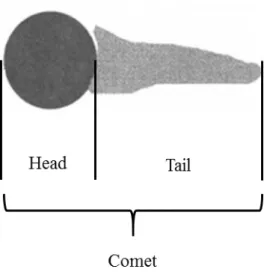
DNA alterations in aquatic organisms have prove to be a very suitable method to evaluate the genotoxic contamination of the environment, allowing the detection of effects after exposure to low concentrations of contaminants, in a variety of aquatic species (Frenzilli et al., 2009; Nacci et al., 1996), such as vertebrates Salmo trutta fario (Belpaeme et al., 1996) and Cyprinus carpio (Pandrangi et al., 1995)). The comet assay is more sensitive than other methods commonly used in genetic ecotoxicology (Frenzilli et al., 2009) and has been successfully used in invertebrates namely: Nereis diversicolor (Catalano et al., 2012; Maranho et al., 2014), including bivalve molluscs (Jha, 2008): Mytilus galloprovincialis (Gomes et al., 2013), Scrobicularia plana (Petridis et al., 2009), Perna viridis (Siu et al., 2004), among others.
Given that, until today, studies with nanoparticles and quantum dots evidence DNA damage using the Comet assay (Gomes et al., 2013; Rocha et al., 2014), the possible genotoxicity of microplastics is an important topic of research, alongside the development of a robust assay that can be used for general screening of anthropogenic impacts.
1.7. Neurotoxicity
Animals are extremely sensitive to environmental contamination, that may affect their neurological and behavioural activities (Costa, 1996; Døving, 1991; Silbergeld, 1993). The main role of acetylcholinesterase (AChE) is the termination of nerve impulse transmission at the cholinergic synapses by rapid hydrolysis of acetylcholine (ACh) into choline and acetic acid (Lackner, 1998; Lionetto et al., 2003).
Its inhibition is directly linked with the mechanisms of toxic action of pollutants (Hernández et al., 1998). Even at low concentrations, these compounds can inhibit AChE, which leads to accumulation of acetylcholine at central cholinergic synapses and at vertebrate neuromuscular junctions (Høy et al., 1991; Sancho et al., 1997). As a result, these disturbances can affect locomotion and equilibrium in exposed organisms (Bretaud et al., 2000).
18
In aquatic organisms there is considerable diversity in the biochemical properties and distribution of AChE as well as in their sensitivity to anticholinesterase agents (Habig & Di Giulio, 1991). Therefore, measurements of acetylcholinesterase activity has been routinely used as a biomarker of exposure to certain groups of contaminants that have the potential to inhibit AChE such as: organophosphate and carbamate insecticides (Grue et al., 1997; Williams & Sova, 1966), pesticides (Davies et al., 1994), herbicides (dos Santos Miron et al., 2005), metals (Garcia et al., 2000; Gill et al., 1990), pharmaceuticals (Luís et al., 2015), between others.
More recently, the acetylcholinesterase activity has also been used to infer the effects of microplastics on cholinergic, neurological and neuromuscular transmission (Avio et al., 2015; dos Santos Norberto, 2014; Ferreira et al., 2016; Oliveira et al., 2012; Oliveira et al., 2013).
1.8. Lipid peroxidation
The most typical reaction induced by reactive oxygen species implies the peroxidation of unsaturated fatty acids (Kappus, 1987). The reaction sequence starts with a radical (e.g., OH.) which removes one proton from the hydrocarbon tail of the fatty acid
and leaves the radical of the acid. This radical experiences isomerization and oxidation with molecular oxygen, producing a peroxy radical of the fatty acid. In turn, peroxy radicals remove protons from other molecules and become hydroperoxides. Since these protons may be from another fatty acid, a new cycle is started. Therefore, lipid peroxidation proceeds via a chain reaction until the chain is interrupted, by either the dimerization of two radicals, or until the proton is removed from a substance which forms relatively stable radicals (radical scavengers). Through this chain reaction, only one initiating radical may lead to the peroxidation of hundreds of fatty acids (Lackner, 1998). The resulting hydroperoxides are unstable and decompose by chain cleavage to a very complex mixture of aldehydes, ketones, alkanes, carboxylic acids and polymerisation products (Esterbauer et al., 1982).
The only mechanism that produces malondialdehyde in biological systems is lipid peroxidation. Malondialdehyde is not the major product of lipid peroxidation however, it is a typical degradation product. This fact coupled to the high sensitivity of the
19
thiobarbituric acid test (described in materials and methods section), have greatly inspired reactive oxygen species research (Lackner, 1998).
Therefore, LPO was found to be suitable as a biomarker of effect (Ahmad et al., 2008; Lackner, 1998; Livingstone, 2001).
1.9. Scrobicularia plana characterization
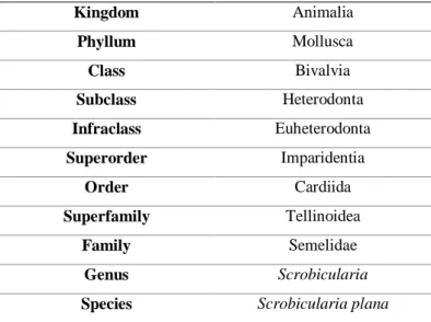
Invertebrates are a very large and diverse group of organisms. They are very useful in monitoring studies with special emphasis on sessile individuals (Dixon et al., 2002). Within this group there is the Bivalvia Class, Phylum Mollusca (Table 1.4), which is composed of approximately 15 000 species, most of them marine (Campbell et al., 1994), including the Scrobicularia plana (da Costa, 1778).
Table 1.4. Scientific classification of Scrobicularia plana (Source: WoRMS http://www.marinespecies.org)
Kingdom Animalia Phyllum Mollusca Class Bivalvia Subclass Heterodonta Infraclass Euheterodonta Superorder Imparidentia Order Cardiida Superfamily Tellinoidea Family Semelidae Genus Scrobicularia
Species Scrobicularia plana
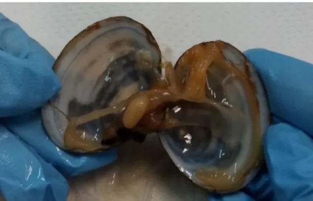
S. plana has an oval, flat shell with an exterior pale yellow grey colour and white in the interior (Figure 1.4). The size vary between 4 to 6 cm length with an outer surface with concentric lines (Campbell et al., 1994). Figure 1.5 shows the internal appearance of S.plana.
20
S. plana is gonochoristic, and sexual maturity occurs 2 to 3 years after settlement, corresponding to a shell length greater than 20 mm (Hughes, 1970; Rodrıguez-Rúa et al., 2003). Regarding the reproduction cycle, S. plana development of gonads occurs from the beginning of February until the end of October, the spawning season is from March to September, and the maximum spawning peak usually occurs in the second half of May and July. October to January represents the inactive reproductive period, during which about more than half of the total population is not sexually determined. This reproductive cycle is influenced by environmental factors, such as water temperature and chlorophyll availability (Rodrıguez-Rúa et al., 2003).
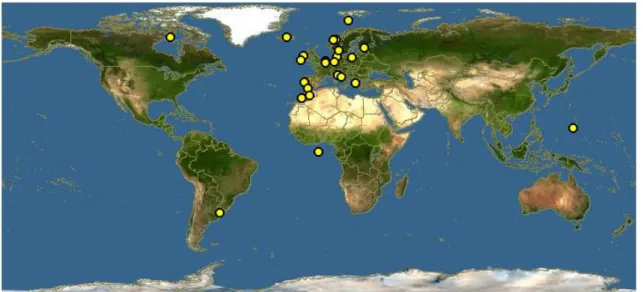
S.plana inhabits the intertidal zone of estuarine muds (Green, 1957), where there is abundance of organic detritus and most pollutants are present (Wootton & Pipe, 2003). This bivalve species is the most representative species of the Atlantic and the Mediterranean coasts. It has a wide geographical distribution: from Norway (in the north) to Senegal (in the south) in the Atlantic ocean, and in all of the Mediterranean, except the Black Sea (Campbell et al., 1994; Parenzan, 1974) (Figure 1.6). Its economic and commercial interest has increased in recent years (FAO, 2014; González De Canales et al., 2009)
Figure 1.5. Internal appearance of S. plana
Figure 04. Specimens of S. plana. A – Interior of the
21
S. plana is a burrowing deposit-feeding bivalve with a filtration efficiency approaching 100% for particles of 4-40 µm, and much of the filtered material is ingested (Hughes, 1969).
1.9.1. S. plana as a bioindicator of environmental contamination
Sentinel species have been used to define the status and the evolution of the quality of the marine environment (Viarengo & Canesi, 1991). Several features make bivalves particularly important as sentinel species: they are sessile, filter-feeders and accumulate particles from the water, allowing to measure stress in their tissues (Canesi et al., 2012). They are resistant to a variety of contaminants and environmental factors (such as temperature or salinity) having the ability to survive in highly stressful environments. They are easily collected and easily maintained in the laboratory. Since they aggregate in large populations, it is possible to repeat sampling over a given period and evaluate the environmental contamination in a given area. As these bivalves have a worldwide distribution (both fresh and salty water), and there is enough information regarding its biology and response to environmental conditions (Canesi et al., 2012; Rocha et al., 2015), the results obtained in experimental studies can be compared.
This bivalve, in direct contact with sediments, through physical contact and ingestion of sediment particles, is a suitable indicator of sediment-associated contaminants (Mouneyrac et al., 2008). Moreover, S. plana forms an important part of
22
the diet of wading birds, crabs and benthic fish (Hughes, 1970) and if contaminants are available, they can be transferred through the food chain.
Concluding, its wide geographic distribution, high tolerance of exposure to contaminants, the type of sedentary life, its low metabolism, the commercial importance, and increasing knowledge about the species, defined it as an excellent candidate to be used in monitoring studies of aquatic ecosystems (Solé et al., 2009).
1.10. Objectives
Since the microplastics mode of action and biological risk are not yet clear, the aim of this study is to investigate the accumulation and mode of action of the polystyrene microparticles in the different tissues of the peppery furrow shell S. plana and assess the potential ecotoxicological risk of this emerging contaminant. Polystyrene (PS) is one of the most largely used plastics worldwide, it is found in the oceans as micro and nano debris and has a considerable impact in marine species, such as bivalves.
The effects of microplastics accumulation in gills and digestive gland of S. plana will be evaluated using a battery of biomarkers of oxidative stress (superoxide dismutase, catalase, glutathione peroxidases), glutathione-S-transferases, genotoxicity (comet assay to evaluate DNA damage), neurotoxicity (acetylcholinesterase activity) and oxidative damage (lipid peroxidation), considered the most appropriate to assess microplastic effects.
The aim of this work was to answer the following specific questions:
1. Do microplastics accumulate in S. plana tissues?
2. Do microplastics have the potential to induce cellular oxidative stress and/or neurotoxicity in S. plana?
3. Are plastic particles responsible for DNA damage in the cells of S. plana?
4. Can this species be a good sentinel to assess the effects of microplastics in marine organisms?