Deception and manipulation: the arms of Leishmania, a
successful parasite
Pedro Cecílio
1, Begoña Pérez-Cabezas
1, Nuno Santarém
1, Joana Maciel
1, Vasco Rodrigues
1and
Anabela Cordeiro da Silva
1,2*
1Parasite Disease Group, Institute for Molecular and Cell Biology (IBMC), University of Porto, Porto, Portugal 2Department of Biological Sciences, Faculty of Pharmacy, University of Porto, Porto, Portugal
Edited by:
Abhay Satoskar, The Ohio State University, USA
Reviewed by:
Hira Nakhasi, US Food and Drug Administration, USA
Diego A. Vargas-Inchaustegui, National Cancer Institute, USA *Correspondence:
Anabela Cordeiro da Silva, Parasite Disease Group, Instituto de Biologia Molecular e Celular (IBMC), Universidade do Porto, Rua do Campo Alegre 823, Porto 4150-180, Portugal e-mail: [email protected]
Leishmania spp. are intracellular parasitic protozoa responsible for a group of neglected
tropical diseases, endemic in 98 countries around the world, called leishmaniasis. These
parasites have a complex digenetic life cycle requiring a susceptible vertebrate host and a
permissive insect vector, which allow their transmission. The clinical manifestations
asso-ciated with leishmaniasis depend on complex interactions between the parasite and the
host immune system. Consequently, leishmaniasis can be manifested as a self-healing
cutaneous affliction or a visceral pathology, being the last one fatal in 85–90% of untreated
cases. As a result of a long host–parasite co-evolutionary process, Leishmania spp.
devel-oped different immunomodulatory strategies that are essential for the establishment of
infection. Only through deception and manipulation of the immune system, Leishmania
spp. can complete its life cycle and survive. The understanding of the mechanisms
asso-ciated with immune evasion and disease progression is essential for the development of
novel therapies and vaccine approaches. Here, we revise how the parasite manipulates cell
death and immune responses to survive and thrive in the shadow of the immune system.
Keywords: Leishmania, immunomodulation, apoptosis, innate immunity, acquired immunity
INTRODUCTION
Parasitism is defined as a “non-mutual symbiotic relationship
between species, where one species, the parasite, benefits at the
expense of the other, the host,” Such relationship occurs
dur-ing leishmaniasis, where the protozoan Leishmania spp. takes
advantage of its mammalian host in order to survive and thrive.
Leishmania is a genus of trypanosomatid protozoa that
com-bines over 30 species, of which 11 have significant medical and
veterinary importance (
1
). These parasites have a complex
dige-netic life cycle, with some particularities, requiring a vertebrate
host and an insect vector. The alimentary tract of female
Phle-botomus spp. and Lutzomyia spp. sandflies is colonized by the
extracellular form of the parasite, the flagellated, and motile
pro-mastigote. Within the insect midgut, Leishmania undergoes several
developmental changes that culminate in the infectious
develop-mental form of the parasite: the metacyclic promastigote. During
the insect blood feeding, the parasite infectious forms are released
into the mammal host dermis and quickly uptaken by mono and
polymorphonuclear (PMN) cells. Ultimately, in the
phagolyso-some of macrophages, promastigotes will differentiate into the
non-motile amastigote form and multiply. The cycle is
com-pleted when the sandfly takes another blood meal, recovering free
amastigotes or infected cells (
1
–
3
).
Leishmaniasis is endemic in 98 countries, 72 of which are
devel-oping nations and 13 correspond to the least developed ones, being
considered by the World Health Organization as a Neglected
Trop-ical Disease (
4
,
5
). Over 350 million people reside in areas with
active parasite transmission (
6
). Annually, an estimated 1.5–2
mil-lion develop symptomatic disease, and approximately 50,000 die,
mostly children (
4
,
7
). Climate changes and population mobility
can contribute to the increase of the vector activity and,
con-sequently of the disease incidence (
8
,
9
). The infection caused
by Leishmania spp. can lead to different clinical manifestations
depending on complex interactions between the parasite and the
host immune response. The disease is normally divided into three
main categories: cutaneous, mucocutaneous, and visceral.
Cuta-neous leishmaniasis is the most extensively studied form of the
disease, usually appearing as a self-healing skin ulcer or
der-mal granuloma that may need several months or years to heal
(
10
). In some cases, these ulcers can become chronic (
11
). While
most Leishmania species cause lesions confined to small areas
of the skin, a few, such as L. braziliensis, cause diffuse lesions
that may even spread to mucosal tissues leading to the
muco-cutaneous form of the disease (
12
). Finally, visceral
leishmania-sis, the most severe leishmaniasis form, is caused by Leishmania
donovani and Leishmania infantum. It is characterized by fever,
cachexia, hepatosplenomegaly and hypergamaglobulinemia and,
when untreated, can be fatal (
13
). In endemic countries,
Leish-mania has gained prominence as an opportunistic pathogen in
HIV positive and other immunocompromised patients (
8
,
14
).
Leishmaniasis is also a major veterinary concern, as dogs are the
main reservoir for the parasite in South America and southwestern
Europe (
15
).
There is no human vaccine available at the moment.
Nonethe-less, prevention of infection through vaccination seems to be a
viable option, since in endemic areas the majority of infected
per-sons do not develop clinical symptoms and previous infection
leads to robust immunity against the parasite (
16
). In the absence
of vaccines, control of the disease relies on prophylaxis and
treat-ment, reviewed elsewhere (
17
,
18
). Treatment options are limited,
present significant toxicity and require, with the exception of oral
miltefosine, administration in ambulatory conditions (
18
). Drug
resistance is also a growing limitation of some anti-leishmanial
therapies (
19
). Therefore, it is essential to develop novel treatment
options and vaccine strategies. Such goal has its cornerstone on the
solid knowledge of the details of parasite infection. For this,
dif-ferent strategies that Leishmania uses to manipulate the immune
system to establish infection will be revised here.
PLAYING WITH DEATH TOWARD THE ESTABLISHMENT AND
MAINTENANCE OF INFECTION
Apoptosis, or programed cell death, is a physiological and
essen-tial process for the maintenance of general cellular homeostasis. In
immunology, this mechanism is indispensable for elimination of
autoreactive immune cells (
20
,
21
) and control of the proliferative
response (
22
,
23
). Programed cell death also plays a key role in
the resolution of infections produced by intracellular pathogens
(
24
). However, and as a result of the continuous host-microbe
co-evolutionary process, Leishmania developed strategies for using
apoptosis to its own benefit.
DEAD PARASITES ARE ESSENTIAL FOR THE SURVIVAL OF FREE PROMASTIGOTES
Parasite cell death, reviewed elsewhere (
25
–
27
), seems to be very
relevant for the deception of the initial immune response. Some
authors described that the presence of apoptotic parasites is
essen-tial for successful infection of mice susceptible to cutaneous
leishmaniasis. Indeed BALB/c mice did not develop disease after
intradermal infection with purified virulent non-apoptotic
para-sites (
28
,
29
). The need for dead parasites in the infective inoculum
is related with the exposure of phosphatidylserine (PS) in the outer
leaflet of the parasite cytoplasmic membrane. The exposure of this
phospholipid enables a silent invasion, inducing the production
of anti-inflammatory cytokines such as TGF-b (
30
,
31
). In fact, a
recent study shows that the administration of a PS-targeting
anti-body after C57Bl/6 mice intradermal infection with L. amazonensis
promastigotes renders the animals more resistant to the infection
(
32
). Thereby, and as represented in Figure 1, the inoculation of
equal proportions of dead and live parasites in the mammalian
host may allow the silent entry of Leishmania into the first cells
recruited to the inoculation site (
28
,
33
).
MODULATING APOPTOSIS OF NEUTROPHILS AT THE INOCULATION SITE
It is accepted that macrophages are the cells predominantly
infected in leishmaniasis. However, they are neither the first nor the
only to be recruited to the site of inoculation. Several evidences
support the early recruitment of neutrophils to the inoculation
site. Two hours after natural infection of C57Bl/6 mice with L.
major, neutrophils are predominantly found (
34
). Such
granu-locyte infiltration was also seen upon intradermal infection of
either BALB/c or C57Bl/6 mice with L. infantum and L. major,
respectively (
35
,
36
), as well as after subcutaneous infection with
L. amazonensis or L. major promastigotes (
37
–
39
). Furthermore in
a murine air pouch model, L. major, and to a lower extent L.
dono-vani, predominantly induced the recruitment of neutrophils 6 h
FIGURE 1 | Silent entry of Leishmania into the host cells. Live and dead
parasites are engulfed by phagocytes. The recognition of the externalized phosphatidylserine present on the cellular membrane of dead parasites induces TGF-b secretion and TNF-a downregulation (1). Neutrophil apoptosis is delayed by Leishmania (2). Both dendritic cells (3) and macrophages (4) remove neutrophil apoptotic bodies carrying Leishmania promastigotes and secrete TGF-b and IL-10. Macrophages (5) can also phagocyte parasites extruded within other macrophage membrane blebs, which in turn promotes the secretion of IL-10.
after infection (
40
,
41
). Interestingly, the air pouch system revealed
that L. major derived extracellular vesicles induced the same type
of cellular recruitment as parasites (
40
). These studies preceded
the description of Wilson et al. who saw neutrophils infiltration
1 h after intradermal inoculation of L. donovani promastigotes in
hamsters (
42
). Although the role of neutrophils during infection is
not consensual, several evidences support the capacity of
Leishma-nia to modulate their life span. Traditionally, neutrophils show a
relatively short life span (
43
), but Leishmania can successfully delay
their programed cell death for up than 24 h, potentially benefiting
from the protection of a safe intracellular niche (
44
). However,
other studies show induction of neutrophil apoptosis after
para-site intake (
35
). These contradictions may be due to differences in
the genetic background of the animal model used (BALB/c versus
C57Bl/6), as well as in the parasite inoculation route (
45
). The
delay in the natural apoptotic process of infected neutrophils was
related to an inhibition of the pro-caspase-3 processing (
44
), and
the consequent diminishment of caspase 3, a well-known
apop-tosis executer in neutrophils (
46
). Moreover, a recent publication
clarified the mechanisms by which L. major contributes for the
neutrophil apoptosis inhibition, showing that the key event is the
activation of the extracellular signal-regulated kinases (ERK1/2)
survival pathway (
47
). Sarkar and colleagues showed that the
par-asite upregulates ERK1/2 phosphorylation, leading to the delay
of neutrophil apoptosis (
48
). Also, this work unveiled additional
players of the apoptotic machinery responsible for neutrophil life
span enhancement. Among these the anti-apoptotic proteins, Bfl-1
and Bcl-2 were upregulated, preventing the release of cytochrome c
from the mitochondria and the downstream activation of caspases.
Additionally, processing of the pro-apoptotic Bid was inhibited
and the Fas expression reduced, preventing apoptosis triggering
(
48
). This delay of neutrophil death may be essential for the arrival
of a sufficient number of antigen-presenting cells (APCs), namely
macrophages, and dendritic cells (DCs), to the inoculation site.
“TROJAN HORSE” STRATEGYAfter being infected, dying neutrophils secrete different
chemo-tactic factors for macrophages (
49
,
50
); cells that then remove
apoptotic neutrophils by phagocytosis and secrete the
anti-inflammatory cytokine TGF-b (50). High amounts of IL-10 and
low amounts of interleukin (IL)-12 may also contribute for the
silent entry of L. major into macrophages (
51
) as shown in
Figure 1. The parasite can, therefore, arrive to its primary host
cell unnoticed and proceed with the infection process, using the
so-called “Trojan horse” strategy (
52
). TGF-b seems to be essential
for the establishment of infection not only by L. major but also
by L. amazonensis, although conclusions about the exploitation of
the “Trojan horse” strategy in this case cannot be withdrawn (
50
,
53
). DCs have also been related with this tactic. Ribeiro-Gomes
et al. recently described in a mouse model of intradermal
infec-tion with L. major that skin resident DCs uptake apoptotic infected
neutrophils and, as a consequence, the activation of
Leishmania-specific CD4
+T cells is prevented somehow (
35
). Other authors
suggested that free parasites silently enter into host cells taking
advantage of nearby neutrophil apoptotic bodies with exposed
PS (
54
).
BUYING TIME BY PROLONGING THE LIFE OF MACROPHAGES
When promastigotes reach macrophages, its definitive cellular
host, a new step of the infective process begins with their
differen-tiation into amastigotes. Therefore, inhibition of apoptosis may be
once more essential for Leishmania to protect its niche, enabling
the differentiation into the amastigote form that is fully adapted to
the phagolysosome. Extensive data exists concerning the capacity
of the parasite to increase the life span of infected macrophages.
The first description was made by Moore and Matlashewski, who
reported that L. donovani infection of murine bone
marrow-derived macrophages (BMM) represses macrophage apoptosis
through a mechanism dependent on the secretion of TNF-a (
55
).
Since then, numerous studies addressed this issue, unveiling some
intracellular mechanisms that could explain the death delay.
Exter-nal ATP is known to trigger death in macrophages when injured
or stressed, by its binding to purinergic receptors of the P2X
fam-ily (
56
,
57
). Interestingly, Kolli et al. showed that L. amazonensis
releases nucleoside diphosphate kinase (NdK), preventing
ATP-induced cytolysis of J774 macrophages (
58
). Further studies are,
however, required to access the relevance of NdK in the context
of infection. The ERK1/2 pathway also plays a role in the
preven-tion of macrophage apoptosis. Kamir and colleagues described
a protein produced by L. major that shows structural homology
with the human macrophage inhibiting factor (MIF) and exerts
similar effects. Indeed this MIF ortholog induced ERK1/2 kinases
activation in a CD74-dependent manner, subsequently resulting
in the inhibition of macrophage apoptosis in vitro (
59
). The
mito-chondrial apoptotic pathway is also modulated by Leishmania.
BMM infected with L. major showed enhanced survival that was
related with the prevention of cytochrome c release by
mito-chondria (
60
), observation possibly explained by the involvement
of an anti-apoptotic signaling pathway (
61
). Ruhland and
col-leagues showed that L. major block macrophage apoptosis through
the phosphatidylinositol 3
0-kinase (PI3K)/protein kinase B (Akt)
signaling pathway. Briefly, Akt phosphorylates the pro-apoptotic
Bad, deactivating it, and preventing the release of mitochondrial
cytochrome c (
62
), which avoids downstream activation of the
effector caspase-3 (
60
). Similar results were obtained with DCs
(
63
,
64
). More recently, it was also shown that apoptosis
trig-gered by oxidative burst is prevented by L. donovani. Although
infected macrophages were capable of ROS production, a
com-plete abrogation of the downstream caspase cascade was observed
due to thioredoxin mediated selective induction of suppressors of
cytokine signaling (SOCS) proteins (
65
). A direct responsibility
of a parasitic protein was not addressed in these studies, but we
cannot exclude the role of phosphoglycans since there are
stud-ies that relate them with apoptosis delay in L. infantum, L. major,
and L. donovani-infected macrophages (
66
,
67
). Notwithstanding,
the parasites capacity to delay macrophage apoptosis is yet to be
shown in vivo.
Although parasites delay macrophage death, they cannot
pre-vent it. However, when an infected macrophage dies, Leishmania
is able to escape. A recent study showed that L. amazonensis
amastigotes are transferred from cell to cell when the donor
host macrophage delivers warning signals of imminent
apopto-sis (Figure 1). Interestingly, that transfer happens without full
exposure of the parasite to the extracellular milieu: the parasites
are extruded from the host macrophages within membrane blebs
rich in phagolysosomal membrane components, which are in turn
phagocytized by nearby macrophages that will then secrete the
infection promoting cytokine IL-10 (
68
).
REMOVAL OF EFFECTOR T CELLS BY APOPTOSIS
Modulation of cell death is also used by parasites as a way to
directly alter the acquired immune response by elimination of
effector cells. Felix de Lima et al. showed that apoptosis levels in
both peripheral blood and spleen T lymphocytes from L. infantum
naturally infected dogs are higher in comparison to control
ani-mals. The authors concluded that immunosuppression associated
with chronic infection is due to accelerated rates of T cell
apop-tosis, which in turn contributes to white pulp disorganization in
the spleen and diminished T cell levels in peripheral blood (
69
,
70
). Furthermore, active human cutaneous leishmaniasis caused
by L. braziliensis was associated with increased apoptosis of CD8
+and CD4
+T cells (
71
). Interestingly, all of these studies linked T
cells apoptosis with active disease. However, the mechanisms are
yet to be unveiled. The death receptors apoptotic pathway may be
involved, as Fas and FasL expression in human splenic lymphocytes
is increased in acute disease (
72
). Furthermore, the correlation
between T cell apoptosis and pathophysiological states was further
accessed using mouse infection models. In fact infection of
suscep-tible, but not resistant mice with L. donovani induced apoptosis of
splenic CD4
+T cells after in vitro stimulation (
73
). In this case, the
mechanisms involved in apoptosis induction, start to be disclosed.
Reckling et al. showed that the pro-apoptotic Bcl-2 family member
Bim possibly has a role in T cell apoptosis in a mouse model of
infection with L. major (
74
). Moreover, in another mouse model
infected with L. donovani, authors concluded that T cell
apop-tosis could be related with downregulation of PKC and ERK1/2
activities. Ser/Thr phosphatase seems to have a major role in the
initiation of this process by dephosphorylation of key molecules
of different T-lymphocyte signaling pathways (
75
).
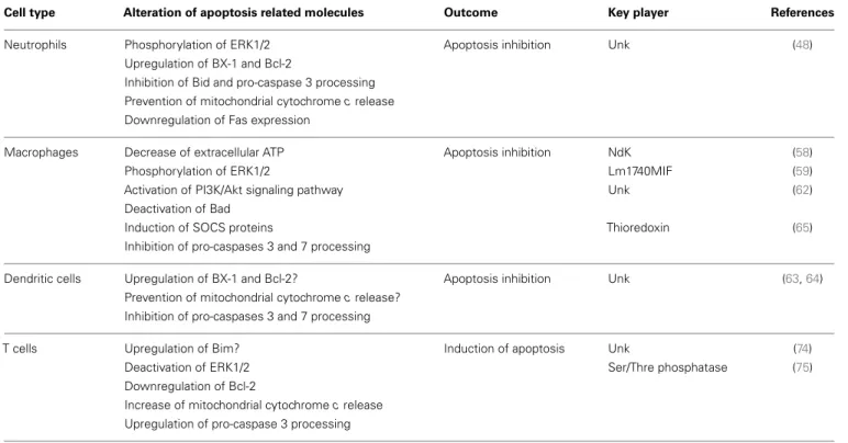
Table 1 resumes the topics described above, overviewing the
modulation of apoptosis by Leishmania in different cell types.
OVERCOMING THE IMMUNE LEISHMANICIDAL MACHINERY
Leishmania is one of the few intracellular pathogens that can live
and replicate inside the harsh environment of a mature
phagolyso-some. Apart from this parasite, only Coxiella brunetti resides
dur-ing its entire replicative cycle inside that cellular compartment,
as reviewed by Voth and Heinzen (
76
), while other
intracellu-lar pathogens that preferentially infect macrophages escape the
phagocytic pathway (
77
). Leishmania must, therefore, cope with
different effector molecules from the innate immune response in
order to survive.
AVOIDING CELL LYSIS AND TAKING ADVANTAGE OF OPSONIZATION
The first challenge Leishmania encounters in the mammalian
host is the complement system (
78
). Traditionally,
promastig-ote complement resistance is associated with two Leishmania
glycocalyx components (
79
): lipophosphoglycan (LPG) and the
metalloprotease leishmanolisin (GP63). Leishmania major
par-asites deficient for both these molecules demonstrated high
complement sensitivity (
80
–
82
). LPG avoids the ultimate step of
the complement cascade through prevention of the attachment
of the C5b-C9-complex to the parasite surface (
83
,
84
). On the
other hand, GP63 inactivates C3b preventing the formation of the
C5 convertase complex (
85
,
86
). Albeit, Dominguez et al. showed
that under physiological conditions 85–100% of L. donovani, L.
infantum, L. major, and L. amazonensis promastigotes are killed
by complement after 2.5 min in human blood (
87
). Yet, it was also
published that as soon as 1 min after L. amazonensis and L.
dono-vani contact with human blood, infected granulocytes were easily
found (
88
). Therefore, it is essential for the parasite to escape the
complement onslaught by quickly entering a phagocytic cell.
Once again Leishmania glycocalyx components are used to
sub-vert the innate immune system enhancing the phagocytosis of the
parasites. Both GP63 and LPG can directly interact with the host
cell surface through binding to the fibronectin receptor and the
mannose/fucose receptor, respectively (
89
–
92
). Moreover, iC3b,
the cleavage product of C3 by GP63, can function as an opsonin
(
85
), and LPG interacts with the early inflammatory C-reactive
protein, which triggers phagocytosis (
93
,
94
). Interestingly, iC3b
is a ligand of the complement receptor 3 (CR3) (
95
), and this
interaction is directly related with the downregulation of IL-12
production by macrophages (
96
). The mechanism by which this
downregulation happens is not known; however, we may not
exclude a toll like receptor (TLR) inhibition since C5a, another
complement component, has a negative impact on the TLR-4
induced IL-12 synthesis (
97
). This may ultimately contribute for
the silent entry of the parasites into the host cells.
Table 1 | Apoptosis modulation during Leishmania infection.
Cell type Alteration of apoptosis related molecules Outcome Key player References
Neutrophils Phosphorylation of ERK1/2 Apoptosis inhibition Unk (48)
Upregulation of BX-1 and Bcl-2
Inhibition of Bid and pro-caspase 3 processing Prevention of mitochondrial cytochrome c release Downregulation of Fas expression
Macrophages Decrease of extracellular ATP Apoptosis inhibition NdK (58)
Phosphorylation of ERK1/2 Lm1740MIF (59)
Activation of PI3K/Akt signaling pathway Unk (62)
Deactivation of Bad
Induction of SOCS proteins Thioredoxin (65)
Inhibition of pro-caspases 3 and 7 processing
Dendritic cells Upregulation of BX-1 and Bcl-2? Apoptosis inhibition Unk (63,64) Prevention of mitochondrial cytochrome c release?
Inhibition of pro-caspases 3 and 7 processing
T cells Upregulation of Bim? Induction of apoptosis Unk (74)
Deactivation of ERK1/2 Ser/Thre phosphatase (75)
Downregulation of Bcl-2
Increase of mitochondrial cytochrome c release Upregulation of pro-caspase 3 processing
Akt, protein kinase B; ERK, extracellular signal-regulated kinases; MIF, macrophage inhibiting factor; NdK, nucleoside diphosphate kinase; SOCS, suppressors of cytokine signaling; Ser/Thr, serine/threonine; Unk, unknown.
TOWARD A SUCCESSFUL DIFFERENTIATION: ALTERATIONS DURING THE PHAGOLYSOSOME MATURATION PROCESS
After promastigote entry into the host cell, Leishmania needs to
differentiate to the amastigote form. Since promastigotes
can-not survive in the harsh environment of the phagolysosome (low
pH, hydrolases), a delay of phagolysosomal fusion was considered
essential for the parasite differentiation process (
98
). Such delay
has been described for L. major, L. infantum, and L. donovani via
mechanisms that may or may not involve LPG (
98
,
99
). However,
with L. mexicana and L. amazonensis, this was not proved (
100
–
103
). For these parasites, the large parasitophorous vacuoles found
in macrophages dilute the hydrolytic enzymes upon lysosome
fusion to a level below their effectiveness, allowing promastigotes
to differentiate without any requirements of fusion delay (
100
). In
the case of L. donovani, it was shown that LPG impairs the
asso-ciation of synaptotagmin V to phagosome membranes, inhibiting
the recruitment of the vesicular proton-ATPase and preventing
their acidification, allowing promastigote to amastigote
differen-tiation (
104
). Leishmania donovani LPG was also associated with
retention of the small GTPase Cdc42 at the phagosome
mem-brane, leading to F-actin accumulation around the phagosome
and presumably interfering with vesicle trafficking and phagosome
maturation (
105
,
106
).
ROLE OF GP63 IN THE DEFENSE AGAINST ANTIMICROBIAL PEPTIDES
Inside a phagolysosome, fully differentiated or not, Leishmania
has to deal with other components of the innate immune system:
the antimicrobial peptides (AMPs). AMPs are structurally diverse
cationic proteins with intrinsic antimicrobial activity, playing
nor-mally by disruption of cell surface membranes resulting in osmotic
lysis of the pathogen. They can be found both intra and
extracel-lularly, and most of them are constitutively produced and secreted
(when applicable) (
107
,
108
). Some human AMPs present activity
against Leishmania. For example, Kulkarni et al. showed that
cathe-licidin, an intracellular AMP present in macrophage lysosomes,
can kill up to 50% of L. major and L. amazonensis parasites (
109
).
The same group showed in a different study that a-defensins,
pro-duced by neutrophils, also kill L. major parasites (
110
). GP63 play
a key role in the defense against these peptides, as it was shown that
gp63 KO promastigotes were efficiently killed in a dose dependent
manner by AMPs (
109
).
COPING WITH REACTIVE OXYGEN AND NITROGEN SPECIES (ROS AND RNS)
Once inside the host cell, ROS and RNS are the cellular major
arms against Leishmania. NO
•is synthesized by nitric oxide
syn-thase (NOS) during the conversion of l-arginine to l-citrulline,
while O
⇧2– and other reactive oxygen species (ROS) are generated
by the membrane-bound NADPH-dependent oxidases (NOX).
These reactive species contribute for the generation of others as
ONOO
-, NO
⇧2, and nitrogen trioxide (
111
). Although NO is
con-sidered the most relevant microbicidal molecule, ROS are also
associated with disease susceptibility since NOX deficient mice
are more susceptible to L. donovani and L. major infection (
112
,
113
). However, unlike what happens with inducible NOS (iNOS)
KO mice, NOX deficient mice eventually control the infection
(
112
–
114
). Therefore, the parasite needs to somehow neutralize
these reactive species and/or prevent their production to avoid
a certain death by oxidative stress. The inflammatory cytokine
TGF-b produced by infected phagocytes shifts the l-arginine
metabolism toward the production of l-ornithine through the
activation of arginase (
115
,
116
). This metabolic shift leads to a
decrease in NO secretion favoring intracellular Leishmania growth
(
117
). Glycocalyx components can also play a role in the
protec-tion of Leishmania parasites from ROS. A genetic rescue of a L.
amazonensis GP63 deficient strain increased its intramacrophage
survival potential, which was probably related with inhibition
of ROS generation (
118
,
119
). In turn, LPG not only prevents
ROS generation through inhibition of NOX recruitment to the
phagosome membrane, but also directly scavenges these reactive
species (
81
,
120
). Glycosylinositolphospholipid (GILP), another
component of the glycocalyx, may also be important during the
amastigote form, suppressing macrophage iNOS expression and,
consequently, NO production (
121
). Finally, we cannot disregard
the intrinsic antioxidant machinery of Leishmania, whose most
important components are trypanothione synthase and
trypan-othione reductase. The last one is essential for the fight against
ROS and NOS, once disruption of the trypanothione reductase
gene renders the parasites susceptible to intracellular killing by
macrophages (
122
). A recent publication shows that L. donovani
activates multiple own enzymatic mechanisms for the
detoxifica-tion of ROS and NOS (
123
). Some of these enzymes have already
been associated with protection against reactive species,
includ-ing the L. infantum peroxiredoxins LicTXNPx and LimTXNPx, L.
major pteridin reductase, and L. donovani superoxide dismutase
(
124
–
126
).
Table 2 discusses the different ways by which components of the
Leishmania glycocalyx prevents parasite killing by innate immune
response.
MODULATING THE IMMUNE RESPONSE THROUGH
ALTERATION OF CYTOKINE AND CHEMOKINE SIGNALING
AND PRODUCTION
Cytokines are cell signaling mediators, which affect cell function
in an autocrine, paracrine, or endocrine manner. Interference
with the normal cytokine production is a powerful weapon that
the parasite can use for the modulation of immune function. It
is generally accepted that production of IL-12 by macrophages
and DCs is associated with resistance against Leishmania. This
cytokine induces naive T cells maturation toward an IFN-g
pro-ducing Th1 phenotype (resistant to infection), which in turn
induce macrophage M1 activation and elimination of parasites
(
127
,
128
). Th2 cytokines, namely IL-4 regarding cutaneous
leish-maniasis and IL-10 and TGF-b in the case of visceral disease,
have been related with disease susceptibility and progression by
induction of an M2 macrophage phenotype (
129
–
131
).
There-fore, parasites seem to modulate the immune response toward
a Th2 phenotype. However, this Th1/Th2 straight polarization
seems only to be observed in some murine models, and
can-not be fully applicable to human diseases (
132
). The Th1/Th2
paradigm (reviewed elsewhere) (
133
,
134
) states that Th1 and
Th2 cells counter-regulate each other. That would imply that
Leishmania-induced polarization of the immune response toward
Table 2 | Glycocalyx components: overcoming innate immune leishmanicidal machinery. Glycocalyx
component
Species Protective role Mechanism References
LPG L. major Inhibition of complement-mediated lysis Prevention of attachment of the C5b-C9-complex (83)
L. donovani L. mexicana
Promotion of phagocytosis to escape the extracellular milieu
Interaction with C-reactive protein and direct binding to phagocytes receptors
(91,93,94)
L. donovani Delay of phagolysosome maturation process Inhibition of the recruitment of vesicular
proton-ATPase
(104)
L. donovani Reduction of leishmanicidal reactive species Inhibition of ROS generation (81,120)
L. major ROS scavenging
GP63 L. major Inhibition of complement-mediated lysis Inactivation of C3b (85,86)
L. infantum L. major L. infantum
Promotion of phagocytosis to escape the extracellular milieu
The C3b inactivation product functions as an opsonin Direct binding to phagocytes receptors
(85,89,92)
L. donovani
L. major Prevention of antimicrobial peptide mediated lysis Proteolytic degradation of the antimicrobial peptides (109)
L. amazonensis Reduction of leishmanicidal reactive species Inhibition of ROS generation (119)
GILP L. major Reduction of leishmanicidal reactive species Suppression of iNOS expression and NO production (121)
GILP, glycosylinositolphospholipid; iNOS, inducible nitric oxide synthase; LPG, lipophosphoglycan; NO, nitric oxide; ROS, reactive oxygen species.
However, what is observed in human disease is a peculiar mixed
cytokine response, variable, depending on the infective species
(
132
,
133
,
135
).
LEISHMANIA MODULATES TLR SIGNALING
Toll like receptors recognize a variety of pathogen-associated
molecular patterns (PAMPs), from proteins to nucleic acids.
Upon engagement, TLRs mediate the activation of different
transcription factors, such as nuclear factor-kB (NF-kB) and
interferon-regulatory factors (IRFs), leading to the production
of inflammatory cytokines (
136
,
137
). Induction of cell
medi-ated immunity (
138
–
140
) and promotion of NO production
(
141
) are other two known TLR triggered responses against
Leish-mania infection. Nevertheless, the parasite developed strategies
that interfere with TLR associated signaling cascades
subvert-ing the traditional pro-inflammatory responses. Ex vivo
exper-iments suggest that TLR-2 performs a minor role in
initiat-ing the synthesis of pro-inflammatory cytokines, namely IL-12,
during mice infection with L. infantum (
142
). Chandra et al.
showed that L. donovani can shift TLR-2 responses toward a
Th2 immune response, with downregulation of IL-12
produc-tion in macrophages, through MAP kinase inactivaproduc-tion (
143
).
The crosstalk between TLR-2 and CCR-5 (which expression is
dependent on the expression of the first one) was also described
as relevant in L. donovani infection, promoting parasite
inter-nalization and inducing a Th2 immune response (
144
).
More-over, the interaction between TLR2 and LPG was shown do
decrease TLR-9 expression leading to a lesser inflammatory
pro-file (
145
). Nevertheless, the interplay between Leishmania and
TLRs is highly complex and needs further clarification, once
there are several reports showing that LPG-TLR interactions can
also result in increase of anti-leishmanial responses by effector
cells (
146
).
The capacity of Leishmania to interact with regulatory proteins
of the host may also be relevant for TLR signaling modulation. As
an example, L. donovani exploits a host negative TLR regulator, the
deubiquitinating enzyme A20, to inhibit the TLR-2-mediated
pro-inflammatory gene expression, consequently suppressing IL-12
and TNF-a production (
147
). It was also described that L.
dono-vani, along with L. mexicana and L. major, uses the macrophage
tyrosine phosphatase SHP-1 to inactivate kinases involved in TLR
signaling (
148
). As happens with TLR-2, Leishmania exploits
host TLR regulators to deal with TLR-4 activation. Gupta et al.
showed that L. donovani parasites alter the ubiquitination
pat-tern of TRAF3, preventing its degradation, which is required
for the effective cytosolic translocation of the TLR-4-anchored
multiprotein complex. As a consequence, NF-kB is silenced
lead-ing to a downregulation of IL-12 and TNF-a production (
149
).
Furthermore, L. amazonensis amastigotes can suppress TLR-4
acti-vation on DCs via rapid degradation of intracellular signaling
proteins (JAK/STAT, NFkB, and IRF) leading to a decrease in
IL-12 production (
150
). The deubiquitinating enzyme A20 also has
a role in the inhibition of the TLR-4-mediated pro-inflammatory
response. However, in this case, the regulation is an indirect
con-sequence of active disease promoted by the high levels of TGF-b
that infected cells produce (
151
). Another “macrophage
imbal-ance” mediated by TLR-4 signaling manipulation was described
by Shweash et al. These authors reported that L. mexicana
pro-mastigotes are able to prolong and enhance PGE
2, NO, and
arginase production through TLR-4, and consequently achieve
the reduction of macrophage released IL-12 (
152
). Finally,
Leish-mania can impair TLR signaling through prevention of receptor
ligand interaction. Here, the player is ectoin-like serine
pepti-dase inhibitor, produced by L. major, which inhibits neutrophil
elastase and consequently prevents TLR-4 activation (
153
,
154
).
Ultimately, TLR-4 signaling inhibition in macrophages induces an
Table 3 | Strategies of TLR signaling modulation by Leishmania: an overview.
TLR Species Key player Mechanism of modulation Reference
TLR 2 L. donovani Unk Shift to Th2 immune response (143)
L. donovani Deubiquitinating enzyme A20 Inhibition of TLR-mediated pro-inflammatory gene expression (147)
L. donovani
L. mexicana SHP-1 Inhibition of TLR-mediated pro-inflammatory gene expression (148)
L. major
L. major LPG Downregulation of TLR-9 expression (145)
TLR-4 L. amazonensis Unk Degradation of intracellular signaling proteins (150)
L. donovani Deubiquitinating enzyme A20/SHP-1 Inhibition of TLR-mediated pro-inflammatory gene expression (151)
L. major Ecotin-like serine peptidase inhibitor Shift to Th2 immune response (154)
L. mexicana Unk Enhancement of PGE2, NO, and arginase production (152)
LPG, lipophosphoglycan; NO, nitric oxide; PGE2, prostaglandin E2SHP, sarcoma homology 2 domain phosphatase-1;Th,T helper;TLR, toll like receptor; Unk, unknown.
M2b phenotype that correlates with higher IL-10 levels and a
Th2-type immune response (
154
). Table 3 collects the data discussed
above.
INFLUENCING CHEMOKINE PRODUCTION
As an intracellular pathogen, Leishmania depends on the initial
recruitment of host cells for successful establishment and
perpet-uation of infection. Chemokines are small proteins that induce
and regulate the migration of immune cells, and their expression
is known to be modulated by Leishmania spp. (
41
,
155
).
Sev-eral studies reported the upregulation of numerous chemokines
(RANTES/CCL5, MIP-1a/CCL3, IP-10/CXCL10, MCP-1/CCL2,
MIP-1b/CCL4, MIP-2/CXCL1, and IL-8/CXCL8) after L. major,
L. donovani, L. tropica, L. infantum, and L. panamensis
inocu-lation (
156
–
161
). Interestingly, few of these chemokines attract
neutrophils, which can be another Leishmania mediated immune
modulation strategy. Although neutrophils may be a possible
vehi-cle for Leishmania, facilitating infection, it was described that
exacerbated neutrophil recruitment is associated with parasite
killing (
162
). On the other way, it was also shown that skin
lesions of L. major infected mice mainly contained Th2
cell-attracting chemokines, such as CCL7 (
163
,
164
). The absence
of Th1 cell-attracting chemokines in these lesions may reflect
the downregulation of the expression of genes linked with Th1
trafficking, such as the ones coding for CXCR3 chemokines
(
165
). Last but not least, it was described that Leishmania may
also profit from malnutrition to impair chemokine secretion
and to establish infection (
158
,
166
). Interestingly, differential
expression of chemokines induced by distinct parasite strains
leads to various infection and disease outcomes. As an
exam-ple, human infection with L. mexicana may lead either to a
self-healing cutaneous form or to a non-healing cutaneous
dis-ease, associated with the increased expression of CCL2 and CCL3,
respectively (
167
). This differential chemokine expression was
also seen in human infection with L. panamensis (
168
), and
may be related with parasite virulence, once in a mouse model
infected with two strains of L. braziliensis (highly virulent
ver-sus less virulent) a differential chemokine expression profile
was observed (
169
). Elaboration of these studies would be of
great interest, particularly regarding the parasite virulence factors
responsible for the induction of the chemokine profiles seen in
non-healing/severe pathologies, which will unveil new parasite
immunomodulatory players.
INTERFERING WITH CYTOKINE PRODUCTION
Although cytokines are important throughout the whole
Leishma-nia infectious process, they are fundamental during the acquired
immunity phase. IL-12 is mainly produced by APCs,
particu-larly by DCs (
170
), and is related with important cytokines that
mediate very different outcomes of Leishmania infection, such
as IFN-g, IL-10, and IL-4. Therefore, the interference with IL-12
is a recurrent phenomenon in Leishmania infection. Leishmania
major was found to deplete cholesterol, inhibiting the assembly
of an IL-12-inducing CD40 signalosome and modifying the cell
effector functions (
171
). Others have reported that L. major
infec-tion directly down-regulates IL-12 producinfec-tion through a CD40
signaling-regulation (
172
). Furthermore, L. mexicana and L.
dono-vani were also found to impair LPS-induced IL-12 production
by BMM through cysteine proteinase mediated NF-kB
degrada-tion (
173
,
174
). Others have correlated IL-12 downregulation with
Leishmania evasion mechanisms, probably through PI3K/Akt
sig-naling pathway modulation (
175
–
179
). In a recent study, Batf3
-/-mice, that lack the major IL-12 producing and cross-presenting
subsets CD8a
+and CD103
+DCs, showed enhanced susceptibility
to L. major infection partially due to reduced IFN-g and increased
IL-4 and IL-10 secretion (
180
). IFN-g is released by Th1 cells
trig-gering the leishmanicidal activity of macrophages via expression
of the inducible NO synthase which, in turn, leads to the killing
of intracellular Leishmania (
181
). Thus, several reports on
pre-vention of IFN-g secretion and/or action by the parasite exist.
Ray et al. showed that infection of macrophages with L. donovani
causes a decrease in the phosphorylation of the IFN-gR-a
sub-unit, which consequently affects the receptor expression (
182
).
Furthermore, GP63 was related with reduction of IFN-g
pro-ducing cells in BALB/c mice infected with L. amazonensis (
183
).
Finally, our group reported that the non-secreted Leishmania
pro-tein LmS3arp is also associated with downregulation of IFN-g
production by splenocytes (
184
). It was described that regulatory
T cells (Tregs) may have a role in the downregulation of IFN-g, in
a murine model infected with L. amazonensis (
185
). However, it
is yet to be unveiled whether and how parasites are able to control
these cells. Furthermore, the role of Tregs in infection progression
and pathology diverges, depending on the infecting Leishmania
species. While Tregs are associated with disease exacerbation and
parasite persistence, in the infection context with L. donovani and
L. major, respectively, in vivo experiments with L. amazonensis
shown that Tregs aid in disease resolution (
185
–
188
).
Addition-ally, Ehrlich et al. demonstrated in vivo that both the transfer of
Tregs to chronically infected animals with L. panamensis, and their
treatment with rIL-2/anti-IL-2 Ab complex for Treg expansion
contributed for disease amelioration, showing the protective role
of Tregs in L. panamensis infection and a possible
immunother-apeutical role of these cells (
189
). The immunosuppressive IL-10
has long been associated to visceral disease pathogenesis (
190
),
being not only important in the establishment of infection but
also during parasite persistence through the direct inhibition of
Th1 cell development, preventing the resolution of the infection
(
191
). In fact, IL-10 receptor blockade or IL-10 KO mice renders
animals resistant to L. donovani infection (
192
,
193
). The major
source of IL-10 in both cutaneous and visceral leishmaniasis is
con-troversial. Some works proposed T regs and Th2 lymphocytes as
the main IL-10 producers (
190
,
194
–
197
), while others claim that
Th1 lymphocytes are the main IL-10 source (
190
,
194
,
198
–
200
).
Notwithstanding, the parasite can also promote IL-10 production
by other cells. For instance, L. braziliensis amastigotes and
pro-mastigotes induce the secretion of this cytokine by PBMCs (
201
).
This IL-10 secretion was shown to be mediated by phagocytosis of
opsonized parasites in an in vivo model of low dose infection with
L. major (
202
) and also with L. amazonensis and L. mexicana (
203
,
204
). The Leishmania secreted protein LiTXN1 is also involved in
the promotion of IL-10 production by spenocytes (
205
). Apart
from IL-10, IL-4 also induces Th2 responses (
206
) and is
par-ticularly involved in the promotion of cutaneous leishmaniasis.
Tabatabaee et al. suggested that L. major secrete
immunosuppres-sive factors that promote IL-4 production by lymphocytes (
207
).
This cytokine was shown to interfere with the synergy of
IFN-g/FasL that contributes to macrophage activation and killing of
intracellular L. major (
208
). There is, however, some
contradic-tory studies showing that IL-4 promotes IL-12 production by bone
marrow-derived DCs (BMDC) and resistance to the disease (
209
,
210
). Hurdayal et al. clearly showed that DC specific IL-4 receptor
alpha (IL-4Ra)-deficient BALB/c mice became hypersusceptible
to L. major infection, due to a decrease in IL-12 and an increase
in IL-10 production by DCs (
211
). These contradictory
observa-tions with IL-4 might be possibly explained by the fact that a low
infection dose with L. major induces a Th2 response in C57BL/6
mice, whereas high doses induce a Th1 response, both dependent
on IL-4 production by lymphocytes (
212
). Considering the fact
that, in average, sandflies transmit not more than 1000 parasites
per bite, an induction of Th2 response might be expected in a real
situation (
213
).
Other cytokines have been studied in the context of Leishmania
infection. IL-17, for instance, has been involved in the outcome of
cutaneous leishmaniasis (
214
–
216
). Although there are not many
studies showing Leishmania modulation of this cytokine, some
clues exist about how this can happen. Castellano et al. showed
that L. amazonensis antigens possibly induce a decrease in the
percentage of CD3
+CD4
+IL-17
+human cells, at least in cases
of HIV/Leishmania co-infection (
217
). Interestingly, patients with
signs of active disease present lower levels of Th17 cytokines (
218
,
219
). Yet, more studies are needed to discover whether
Leishma-nia can directly modulate IL-17 production or if it acts on other
interlinked cytokines such as IL-6 and IL-23 (
201
,
216
). IL-1b
was also shown to influence the clinical course of leishmaniasis,
and is strictly related with inflammasome activation, a general
but powerful antimicrobial strategy in innate immunity (
220
).
A recent study showed that Leishmania can prevent
caspase-1-dependent IL-1b activation through a C-type lectin (SIGNR3)
mediated signaling process, which consequently favors parasite
persistence (
221
). The parasite key player responsible for this
sig-naling modulation is, however, yet unknown. Finally, IL-13, IL-21,
and IL-27 may also have a role in leishmaniasis, either preventing
or inducing pathology (
222
–
225
).
IMPAIRING CELLULAR FUNCTION
Leishmania is able to control the acquired immunity through
the impairment of effector cells function. Antigen processing and
presentation by APCs is necessary for the efficient priming of
effec-tor T cells which, in turn, will generate a directed and specific
immune response (
226
). Through phagocytosis of parasite debris
or intracellular parasite degradation, APCs process and present
Leishmania antigens (
227
). Both major histocompatibility
com-plex (MHC) I and MHC II antigen presentation are related with
Leishmania elimination, although only the second one is
essen-tial for complete parasite clearance (
212
,
228
). Leishmania can
interfere with antigen processing and presentation, consequently
modulating once again the immune function.
LEISHMANIA INTERFERES WITH ANTIGEN PRESENTATION BY
PROFESSIONAL CELLS
In 1987, Reiner et al. described that L. donovani decreases
macrophage expression of both MHC I and MHC II molecules
(
229
). Others have also reported a L. major related
downregula-tion of MHC molecules in DCs (
230
), which can be mediated by
direct parasite internalization of these molecules (
231
–
233
).
Inter-estingly, L. donovani extracellular vesicles were shown to inhibit
MHC-II expression in human monocyte-derived DCs (
234
).
Fur-thermore, both L. pifanoi and L. amazonensis amastigotes interfere
with the macrophage antigen processing process by sequestration
of antigens from the MHC II pathway, through a mechanism
involving targeted vacuolar fusion (
235
,
236
). However,
preven-tion of surface-expressed MHC class II-peptide complexes is not
the only way by which the parasite impairs antigen presentation
(Figure 2). L. donovani was shown to interfere with BMM
anti-gen presentation by modulating the capacity of surface MHC class
II-peptide complexes to engage the T cell receptor (TCR) (
237
).
An increase in the infected cell membrane fluidity by
choles-terol depletion and ceramide generation may justify this inefficient
engagement (
238
,
239
). Adhesion molecules are also important in
the process of antigen presentation. They help during the initiation
of contact between APCs and T cells, required for the subsequent
formation of the immunological synapse. Bimal et al. reported
that particularly CD4
+, but also CD8
+T cells, from patients
with active visceral leishmaniasis caused by L. donovani express
FIGURE 2 | Leishmania interferes on MHC II antigen presentation process. Leishmania impairs the antigen presentation process through
several mechanisms. The parasite is responsible for the downregulation of MHC II in APC (1), sequestration of antigens from the MHC II pathway (2), limitation of the MHC II-peptide-TCR engagement (3), and down-regulation of co-stimulatory (4), and adhesion molecules (5) on APCs and
lymphocytes, respectively.
less CD2 than the ones from healthy subjects (
240
). In vitro and
in vivo studies must, however, be performed to confirm that this
downregulation of CD2 in CD4
+T cells is caused directly by the
parasite. Co-stimulatory molecules are necessary for the full
acti-vation of T cells by APCs, which expression can be downregulated
by Leishmania. For instance, Kaye et al. showed that BMM infected
with L. donovani expressed lower levels of co-stimulatory
mole-cules B7.1 and heat stable antigen than the non-infected controls
(
241
). Mbow et al. also reported that Langerhan cells of BALB/c
mice infected with L. major showed a down-regulation of B7.1
expression (
242
).
LEISHMANIA-INDUCED CELLULAR ANERGY AND EXHAUSTION
The lack of co-stimulatory molecules on APCs, particularly in
DCs, can be a consequence of another immune modulation
strategy used by Leishmania, the inhibition of cell
matura-tion/activation. The induction of cellular unresponsiveness or
anergy is the ultimate weapon that Leishmania uses in the fight
against the immune system. Impairment of APC function was
reported by our group. Briefly, BMDC infection with L.
infan-tum promastigotes counteracts LPS-triggered activation. Parasites
avoided the upregulation of transcription and surface expression
of CD40 and CD86 co-stimulatory molecules on BMDC, through
activation of the PI3k/Akt pathway and the impairment of
NF-kB transcription factor (
243
). This DCs activation/maturation
arrest was also described for L. amazonensis infection on mice
and human cells (
150
,
217
,
231
,
244
). Leishmania has also been
associated with T cell exhaustion (
245
). Gautam et al. described
that IFN-g production by CD8
+effector cells was absent in active
human visceral leishmaniasis. These cells expressed elevated levels
of Cytotoxic T Lymphocytes Antigen 4 (CTLA-4) and programed
death protein 1 (PD1) (
246
), negative regulators of T cell
activa-tion associated with T cell anergy and exhausactiva-tion (
247
). Similar
results were also reported by Esch and colleagues, regarding not
only CD8, but also CD4 T cells (
248
). This topic was recently
reviewed by our group regarding Leishmania and other parasitic
infections (
249
).
CONCLUSION
Remarkable progresses were made in the past years in the
knowl-edge of immunomodulation by Leishmania. As a result of a long
parasite-host co-evolutionary process, this organism can escape
or fight the immune system using diverse and complex
strate-gies. However, the knowledge produced is sometimes dispersed
and contradictory, reflecting several variables such as infecting
species and different infection models. Notwithstanding, it is now
clear that the parasite can modulate cell death, alter the maturation
process of the phagolysosome, modulate cytokine, and chemokine
production by host cells, and impair cell function, in order to
silently enter in host cells and successfully differentiate and infect.
Furthermore, Leishmania released material seems to have by itself
some immunomodulatory potential. Therefore, the study of the
parasite exoproteome may contribute for the discovery and
charac-terization of the yet unknown arms that the parasite uses to achieve
victory against the immune system. The unraveling of the agents
responsible for this modulation will help us to define the
require-ments for infection and disease. This will ultimately become the
cornerstone that will contribute to develop novel strategies to fight
the disease. Although not discussed in this review, but not less
important, the pressure that the parasite exerts in the host cells
metabolism is now an area of growing interest. The nascent field
of immunometabolism will also contribute significantly for the
full understanding of the infectious process.
ACKNOWLEDGMENTS
This work was funded by FEDER funds through the
Opera-tional Competitiveness Programme – COMPETE and by NaOpera-tional
Funds through FCT – Fundação para a Ciência e a Tecnologia
under the project FCOMP-01-0124-FEDER-019648
(PTDC/BIA-MIC/118644/2010). Pedro Cecílio, Begoña Pérez-Cabezas, and
Nuno Santarem are supported by fellowships from the European
Community’s Seventh Framework Programme under grant
agree-ments No. 603181 (Project MuLeVaClin), No. 603240-2 (Project
NMTryPI), and No. 602773 (Project KINDRED), respectively.
Joana Maciel is supported by a post-doctoral fellowship from
the FCT project grant No. PTDC/BIA-MIC/118644/2010. Vasco
Rodrigues is supported by a doctoral fellowship from FCT code
SFRH/BD/64064/2009. The funders had no role in study design,
data collection, and analysis, decision to publish, or
prepara-tion of the manuscript. The authors would like to acknowledge
COST Action BM1202: European Network on Microvesicles and
Exosomes in Health and Disease (Me-HaD).
REFERENCES
1. Bates PA. Transmission of Leishmania metacyclic promastigotes by phle-botomine sand flies. Int J Parasitol (2007) 37(10):1097–106. doi:10.1016/j. ijpara.2007.04.003
2. Beattie L, Kaye PM. Leishmania-host interactions: what has imaging taught us?
3. Kaye P, Scott P. Leishmaniasis: complexity at the host-pathogen interface. Nat
Rev Microbiol (2011) 9(8):604–15. doi:10.1038/nrmicro2608
4. WHO. Control of Leishmaniasis: Report of a meeting of the WHO Expert
Com-mittee on the Control of Leishmaniasis. WHO Technical Report Series n 949.
Switzerland: Published by World Health Organization (2010).
5. Sinha PK, Pandey K, Bhattacharya SK. Diagnosis & management of
Leishma-nia/HIV co-infection. Indian J Med Res (2005) 121(4):407–14.
6. Murray HW, Berman JD, Davies CR, Saravia NG. Advances in leishmaniasis.
Lancet (2005) 366(9496):1561–77. doi:10.1016/S0140-6736(05)67629-5
7. Desjeux P. Leishmaniasis: current situation and new perspectives. Comp
Immunol Microbiol Infect Dis (2004) 27(5):305–18. doi:10.1016/j.cimid.2004.
03.004
8. Antinori S, Schifanella L, Corbellino M. Leishmaniasis: new insights from an old and neglected disease. Eur J Clin Microbiol Infect Dis (2012) 31(2):109–18. doi:10.1007/s10096-011-1276-0
9. Campino LMC. Epidemiologia das leishmanioses em Portugal. Acta Med Port (2010) 23:859–64.
10. Salman SM, Rubeiz NG, Kibbi AG. Cutaneous leishmaniasis: clinical fea-tures and diagnosis. Clin Dermatol (1999) 17(3):291–6. doi:10.1016/S0738-081X(99)00047-4
11. Kroidl A, Kroidl I, Bretzel G, Loscher T. Non-healing old world cutaneous leishmaniasis caused by L. infantum in a patient from Spain. BMC Infect Dis (2014) 14:206. doi:10.1186/1471-2334-14-206
12. Strazzulla A, Cocuzza S, Pinzone MR, Postorino MC, Cosentino S, Serra A, et al. Mucosal leishmaniasis: an underestimated presentation of a neglected disease. Biomed Res Int (2013) 2013:805108. doi:10.1155/2013/805108 13. Ready PD. Epidemiology of visceral leishmaniasis. Clin Epidemiol (2014)
6:147–54. doi:10.2147/CLEP.S44267
14. Okwor I, Uzonna JE. The immunology of Leishmania/HIV co-infection.
Immunol Res (2013) 56(1):163–71. doi:10.1007/s12026-013-8389-8
15. Moreno J, Alvar J. Canine leishmaniasis: epidemiological risk and the exper-imental model. Trends Parasitol (2002) 18(9):399–405. doi:10.1016/S1471-4922(02)02347-4
16. Evans KJ, Kedzierski L. Development of vaccines against visceral Leishmaniasis.
Re Dai Yi Xue Za Zhi (2012) 2012:892817. doi:10.1155/2012/892817
17. Stockdale L, Newton R. A review of preventative methods against human leish-maniasis infection. PLoS Negl Trop Dis (2013) 7(6):e2278. doi:10.1371/journal. pntd.0002278
18. van Griensven J, Balasegaram M, Meheus F, Alvar J, Lynen L, Boelaert M. Combination therapy for visceral leishmaniasis. Lancet Infect Dis (2010) 10(3):184–94. doi:10.1016/S1473-3099(10)70011-6
19. Yasinzai M, Khan M, Nadhman A, Shahnaz G. Drug resistance in leishmaniasis: current drug-delivery systems and future perspectives. Fut Med Chem (2013) 5(15):1877–88. doi:10.4155/fmc.13.143
20. Feig C, Peter ME. How apoptosis got the immune system in shape. Eur J
Immunol (2007) 37(Suppl 1):S61–70. doi:10.1002/eji.200737462
21. Kappler JW, Roehm N, Marrack P. T cell tolerance by clonal elimination in the thymus. Cell (1987) 49(2):273–80. doi:10.1016/0092-8674(87)90568-X 22. Alderson MR, Tough TW, Davis-Smith T, Braddy S, Falk B, Schooley KA, et al.
Fas ligand mediates activation-induced cell death in human T lymphocytes. J
Exp Med (1995) 181(1):71–7. doi:10.1084/jem.181.1.71
23. Dhein J, Walczak H, Baumler C, Debatin KM, Krammer PH. Autocrine T-cell suicide mediated by APO-1/(Fas/CD95). Nature (1995) 373(6513):438–41. doi:10.1038/373438a0
24. Mattner J, Donhauser N, Werner-Felmayer G, Bogdan C. NKT cells mediate organ-specific resistance against Leishmania major infection. Microbes Infect (2006) 8(2):354–62. doi:10.1016/j.micinf.2005.07.002
25. Kaczanowski S, Sajid M, Reece SE. Evolution of apoptosis-like programmed cell death in unicellular protozoan parasites. Parasit Vectors (2011) 4:44. doi:10.1186/1756-3305-4-44
26. Lee N, Bertholet S, Debrabant A, Muller J, Duncan R, Nakhasi HL. Programmed cell death in the unicellular protozoan parasite Leishmania. Cell Death Differ (2002) 9(1):53–64. doi:10.1038/sj.cdd.4400952
27. Proto WR, Coombs GH, Mottram JC. Cell death in parasitic protozoa: reg-ulated or incidental? Nat Rev Microbiol (2013) 11(1):58–66. doi:10.1038/ nrmicro2929
28. van Zandbergen G, Bollinger A, Wenzel A, Kamhawi S, Voll R, Klinger M, et al. Leishmania disease development depends on the presence of apoptotic
promastigotes in the virulent inoculum. Proc Natl Acad Sci U S A (2006) 103(37):13837–42. doi:10.1073/pnas.0600843103
29. Wanderley JL, Pinto da Silva LH, Deolindo P, Soong L, Borges VM, Prates DB, et al. Cooperation between apoptotic and viable metacyclics enhances the pathogenesis of Leishmaniasis. PLoS One (2009) 4(5):e5733. doi:10.1371/ journal.pone.0005733
30. Fadok VA, Bratton DL, Konowal A, Freed PW, Westcott JY, Henson PM. Macrophages that have ingested apoptotic cells in vitro inhibit proinflamma-tory cytokine production through autocrine/paracrine mechanisms involving TGF-beta, PGE2, and PAF. J Clin Investigat (1998) 101(4):890–8. doi:10.1172/ JCI1112
31. Ravichandran KS. Find-me and eat-me signals in apoptotic cell clearance: progress and conundrums. J Exp Med (2010) 207(9):1807–17. doi:10.1084/ jem.20101157
32. Wanderley JL, Thorpe PE, Barcinski MA, Soong L. Phosphatidylserine expo-sure on the surface of Leishmania amazonensis amastigotes modulates in vivo infection and dendritic cell function. Parasite Immunol (2013) 35(3–4):109–19. doi:10.1111/pim.12019
33. van Zandbergen G, Hermann N, Laufs H, Solbach W, Laskay T.
Leish-mania promastigotes release a granulocyte chemotactic factor and induce
interleukin-8 release but inhibit gamma interferon-inducible protein 10 pro-duction by neutrophil granulocytes. Infect Immun (2002) 70(8):4177–84. doi:10.1128/IAI.70.8.4177-4184.2002
34. Peters NC, Egen JG, Secundino N, Debrabant A, Kimblin N, Kamhawi S, et al. In vivo imaging reveals an essential role for neutrophils in leishmani-asis transmitted by sand flies. Science (2008) 321(5891):970–4. doi:10.1126/ science.1159194
35. Ribeiro-Gomes FL, Peters NC, Debrabant A, Sacks DL. Efficient capture of infected neutrophils by dendritic cells in the skin inhibits the early anti-leishmania response. PLoS Pathog (2012) 8(2):e1002536. doi:10.1371/journal. ppat.1002536
36. Thalhofer CJ, Chen Y, Sudan B, Love-Homan L, Wilson ME. Leukocytes infil-trate the skin and draining lymph nodes in response to the protozoan
Leish-mania infantum chagasi. Infect Immun (2011) 79(1):108–17. doi:10.1128/IAI.
00338-10
37. Beil WJ, Meinardus-Hager G, Neugebauer DC, Sorg C. Differences in the onset of the inflammatory response to cutaneous leishmaniasis in resistant and sus-ceptible mice. J Leukoc Biol (1992) 52(2):135–42.
38. Muller K, van Zandbergen G, Hansen B, Laufs H, Jahnke N, Solbach W, et al. Chemokines, natural killer cells and granulocytes in the early course of
Leish-mania major infection in mice. Med Microbiol Immunol (2001) 190(1–2):73–6.
doi:10.1007/s004300100084
39. Pompeu ML, Freitas LA, Santos ML, Khouri M, Barral-Netto M. Granu-locytes in the inflammatory process of BALB/c mice infected by
Leishma-nia amazonensis. A quantitative approach. Acta Trop (1991) 48(3):185–93.
doi:10.1016/0001-706X(91)90046-M
40. Hassani K, Shio MT, Martel C, Faubert D, Olivier M. Absence of metallo-protease GP63 alters the protein content of Leishmania exosomes. PLoS One (2014) 9(4):e95007. doi:10.1371/journal.pone.0095007
41. Matte C, Olivier M. Leishmania-induced cellular recruitment during the early inflammatory response: modulation of proinflammatory mediators. J Infect
Dis (2002) 185(5):673–81. doi:10.1086/339260
42. Wilson ME, Innes DJ, Sousa AD, Pearson RD. Early histopathology of exper-imental infection with Leishmania donovani in hamsters. J Parasitol (1987) 73(1):55–63. doi:10.2307/3282344
43. Geering B, Simon HU. Peculiarities of cell death mechanisms in neutrophils.
Cell Death Differ (2011) 18(9):1457–69. doi:10.1038/cdd.2011.75
44. Aga E, Katschinski DM, van Zandbergen G, Laufs H, Hansen B, Muller K, et al. Inhibition of the spontaneous apoptosis of neutrophil granulocytes by the intracellular parasite Leishmania major. J Immunol (2002) 169(2):898–905. doi:10.4049/jimmunol.169.2.898
45. Allenbach C, Zufferey C, Perez C, Launois P, Mueller C, Tacchini-Cottier F. Macrophages induce neutrophil apoptosis through membrane TNF, a process amplified by Leishmania major. J Immunol (2006) 176(11):6656–64. doi:10.4049/jimmunol.176.11.6656
46. Santos-Beneit AM, Mollinedo F. Expression of genes involved in initiation, regulation, and execution of apoptosis in human neutrophils and during neu-trophil differentiation of HL-60 cells. J Leukoc Biol (2000) 67(5):712–24.