Liliana Manuela Bento Lopes
Licenciada em Ciências Biomédicas
Role of the endocytic recycling pathway
in lysosome exocytosis
Dissertação para obtenção do Grau de Mestre em Genética
Molecular e Biomedicina
Orientador: Prof. Dr. Duarte C. Barral
Investigador Principal no CEDOC, Chronic Diseases
Research Centre, NOVA Medical School|Faculdade de
Ciências Médicas, Universidade NOVA de Lisboa
Júri:
Presidente: Doutora Margarida Casal Ribeiro Castro Caldas Braga, Professora Auxiliar da Faculdade de Ciências e
Tecnologia da Universidade Nova de Lisboa
Arguente: Doutora Júlia Carvalho Costa, Investigadora Principal do Instituto de Tecnologia Química e Biológica da Universidade Nova de Lisboa
Vogal: Doutor Duarte Custal Ferreira Barral, Investigador Principal no CEDOC, Chronic Diseases Research Centre, NOVA Medical School|Faculdade de Ciências
Médicas, Universidade NOVA de Lisboa
i
Role of the endocytic recycling pathway in lysosome exocytosis
Copyright© Liliana Manuela Bento Lopes, FCT/UNL, UNL
A Faculdade de Ciências e Tecnologia e a Universidade Nova de Lisboa têm o direito, perpétuo e sem limites geográficos, de arquivar e publicar esta dissertação através de exemplares impressos reproduzidos em papel ou de forma digital, ou por qualquer outro meio conhecido ou que venha a ser inventado, e de a divulgar através de repositórios científicos e de admitir a sua cópia e distribuição com objectivos educacionais ou de investigação, não comerciais, desde que seja dado crédito ao autor e editor.
i
Acknowledgements
Ao ultrapassar mais uma etapa bastante gratificante da minha vida, não poderia deixar de agradecer às pessoas que me acompanharam e permitiram que crescesse profissionalmente e pessoalmente.
Em primeiro lugar, gostaria de agradecer ao Dr. Duarte Barral, por ter depositado a sua confiança em mim e me ter dado a oportunidade de trabalhar no seu laboratório. Estou muito grata por ter tido a oportunidade de aprender com alguém com tão vasto conhecimento científico, espírito crítico, rigor e dedicação à investigação. Obrigada pela sua preocupação e por querer acompanhar sempre o desenrolar do meu trabalho, por todos os conselhos e principalmente por ter acreditado em mim.
Em segundo lugar, quero agradecer imenso à Cristina Escrevente por todo o conhecimento que me transmitiu, pela dedicação que sempre demonstrou, por se preocupar comigo, por nunca me deixar desamparada, pelos conselhos e pela tua amizade. Além disso és para mim uma inspiração e um grande exemplo de trabalho, rigor, organização e dedicação. És sem dúvida uma excelente cientista e uma pessoa espetacular. Sou uma sortuda por ter tido o privilégio de trabalhar e aprender contigo. Muito obrigada por tudo!
Gostava de agradecer ao Hugo por tudo o que me ensinou e ensina. És como um modelo a seguir para mim, pois admiro a tua força de vontade e fome de conhecimento. Quero agradecer-te por me teres recebido tão bem e me ensinares com toda a dedicação e rigor. Obrigado por me ajudares a ser melhor, por todos os conselhos, pela confiança que me transmites e por estares sempre a torcer por mim. Obrigado por seres quem és, por seres divertido e tornares o laboratório mais animado.
Não poderia deixar de agradecer à Cristina Casalou, por se ter disponibilizado a ajudar-me sempre que foi necessário, transmitindo conselhos e conhecimentos tão importantes. Obrigada pelo teu apoio, companheirismo e boa disposição. Gostava também de agradecer à Cecília, pelos conselhos e pela ajuda que sempre me disponibilizou, à Xana e ao Francisco pelo companheirismo e por nos fazerem rir quando menos esperamos. Não me poderia esquecer de deixar uma palavra à Ana Portelinha que, apesar do pouco tempo que privámos, me marcou por me ter recebido tão bem, pelos conselhos, pelo exemplo e pela sua amizade.
O meu muito obrigado à nossa “vizinha” Tatiana pela amizade, pelos conselhos, pela ajuda que sempre me disponibilizaste e pela paciência em me responder a todas a questões relativas ao mestrado. À Diana, por ser das pessoas que mais anima o laboratório e estar sempre disponível para ajudar e motivar todos, obrigada por seres assim.
Agradeço também a todas as pessoas do CEDOC que me acolherem e me fizeram sentir parte de uma família em que a entreajuda e união são palavras de ordem.
Gostaria de deixar uma palavra de agradecimento à coordenadora do mestrado, Prof. Paula Gonçalves e ao DCV pela prontidão nas respostas às dúvidas colocadas.
ii
Gostaria de agradecer do fundo do coração à minha mãe, que é um dos pilares mais importantes da minha vida. Obrigada por lutares com todas as forças que tens e fazeres de tudo para que nada nos falte. Tenho muito orgulho em ti. Obrigada por me apoiares, me dares força, te preocupares comigo e por aquele mimo que só tu sabes quando e como dar. Cada passo que dou, devo-o a ti, ao teu esforço, à tua luta e espero um dia poder recompensar-te por tudo o que fizeste e fazes por mim. Gosto muito de ti e espero que tenhas orgulho em mim.
Agradeço também à minha “pequena” irmã Daniela, por ser a minha companheira de todas as horas e aturar o meu mau humor quando não estou tão bem. Obrigada por me ajudares a ultrapassar todos os obstáculos, por me dares conselhos, por me fazeres rir, pelo mimo e principalmente por me compreenderes e por estares sempre aí quando preciso. Sou uma sortuda por te ter como irmã e tenho um orgulho gigante em ti. És uma das pessoas mais importantes da minha vida, sabes que gosto muito de ti e que podes sempre contar comigo para tudo o que precisares.
O meu muito obrigado para toda a minha família especialmente para os meus tios Augusto, Avelina, Salomé e Adelino, que estão perto e sempre me ajudaram e apoiaram em tudo, e para os meus tios Virgínia, Moisés, Maria e Roberto, que apesar de longe, fazem-me sentir todo o vosso apoio e força. A todos os meus primos pelos conselhos, animação e força que sempre me deram e pela união que existe entre nós, apesar de nos vermos tão poucas vezes. Gosto muito de todos vocês. Gostaria ainda de deixar uma palavra especial de agradecimento ao meu padrinho pelo apoio e força que me tem dado na minha formação académica.
Finalmente, mas não menos importante, gostava de agradecer ao meu namorado João, por ser a pessoa que é e me ter acompanhado durante todos estes anos, com carinho, preocupação, apoio e companheirismo apesar de todos os maus momentos. Obrigada pelos conselhos, por me fazeres sentir especial, por me ajudares a descomprimir do stress e seres a lufada de ar fresco que preciso para continuar a lutar pelos meus objetivos. Sabes que és muito importante para mim e espero poder contar contigo e ter-te sempre presente em todas as conquistas da minha vida. Gosto muito de ti. Quero agradecer também aos teus pais, Trindade e Manuel, e ao teu irmão José, por me receberem sempre de braços abertos e me tratarem como se fosse vossa filha/irmã. Obrigada por todos os momentos que passámos juntos e principalmente pelos preciosos testemunhos e conselhos sobre a escola da vida. Gosto muito de vocês e têm um lugar muito especial no meu coração.
iii
Abstract
Until recently, lysosomes were considered as the end-point of the endocytic pathway, to where cargo is delivered to be degraded. However, several studies showed that conventional lysosomes can undergo regulated exocytosis in response to an increase in intracellular calcium concentration, an important process for plasma membrane repair and secretion of lysosomal contents. Nevertheless, the molecular machinery involved in lysosome transport and fusion with the plasma membrane is not fully understood. Our group found recently that the small GTPases Rab11a and Rab11b are essential for calcium-triggered lysosome exocytosis in HeLa cells.
Therefore, we aim to find the Rab11a/b effectors that mediate lysosome exocytosis process. To this end, we silenced several known effectors such as Rab11 family of interacting proteins (FIPs), Myosin Va/b or subunits of the exocyst tethering complex (Sec8, Sec15 and Exo70). After stimulation with the calcium ionophore ionomycin, we investigated the cell surface expression levels of the late endosome/lysosome marker LAMP1, as well as the release of the lysosomal enzyme β-hexosaminidase. We found that the silencing of Sec15 impairs lysosome exocytosis, while in the absence of FIP1-C or FIP2 there is an increase in LAMP1 cell surface levels and β-hexosaminidase release. Moreover, we confirmed the interaction and co-localization of Rab11a/b with the effector proteins studied using co-immunoprecipitation assays and confocal immunofluorescence microscopy, respectively. Finally, using live cell imaging, we found that Rab11-positive vesicles interact transiently with late endosomes/lysosomes near the plasma membrane, upon ionomycin stimulation.
Thus, our results provide new insights into the role of Rab11 and its effectors in the regulation of conventional lysosome exocytosis.
v
Resumo
Até há pouco tempo os lisossomas eram apenas conhecidos como organelos degradativos, para onde convergem moléculas para serem degradadas. No entanto, vários estudos mostraram que os lisossomas convencionais também podem ser exocitados de uma forma regulada, em resposta ao aumento da concentração intracelular de cálcio, um evento importante para a reparação da membrana plasmática e secreção do conteúdo lisossomal. Porém, a maquinaria molecular envolvida no transporte e fusão dos lisossomas com a membrana plasmáticas ainda não é bem conhecida. O nosso grupo descobriu que as pequenas GTPases Rab11a e Rab11b são essenciais na exocitose de lisossomas induzida por cálcio, em células HeLa.
Por este motivo, o nosso objetivo é encontrar efetores das Rab11a/b que medeiam a exocitose de lisossomas. Para isso, silenciámos vários efetores conhecidos como a família de proteínas que interagem com a Rab11 (FIPs), Miosina Va/b ou subunidades do complexo exocisto (Sec8, Sec15 e Exo70). Após estimulação com o ionóforo de cálcio ionomicina, investigámos os níveis de expressão do marcador de endossomas tardios/lisossomas LAMP1 à superfície da célula, bem como a libertação da enzima lisossomal β-hexosaminidase. Verificámos que o silenciamento da subunidade Sec15 diminui a exocitose de lisossomas, e que a ausência de FIP1-C ou FIP2 leva ao aumento dos níveis de expressão de LAMP1 à superfície da célula e da libertação de β-hexosaminidase. Além disso, confirmámos a interação e co-localização das Rab11a/b com as proteínas efetoras estudadas, usando ensaios de co-imunoprecipitação e microscopia confocal de imunofluorescência, respetivamente. Finalmente, usando microscopia em células vivas, verificámos que vesículas Rab11 positivas interagem transientemente com endossomas tardios/lisossomas perto da membrana plasmática, após estimulação com ionomicina.
Desta forma, os nossos resultados providenciam novos dados sobre o papel da Rab11 e dos seus efetores na regulação da exocitose de lisossomas convencionais.
vii
Index
Acknowledgements i Abstract iii Resumo v Index vii Index of Figures ix Index of Tables xi Abbreviations xiii I. Introduction 1 1. Vesicular trafficking 11.1. Rab GTPase family 3
1.1.1. Rab11 subfamily 6
1.1.2. Rab11 effectors 7
1.1.2.1. Rab11 family of interacting proteins (FIPs) 7
1.1.2.2. Class V Myosins 9
1.1.2.3. Exocyst complex 9
2. Lysosomes 11
2.1 Regulated exocytosis of conventional lysosomes 12 2.2 Diseases of lysosomes and lysosome-related organelles 15
3. Previous results 17
4. Objectives 19
II. Materials and Methods 21
1. Cell Culture 21
2. Gene silencing 21
3. Cell transfection 22
4. RNA extraction, cDNA production and real-time quantitative PCR 22
5. Immunoprecipitation 23
6. Immunoblotting 24
7. β-hexosaminidase release assay 25
viii
9. Immunofluorescence Microscopy 26
10. Live cell-imaging 27
11. Statistical analysis 27
III. Results 29
1. Identification of Rab11a/b effectors required for lysosome exocytosis 29 2. Interaction of Rab11a/b with their effectors 34 3. Intracellular localization of Rab11a/b and their effectors 36 4. Intracellular localization of Rab11a/b and lysosomes in HeLa cells 42
IV. Discussion 47
ix
Index of Figures
I. Introduction 1
Figure I.1 - Vesicular trafficking steps. 2
Figure I.2 - Intracellular trafficking pathways. 3 Figure I.3 - Localization and function of Rab GTPases. 4
Figura I.4 - Rab GTPases cycle. 5
Figure I.5 - Rab11-regulated vesicle transport processes. 7 Figure I.6 – Structure of Rab11-family of interacting proteins. 8 Figure I.7 - Rab11 interaction with the exocyst complex. 10 Figure I.8 - Biological functions that require lysosome exocytosis. 13 Figure I.9 - LAMP1 cell surface expression levels and β-hexosaminidase release in HeLa cells
silenced for Rab11a, Rab11b or both. 18
III. Results 28
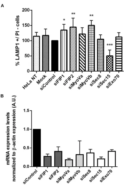
Figure III.1 - Ionomycin treatment increases LAMP1 cell surface expression in HeLa cells. 30 Figure III.2 - LAMP1 cell surface expression levels in HeLa cells silenced for FIP1-C, FIP2, Myosin
Va, Myosin Vb, Sec8, Sec15 or Exo70. 31
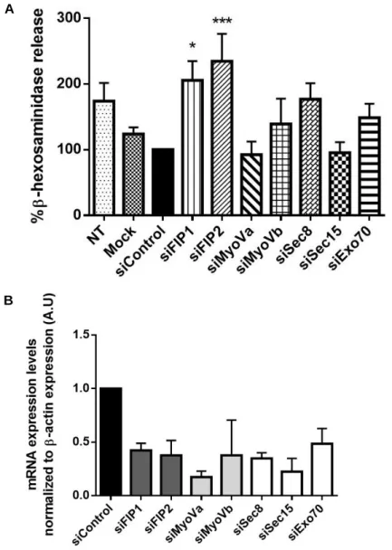
Figure III.3 - Ionomycin treatment increases β-hexosaminidase release in HeLa cells. 32 Figure III.4 - β-hexosaminidase release in HeLa cells silenced for FIP1-C, FIP2, Myosin Va, Myosin
Vb, Sec8, Sec15 or Exo70. 33
Figure III.5 - Co-immunoprecipitation of endogenous Rab11a and Rab11b with different exocyst
complex subunits in HeLa cells. 34
Figure III.6 - Co-immunoprecipitation of Rab11a- or Rab11b-mCherry with Sec15-GFP in HeLa
cells. 35
Figure III.7 - Co-immunoprecipitation of endogenous Rab11a and Rab11b with FIP2 in HeLa
cells. 35
Figure III.8 - A. Intracellular localization of endogenous and transfected Rab11a and Rab11b. 37 Figure III.9 - Intracellular localization of Rab11a and FIP1-C, FIP2 or Myosin Va. 38 Figure III.10 - Intracellular localization of Rab11b and FIP1-C, FIP2 or Myosin Va. 39 Figure III.11 - Intracellular localization of Rab11a and exocyst subunits Sec15, Sec8 or Exo70. 40 Figure III.12 - Intracellular localization of Rab11b and exocyst subunits Sec15, Sec8 or Exo70. 41 Figure III.13 - Intracellular localization of Rab11 and LAMP1 in HeLa cells. 42
x
Figure III.14 - Rab11a and lysosomes co-localize transiently at the cell tips upon ionomycin
stimulation. 44
Figura III.15 - Rab11b and lysosomes co-localize transiently at the cell tips upon ionomycin
xi
Index of Tables
II. Materials and Methods 20
Table II.1 - siRNA’s used for silencing Rab11 effectors.
21
Table II.2 - DNA plasmids used to transfect HeLa cells.
22
Table II.3 - Primers used in qRT-PCR assays.
23
xiii
Abbreviations
ANOVA Analysis of variance BCA Bicinchoninic acid assay
BLOC-1 Biogenesis of lysosome-related organelles complex 1 BORC BLOC-1 related complex
CHO Chinese hamster ovary CHS Chediak-Higashi syndrome CTL Cytotoxic T-lymphocytes
DMEM Dulbecco’s modified Eagle’s medium DTT Dithiothreitol
EE Early endosomes
EDTA Ethylenediamine tetraacetic acid EGTA Ethylene glycol tetraacetic acid ER Endoplasmic reticulum
ERC Endocytic recycling compartment FBS Fetal bovine serum
FIP Rab11 family of interacting protein FRET Fluorescence resonance energy transfer GAP GTPase-activating protein
GDF GDI-displacement factor GDI GDP-dissociation inhibitor GDP Guanosine diphosphate
GEF Guanine nucleotide exchange factor GFP Green fluorescent protein
GS Griscelli-syndrome GTP Guanosine triphosphate HBSS Hanks balanced salt solution HCl Hydrochloric acid
HEPES 4-(2-hydroxyethyl)-1-piperazineethanesulfonic acid HPS Hermansky-Pudlak syndrome
HRP Horseradish peroxidase
IB Immunoblotting
IP Immunoprecipitation KIF5B Kinesin family member 5B
LAMP Lysosome-associated membrane glycoprotein
LE Late endosomes
LIMP Lysosomal integral membrane protein LROs Lysosome-related organelles
xiv
MFI Mean fluorescence intensity MgCl2 Magnesium chloride
MHC Major histocompatibility complex MTOC Microtubule-organizing center
Myo Myosin
NaCl Sodium chloride Neu1 Neuraminidase 1 NMHC IIA Non-muscle myosin IIA
NRK Normal rat kidney epithelial cells
PM Plasma membrane
PAGE Polyacrylamide gel electrophoresis PBS Phosphate-buffered saline
PFA Paraformaldehyde PVDF Polyvinylidene difluoride
qRT-PCR Quantitative real-time polymerase chain reaction Rab Ras-like in brain
RBD Rab-binding domain RE Recycling endosomes REP Rab escort protein
RIPA Radioimmunoprecipitation assay buffer RNA Ribonucleic acid
RT Room temperature
SD Standard deviation SDS Sodium dodecyl sulfate SE Sorting endosomes
SEM Standard error of the mean siRNA Small interfering RNA shRNA Short hairpin RNA
Slp4-a Synaptotagmin-like protein 4a
SNARE Soluble N-ethylmaleimide sensitive fusion attachment receptor SNAP-23 Synaptosome associated protein 23
Syt VII Synaptotagmin VII
TGN trans-Golgi network
TFEB Transcription factor EB TfR Transferrin receptor
TRPML Transient receptor potential cation channel, mucolipin subfamily VAMP7 Vesicle associated membrane protein 7
1
I.
Introduction
1. Vesicular trafficking
The evolution of eukaryotic cells led to an increase in intracellular complexity, particularly in the compartmentalization of biological functions into organelles and the need of communication between them in a specific and regulated manner. Such communication is achieved through vesicular transport pathways, between donor and acceptor compartments, which involves four distinct steps.
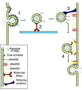
The first step is the budding of a vesicle with specific cargo from a donor compartment (Figure I.1, step 1). This process is facilitated by coat proteins that are recruited from the cytosol to the budding site. Moreover, coat proteins bind to adaptor proteins that assist in cargo selection by recognizing sorting signals present in the cytosolic domains of transmembrane proteins (Bonifacino and Lippincott-Schwartz, 2003). Clathrin-coated vesicles, composed of the coat clathrin, were the first to be described and are responsible for the uptake of extracellular molecules, as well as the transport from the trans-Golgi network (TGN) to endosomes (Bonifacino and Lippincott-Schwartz, 2003; Cai et al., 2007). However, there are other types of coated vesicles, like COP I/II-coated vesicles, which are involved in trafficking between the endoplasmic reticulum (ER) and the Golgi complex (Bonifacino and Lippincott-Schwartz, 2003; Cai et al., 2007). In the following step, vesicles are uncoated and transported to the acceptor compartment via the actin and microtubule cytoskeletons (Figure I.1, step 2). This transport is ensured by actin-dependent motor proteins, such as Myosins (Hartman and Spudich, 2012), as well as kinesins and dyneins, which transport vesicles along microtubules (Cooper, 2000). While kinesins direct vesicular transport towards the cell periphery, dyneins transport cargo from periphery to the cell center (Cooper, 2000). Finally, the tethering and fusion of vesicles to specific acceptor compartments are mediated by tethers, such as the exocyst (Figure I.1, step 3), and the soluble N-ethylmaleimide-sensitive-factor attachment protein receptors (SNAREs), respectively. SNAREs are present both in vesicles (v-SNAREs) and target membranes (t-SNAREs), allowing the fusion of vesicles to specific acceptor compartments and the release of cargo (Figure I.1, step 4) (Hong and Lev, 2014). Additionally, there are other regulatory components that modulate the action of SNAREs, namely Synaptotagmins and Ras small GTPases.
2
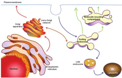
In eukaryotic cells, there are two main pathways of vesicular trafficking, namely the endocytic and the exocytic/secretory pathways (Figure I.2). In the endocytic pathway, cargo is internalized via clathrin-dependent or inclathrin-dependent mechanisms and delivered to early/sorting endosomes (EE/SE). These mature and can direct cargo to different routes. Cargo can be directed to late endosomes (LE) and lysosomes for degradation. Moreover, cargo can follow a fast recycling pathway from EE to the plasma membrane (PM) or a slow recycling pathway through the endocytic recycling compartment (ERC), located in the perinuclear region (Ijzendoorn 2006; Si et al., 2009). The secretory pathway is followed by newly synthesized cargo, which is transported from the ER to the Golgi apparatus and from there to endosomes to the PM. Exocytosis is the final step of the secretory pathway, which refers to the vesicle fusion with the PM, allowing the release of their contents to the extracellular space. Exocytosis can be constitutive or regulated. Constitutive exocytosis is the result of a steady-state trafficking of secretory vesicles from the ER to the PM. In the case of regulated exocytosis, an external stimulus is required to induce exocytosis. For example, the increase in intracellular calcium (Ca2+) concentration is known to
trigger exocytic events in specialized secretory cells, such as endocrine and neuronal cells (Shandala
et al., 2012).
Figure I.1 - Vesicular trafficking steps. 1- Budding of a vesicle from the donor compartment; 2- Transport along
the cytoskeleton; 3-Tethering and docking to the target compartment; 4- Fusion and release of vesicle content. Taken from Prekeris, 2003.
Figure I.2 - Vesicular trafficking steps. 1- Budding of a vesicle from the donor compartment; 2- Transport along
cytoskeleton; 3-Tethering and docking to the target compartment; 4- Fusion and release of vesicle content. Taken from Prekeris, 2003.
3 All steps of membrane trafficking need to be tightly regulated to ensure the correct communication between organelles. Several groups of molecules confer spatial and temporal regulation to all membrane trafficking steps. Prominent among them are the Rab small GTPases (Prekeris, 2003; Li et
al., 2013).
Rab GTPase family
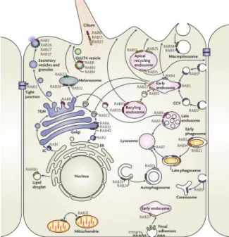
Rab small GTPases are master regulators of all steps of vesicular transport, maintaining compartment identity and controlling endocytic and exocyitc pathways (Figure I.3) (Schwartz et al., 2008). These are small proteins with 20-30 kDa that in most cases belong to the Ras superfamily, which is composed of five main families: Rat sarcoma (Ras), Ras homologous (Rho), ADP-ribosylation factor (Arf), Ras-like nuclear (Ran) and Ras-like in brain (Rab) (Bhuin and Roy, 2014; Reiner and Lundquist, 2016).
Rab GTPases are evolutionary conserved and ubiquitously expressed in all eukaryotic cells. However, there are some Rabs that are expressed only in certain tissues or have cell-type specific functions (Stenmark and Olkkonen, 2001; Stenmark, 2009). To date, around 65 Rabs were identified in humans. Rab proteins have similar structures and highly conserved regions, including a nucleotide binding domain, that can bind to guanosine triphosphate (GTP) or guanosine diphosphate (GDP). When they are bound to GTP they are considered active and when they are bound to GDP they are inactive. Figure I.2 - Intracellular trafficking pathways. Endocytic pathway (blue arrows): Cargo is internalized and
delivered to lysosomes through early/sorting endosomes and late endosomes. Recycling pathway (green arrows): Cargo can be recycled back to the plasma membrane through the endocytic recycling compartment (slow recycling) or directly from sorting endosomes (fast recycling). Exocytic pathway (red arrows): Vesicular transport of newly synthesized cargo from endplasmic reticulum to PM, through the Golgi apparatus. Adapted from Seixas et al., 2013.
4
However, Rab proteins are also composed by a variable region, localized at the carboxyl (C)-terminal, which has been implicated in targeting to specific subcellular locations (Stenmark and Olkkonen, 2001; Mitra et al., 2011). Based on specific sequence motifs, Rab GTPases can be divided into several subfamilies, such as, Rab1, Rab3, Rab5, Rab6, Rab8, Rab11, Rab22, Rab27 and Rab40. However, there are some Rab small GTPases that lack a subfamily-specific motif and cannot be grouped into subfamilies, namely Rab20, Rab17, Rab41 and Rab33 (Pereira-Leal and Seabra, 2000; Stenmark and Olkkonen, 2001). Moreover, several Rab proteins, like Rab11 (Junutula et al., 2004) and Rab27 (Barral
et al., 2002), have different isoforms that can perform partially redundant functions.
The subcellular localization of each Rab protein is directly related to its function (Figure I.3). For instance, Rab1 and Rab2 are located at the ER and Golgi apparatus, respectively, regulating ER-to-Golgi trafficking (Stenmark, 2009; Bhuin and Roy, 2014). Rab6 and Rab8 are located at the ER-to-Golgi apparatus and regulate the transport of newly synthesized membrane proteins from the TGN to the PM (Stenmark, 2009; Bhuin and Roy, 2014).Rab3, Rab27 and Rab37, localize to secretory vesicles, and are involved in several regulated secretory events (Stenmark, 2009). Rab22 is located in vesicles involved in trafficking between the TGN and EEs.Rab5 localizes to EEs and regulates EE homotypic fusion. Rab11 localizes to the ERC and regulates slow recycling, while Rab4 localizes to sorting endosomes and is involved in the fast recycling pathway (Stenmark, 2009; Bhuin and Roy, 2014). Rab7 localizes to LEs and regulates their maturation and the interaction with lysosomes. Rab9 is also important for LE function, since it regulates the trafficking between the TGN and LEs (Stenmark, 2009). Moreover, it has been reported that different Rab proteins can localize in the same organelle, defining distinct microdomains and increasing the complexity of their regulatory functions (Stenmark and Olkkonen, 2001). For example, endosomes can be simultaneously positive for Rab5, Rab4 and Rab11, which create different microdomains, and lead to the sorting of cargo into different pathways. LEs can also be enriched in both Rab7 and Rab9 defining different steps of late endosome maturation (Stenmark, 2009; Pfeffer, 2013).
Figure I.3 - Localization and function of Rab GTPases. Rab proteins are localized to all intracellular membranes,
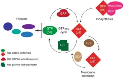
5 Rab proteins function as molecular switches, cycling between a GTP-bound and GDP-bound state (Figure I.4) (Schwartz et al., 2008). Depending on the nucleotide bound, GTPases adopt different conformations in mainly two regions called “switch I” and “switch II”, which are localized at the N-terminal. Moreover, the different conformations determine the interaction of the Rabs with regulatory proteins (Wennerberg et al., 2005; Stenmark, 2009). The cycling of GTPases is regulated by guanine-nucleotide-exchange factors (GEFs) and GTPase-activating proteins (GAPs). GEFs catalyze the exchange of GDP by GTP, activating Rabs, while GAPs catalyze the intrinsic guanosine triphosphatase (GTPase) activity of Rabs, which leads to the hydrolysis of GTP, and the inactivation of the proteins (Park, 2013; Pfeffer, 2013). The regulation of the nucleotide binding state of Rabs is intrinsically coupled to their reversible association with membranes. Newly synthesized Rabs are inactive and have low affinity to membranes. To allow their association with membranes, Rab-escort protein (REP) binds to newly synthesized Rabs and recruits Rab geranylgeranyltransferase (RabGGTase). This enzyme catalyzes a post-translational modification known as prenylation, adding hydrophobic geranylgeranyl groups to the C-terminal of Rab proteins. Rabs are then anchored to membranes and activated by GEFs, which allow them to recruit specific effectors to perform their downstream functions (Mitra et al., 2011). After exerting their function Rabs, are inactivated by GAPs and then recognized by guanine-nucleotide dissociation inhibitors (GDI), which are responsible for membrane extraction and stabilization of Rab proteins in their inactive state. Before inactive Rabs can be presented again to membranes, they need to be dissociated from GDI, in a step mediated by GDI displacement factors (GDF) (Schwartz et
al., 2008; Mitra et al., 2011; Seixas et al,. 2013).
Figura I.4 - Rab GTPases cycle. Rab small GTPases interconvert cyclically between an active GTP-bound and
an inactive GDP-bound state. Several regulators are involved in both activation/inactivation and association/dissociation with membranes Taken from Brighouse et al., 2010.
6
Rab small GTPases perform their functions through the direct or indirect interaction with effector proteins. They are able to regulate recruitment and function of coat, motor and tethering proteins and also other molecules involved in vesicular trafficking (Pfeffer, 2013), allowing the control of many molecular events at a restricted localization.
1.1.1. Rab11 subfamily
The Rab11 subfamily of Rab GTPases is evolutionary conserved and comprise three members (isoforms): Rab11a, Rab11b and Rab11c, also known as Rab25. Rab11a and Rab11b are closely related proteins sharing 90% amino acid identity, while only 60% of homology was found between Rab11a/b and Rab25 (Khvotchev et al., 2003; Kelly et al., 2012). In mammalians, Rab11 tissue expression is variable, according to the isoform. Rab11a is the best characterized and is ubiquitously expressed, whereas Rab11b is restricted to the brain, testis and heart. Rab25 is mostly expressed in epithelial tissues (Welz et al., 2014) and has been associated with cancer. Indeed, Rab25 was found to be overexpressed in several cancers, such as breast and ovarian, acting as a tumor promotor (Kelly et
al., 2012), but was also identified as a tumor suppressor in other cancers, like colon carcinoma (Welz et al., 2014).
Rab11 proteins are key regulators of specific intracellular trafficking steps. Rab11 proteins localize primarily to the ERC in the perinuclear region and in recycling endosomes (RE) in the cytoplasm (Welz
et al., 2014). Rab11 is also found in the TGN and post Golgi-vesicles (Kelly et al., 2012; Welz et al.,
2014). The major role of Rab11 GTPase is the coordination of the slow endocytic recycling pathway (Ullrich et al., 1996), from the ERC to the PM (Figure I.5) (Welz et al., 2014). In fact, the recycling of several PM receptors, such as the transferrin receptor (TfR) (Ren et al., 1998), β2-adrenergic receptors (Moore et al., 2004), cannabinoid receptor 2 (Grimsey et al., 2011) or fibroblast growth factor receptor 4 (Haugsten et al., 2014) are regulated by Rab11 GTPases. Rab11 is also involved in the regulation of several exocytic processes at the TGN, like the budding of a subset of TGN vesicles (Figure I.5) (Urbé
et al., 1993) (Chen et al., 1998). Additionally, Rab11 plays other relevant cellular functions, such as the
delivery of cargo to the cleavage furrow/midbody during cytokinesis, as well as cell migration, ciliogenesis, neuritogenesis and oogenesis (Figure I.5) (Welz et al., 2014). In recent years, Rab11 was also shown to be associated with constitutive and regulated exocytosis in specialized cells, such as neurons (Khvotchev et al., 2003), skin melanocytes (Tarafder et al., 2014) and cytotoxic T-lymphocytes (CTL) (Sluijs et al., 2013).
7 Similar to other small GTPases, Rab11 switches between an active GTP-bound and an inactive GDP-bound form, through the action of specific GAPs and GEFs. To date, three Rab11 GAPs (TBC1D11, TBC1D15 and Evi5) and one Rab11 GEF (Crag) (Welz et al., 2014) were described. In its active form, Rab11 is able to recruit specific effector proteins in order to fulfill its functions. Several Rab11 effector proteins were already identified, namely Rab11-family of interacting proteins (FIPs) (Horgan and McCaffrey, 2009), Rabphilin-11/Rab11BP (Mammoto et al., 2000), motor proteins such as Myosin Va and Myosin Vb (Lapierre et al., 2001; Lindsay et al., 2013), phosphoinositide 4-kinase β (Graaf et al., 2004), the Rab8-GEF, Rabin8 (Knodler et al, 2010) and the exocyst complex subunit Sec15 (Zhang et al., 2004). Rab11 can interact with multiple effector proteins at the same time (Vetter
et al., 2015) or it can share effectors with other Rab GTPases to coordinate specific intracellular
pathways.
1.1.2. Rab11 effectors
1.1.2.1.
Rab11 family of interacting proteins (FIPs)
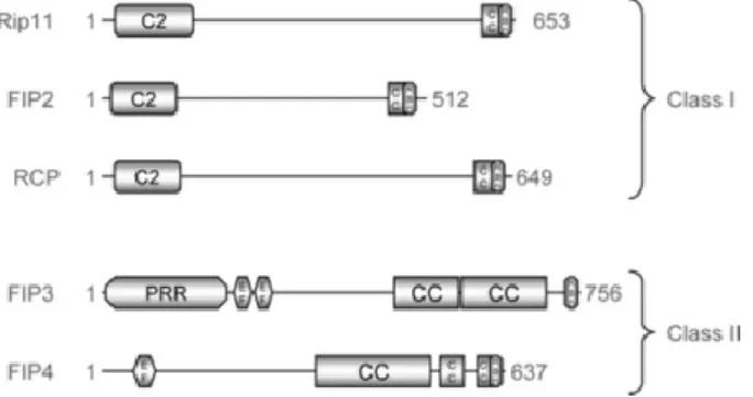
Among the Rab11 effector proteins identified so far, the Rab11-family of interacting proteins (FIPs) are the best characterized. FIPs are encoded by five genes (FIP1, FIP2, FIP3, FIP4 and FIP5). Alternative splicing was also reported, originating different isoforms for each FIP, like, FIP1-A, FIP1-B and FIP1-C. They are an evolutionarily conserved family, ubiquitously expressed and known to interact Figure I.5 - Rab11-regulated vesicle transport processes. The major function of Rab11 proteins is the
coordination of the endocytic recycling pathway. Rab11 GTPases were found to have an important role in several cell functions, namely ciliogenesis, cytokinesis, neuritogenesis and oogeneis.Taken from Welz et al., 2014.
8
with different Rabs and Arfs. The interaction of FIPs with Rab proteins, namely with Rab11, is mediated by a twenty amino acid C-terminal conserved domain called Rab-binding domain (RBD) (Horgan and McCaffrey, 2009; Kelly et al., 2012). FIPs are divided into two classes (Horgan and McCaffrey, 2009): Class I, which includes FIP1C (Rab-coupling protein (RCP)), FIP2 and FIP5 or Rip11/pp75/Gaf1 and class II, composed by FIP3 or Eferin/Arfophilin-1 and FIP4 or Arfophilin-2.
Class I FIPs (FIP1/RCP, FIP2 and FIP5) contain a Ca2+-binding C2 domain near the N-terminal
(Figure I.6), which interacts with phospholipids and targets Rab11-FIP complexes to endomembranes (Prekeris, 2003; Horgan and McCaffrey, 2009). FIP1/RCP is the most studied FIP1 transcript, and it was first described to interact with Rab11 and Rab14 in yeast (Prekeris, 2003). Rab11-FIP1 interaction is known to play a key role in endocytic sorting, trafficking of receptors, transport between ERC and TGN and also in recycling of PM receptors (Horgan and McCaffrey, 2009; Baetz et al., 2013). FIP2 has been associated with endosomal recycling processes (Kelly et al., 2012), where it functions as an adaptor protein between Rab11 and the motor protein Myosin Vb. The Rab11-FIP2-Myosin Vb complex is involved in the recycling of several receptors, channels and transporter proteins, thus regulating vesicle motility towards the cell periphery (Kelly et al., 2012; Baetz et al., 2013). FIP5 co-localizes with Rab11 at RE, and is known to regulate protein recycling to the PM (Schonteich et al, 2008; Jing and Prekeris, 2009). FIP5 was also found to regulate insulin granule exocytosis (Sugawara et al., 2009).
Class II FIPs (FIP3 and FIP4) contain two EF-hands and a proline-rich region at the N-terminal (Figure I.6), which function as Ca2+-binding domains (Prekeris, 2003). Besides interacting with Rab11,
FIP3 and FIP4 can also interact with Arf proteins, namely Arf5 and Arf6 (Prekeris, 2003). Both class II FIPs localize to the cleavage furrow/midbody during cell division (Horgan and McCaffrey, 2009; Baetz
et al., 2013), and are involved in the delivery of membranes to this location, which requires the
integration of signals both from Rab and Arf regulated pathways (Prekeris, 2003; Horgan and McCaffrey, 2009). FIP3 plays a role in the late stages of cytokinesis, namely in abscission, while FIP4 was described to be important in the targeting of molecules to the cleavage furrow (Kelly et al., 2012).
Figure I.6 – Structure of Rab11-family of interacting proteins. Class I FIPs share a Ca2+-binding C2-domain
near the N-terminal, while class II FIPs contain two EF-hands and a proline-rich regions at the N-terminal.Taken from Horgan and McCaffrey, 2009
9 The formation of Rab11-FIP protein complexes defines discrete membrane subdomains, that regulate several vesicular trafficking steps (Prekeris, 2003; Baetz et al., 2013). Furthermore, the interaction of Rab11 with FIPs and other molecules, such as motor proteins, seems to be important to ensure the correct subcellular localization of Rab11 proteins (Prekeris, 2003).
1.1.2.2.
Class V Myosins
The subfamily of Class V unconventional Myosins is composed by Myosin Va (Myo Va), Myosin Vb (Myo Vb) and Myosin Vc (Myo Vc), each of them displaying different tissue distribution and regulating specific membrane traffic events (Trybus, 2008). Class V Myosins are responsible for cargo transport along the actin cytoskeleton (Trybus, 2008) and are known to interact direct and indirectly with small GTPases, controlling their localization and recruitment to specific membranes (Lapierre et al., 2001; Lindsay et al., 2013). Myo Va is expressed mainly in the brain and melanocytes, where the complex Myo Va-Melanophilin-Rab27a was shown to control melanosome tethering at the cell periphery (Strom
et al., 2002). Myo Vb is primarily expressed in epithelial cells, where it has been linked to RE movement
and recycling of PM receptors, in complex with Rab11 and FIP2 (Hales et al., 2002; Schafer et al., 2013). In non-polarized cells, recycling of PM receptors is also regulated by Myo Vb, which mediates Rab11a-positive RE movement to the cell periphery (Lapierre et al., 2001). Rab11a-Myo Vb interaction, together with Rab8a, was also described to be involved in promoting regulated exocytosis in bladder umbrella cells (Khandelwal et al., 2013). Myo Vc is known to be involved in secretory granule trafficking (Jacobs
et al., 2009) and interacts with Rab32 and Rab38 during the biogenesis and secretion of melanosomes
(Bultema et al., 2014).
1.1.2.3.
Exocyst complex
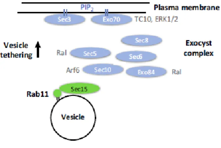
The evolutionarily conserved octameric protein complex, known as exocyst is composed of eight subunits: Sec3, Sec5, Sec6, Sec8, Sec10, Sec15, Exo70 and Exo84 (Figure I.7) (T.Shandala et al., 2012; B. Wu and Guo, 2015). In Drosophila melanogaster, the exocyst was described as an effector of different Rab GTPases, namely Rab3, Rab8, Rab11 and Rab27 (Wu et al., 2005). Although the complex is known to function only when all subunits are present, some reports suggest the existence of sub-complexes, namely Sec5-Exo84 and Sec10-Sec15, with unknown functions (Zhang et al., 2004; Heider and Munson, 2012).
The exocyst complex is known to be recruited to sites with active exocytosis to regulate SNARE function and the tethering of secretory vesicles (Wu and Guo, 2015). Moreover, this protein complex was shown to be important in several other cellular functions, besides its function as vesicle tethers
10
(Heider and Munson, 2012; B. Wu and Guo, 2015). For example, the exocyst is necessary to ensure the correct localization of membrane-bound vesicles to specific target sites at the appropriate moment, for example during cytokinesis (Heider and Munson, 2012). Moreover, studies in yeast reported an interaction between the exocyst and the actin cytoskeleton, suggesting a role for the exocyst complex in vesicular transport (Heider and Munson, 2012).
In mammalian cells, Sec15 was shown to interact directly with Rab11 in a GTP-dependent manner and to co-localize with Rab11 to the perinuclear region (Zhang et al., 2004). Rab11-Sec15 interaction might be important for endocytic recycling, directing recycling vesicles to the PM (Heider and Munson, 2012; B. Wu and Guo, 2015). While Sec15 regulates the vesicular transport to the cell periphery, other subunits like Sec3 and Exo70 regulate the assembly of the complex at the PM (T.Shandala et al., 2012). In fact, it was shown that Rab11 and the exocyst complex participate in the exocytosis of recycling vesicles at the PM (Takahashi et al., 2012).
The interaction of Rab11 with different effector proteins, can explain the specific functions of Rab11 in different intracellular traffic pathways and the pleiotropic functions of this subfamily of Rab proteins. Figure I.7 - Rab11 interaction with the exocyst complex. - Direct binding of Sec15 to Rab11 promotes vesicular
transport to the cell periphery, leading to the assembly of the exocyst complex and tethering of vesicle to the PM. Taken from Welz et al., 2014.
11
2. Lysosomes
Intracellular homeostasis is achieved by balancing what is endocytosed and exocytosed, as well as through what is newly-synthesized and degraded. In cells, lysosomes are the major organelles responsible for the degradation of several biomolecules, such as proteins, lipids and nucleic acids; (Mullins and Bonifacino 2001; Samie and Xu, 2014). Lysosomes are described as dense, acidic membrane-bound compartments, delimited by a single membrane. They accumulate in the perinuclear region of the cell but are also found dispersed in the cytoplasm and near the PM.
Lysosomes are composed by a specific set of hydrolases that include sulphatases, phosphatases, lipases, proteases, carbohydrases and glycosidases (Mullins and Bonifacino, 2001; Samie and Xu, 2014). The majority of the lysosomal enzymes requires an acidic luminal pH (pH 4.6-5) to be active. This pH is established and maintained by a proton pump, known as vacuolar-ATPase (v-ATPase), located in the lysosomal membrane (Samie and Xu, 2014). Lysosomes also contain several highly glycosylated proteins known as lysosome-associated membrane proteins (LAMP) 1, 2 and 3, lysosomal integral membrane protein (LIMP), which have important roles in lysosomal function, by mediating the interaction with other organelles (Luzio et al., 2000; Mullins and Bonifacino, 2001; Eskelinen, 2006). These proteins are commonly used as lysosomal markers, since they are major components of lysosomal membrane.
Material destined for degradation is delivered to lysosomes via the endocytic/phagocytic and autophagic pathways (Li et al., 2013). Endocytosis starts with the uptake of PM receptors or extracellular soluble cargo into endocytic vesicles. These vesicles undergo maturation into EEs/SEs and through sorting processes cargo can be delivered to LEs (Li et al., 2013), which eventually fuse with lysosomes (Luzio et al., 2000; Luzio et al., 2007), in a process mediated by the small GTPase Rab7 (Bucci et al., 2000; Hyttinen et al., 2013). In parallel with the endocytic pathway, the autophagic pathway also delivers cytoplasmic components, such as misfolded proteins and dysfunctional organelles for turnover in the lysosomes (Hyttinen et al., 2013).
Some cells contain specialized lysosomes, known as lysosome-related organelles (LROs), which share common features with lysosomes, including the biogenesis pathway, low pH, and the presence of hydrolytic enzymes and highly glycosylated membrane proteins (Mullins and Bonifacino, 2001; Luzio
et al., 2014). However, LROs differ from conventional lysosomes in morphology, including in size, and
in their biochemical composition (Blott and Griffiths, 2002; Sluijs et al., 2013), thus reflecting their divergent functions. While the major function of conventional lysosomes is the degradation of biomolecules, LROs have specific functions depending on the cell type. LROs can also undergo regulated exocytosis in response to stimuli, and hence they are also known as secretory lysosomes (Blott and Griffiths, 2002).
LROs are present in different cell types, including the hematopoietic cell lineages (Raposo et al., 2002). Platelets have dense granules and α-granules that are responsible for the secretion of molecules essential for blood clotting. Natural killer cells and cytotoxic T-lymphocytes have lytic granules that play a key role in the elimination of infected cells. Antigen-presenting cells, like dendritic cells, contain major
12
histocompatibility complex (MHC) class II compartments, where antigens are processed. Inflammatory mediators are stored in basophilic granules present in basophils and mast cells, while the defense against parasites and bacteria is mediated by specific granules in eosinophils and azurophilic granules in neutrophils (Raposo et al., 2002; Seixas et al., 2013).
However, LROs are also found in other cell types, for example in osteoclasts, they are responsible for bone resorption and remodeling (Raposo et al., 2002). In melanocytes, retinal and iris pigmented epithelial cells, melanosomes, are the LROs responsible for synthesis and storage of the pigment melanin (Wasmeier et al., 2008). Lung epithelial type II cells contain lamellar bodies, that store surfactants, which are required to avoid alveolar collapse. Finally endothelial cells secrete hemostatic and proinflammatory factors that are stored in Weibel-Palade bodies (Raposo et al., 2002; Seixas et al., 2013).
Regulated exocytosis of conventional lysosomes
Until recently, lysosomes were considered the endpoint of the endocytic and autophagic pathways, to where cargo and organelles converge to be degraded. However, in recent years, several reports have shown that lysosomes are dynamic organelles (Luzio et al., 2014; Samie and Xu 2014) that can undergo Ca2+-regulated exocytosis, in a mechanism similar to LROs, in non-specialized cells such as fibroblasts
and epithelial cells (Rodríguez et al., 1997; Andrews, 2000). Several studies have shown the importance of lysosome exocytosis in different cellular functions, namely, in bone resorption, neurite outgrowth, secretion and PM repair (Figure I.8) (Samie and Xu, 2014). PM repair is a very important mechanism to maintain the integrity of cellular membrane and therefore cell survival. PM disruptions are frequent in mechanically-active tissues, like muscle cells (Andrews, 2002) or during pathogen infection. For example, the mechanism of Trypanosoma cruzi cell invasion is known to be dependent on lysosome exocytosis (Rodríguez et al., 1997).
Interestingly, only a small fraction of the lysosome population is able to undergo regulated exocytosis, pointing to an heterogeneous spatial distribution of lysosomes in the cell (Rodríguez et al., 1997). Recently two distinc lysosomal populations have been described (Johnson et al., 2016; Gowrishankar and Ferguson, 2016). One population that localizes at the perinuclear region and is more acidic and a second pool, less acidic that localizes close to the PM (Johnson et al., 2016; Gowrishankar and Ferguson, 2016). Since lysosomes that are closer to the PM are the ones prone to fuse (Jaiswal et
al., 2002), it is likely that the luminal pH of the different population of lysosomes is important for the
lysosomal exocytosis process (Johnson et al., 2016; Gowrishankar and Ferguson, 2016). In other words, a less acidic pH favors the exocytic process.
13 Although lysosomal exocytosis was initially observed in Chinese Hamster Ovary (CHO) and Normal Rat Kidney Epithelial Cells (NRK) fibroblasts it is now accepted as an ubiquitous mechanism present in all cell types (Andrews 2000). Despite the physiological importance of conventional lysosome exocytosis, the molecular mechanism by which it occurs is commonly compared to the synaptic vesicle and LRO exocytosis mechanisms, but is far from being fully understood. Lysosome exocytosis depends on stimulation and requires specific machinery necessary for the transport of lysosomes from the perinuclear region to the cell periphery, for the tethering and docking of lysosomes to the PM and for the fusion and release of the lysosomal content to the extracellular space, in a mechanism that has to be tightly regulated.
Lysosomes accumulate at the perinuclear region, around the microtubule-organizing center (MTOC) (Matteoni and Kreis, 1987). Lysosomal long range transport from the perinuclear region to the cell periphery was shown to be a Ca2+-independent mechanism (Jaiswal et al, 2002) and occurs in a
bidirectional manner along microtubules. Moreover, this type of transport is mediated by motor proteins such as kinesins and dyneins (Matteoni and Kreis, 1987). In MCF-7 breast cancer cells, kinesin 5B (KIF5B) was found to associate with lysosomes and the silencing of KIF5B induces peripheral aggregations of lysosomes (Cardoso et al., 2009). Also, lysosome localization at the cell periphery is known to be mediated by the Arf small GTPase Arl8b, SifA kinesin-interacting protein (SKIP) and kinesin-1 (Hofmann and Munro, 2006; Bagshaw et al, 2006; Rosa-Ferreira and Munro, 2011). Recently, BLOC-1-related complex (BORC) was described to have an important role in the recruitment of Arl8b to the lysosomal membrane, enabling the coupling with SKIP-kinesin complex and the movement to the periphery (Pu et al., 2015). Additionally, Rab7, is known to bind to FYVE and coiled-coil domain-containing 1 protein (FYCO 1), mediating kinesin-dependent transport of lysosomes towards the periphery (Pankiv et al., 2010).
Due to the presence of a cortical actin meshwork beneath the PM, lysosomes also undergo short range movement in a process that is likely mediated by actin motors. Recently, our group showed that Rab3a, which is involved in exocytic processes, recruits the actin motor non-muscle myosin heavy chain Figure I.8 - Biological functions that require lysosome exocytosis. - Lysosome exocytosis is an important
mechanism in all cell types. It is involved in several cellular functions, namely PM repair, secretion, phagocytosis and neurite outgrowth. Taken from Samie and Xu, 2014.
14
IIA (NMHC IIA) to lysosomes. This interaction allows lysosomes to travel through the cortical actin and be positioned closer to the PM (Encarnação et al., 2016). After this step, lysosomes must be tethered and docked to the PM. The Rab3a effector protein synaptotagmin-like protein 4-a (Slp4-a) is likely involved in remodeling cortical actin to allow lysosomes to tether and dock to the PM (Encarnação et
al., 2016).
SNARE proteins are involved in the final steps of lysosomes exocytosis, more precisely in lysosome fusion with the PM. The vesicle-associated membrane protein 7 (VAMP7) localizes in the lysosomal membrane and interacts with Syntaxin-4 and synaptosome-associated protein of 23 kDa (SNAP-23) on the PM, forming a trans-SNARE complex (Rao et al., 2004), which primes the lysosomes to fuse with the PM. The fusion of lysosomes with the PM is triggered by an increase in intracellular Ca2+
concentration that can be induced by membrane disruption, or artificially by Ca2+ ionophores (Rodríguez et al., 1997; Andrews 2000; Reddy et al., 2001). Synaptotagmin VII (Syt VII), a high affinity Ca2+ sensor,
was found to regulate this step. Syt VII is ubiquitously expressed, localizes to lysosomes and was shown to bind to Syntaxin-4 at low Ca2+ concentrations, promoting membrane fusion and lysosomal exocytosis
(Martinez et al., 2000; Andrews, 2000). However, the precise mechanism by which Syt VII promotes lysosome fusion with PM is still not clear.
The Ca2+ channels that mediate Ca2+- triggered lysosome exocytosis are not fully known, but the
transient receptor potential channels (TRPML), namely TRPML1, seem to be involved (Samie and Xu, 2014). Additionally, overexpression of the transcription factor TFEB, which regulates the expression of several lysosomal genes such as TRPML1, was shown to increase the docking and fusion of lysosomes with the PM (Medina et al., 2011; Settembre et al., 2013; Samie and Xu, 2014), suggesting a role for TFEB in lysosome exocytosis. Importantly, this role might not be restricted to the regulation of TRPML1 but it can also be involved in the positioning of lysosomes near the PM, by increasing the expression and activity of tethering and motor proteins (Medina et al., 2011; Samie and Xu, 2014).
Lysosome exocytosis was found to be negatively regulated by Neuraminidase 1 (Neu1). Neu1 is responsible for post-translational modifications in lysosomal proteins, namely LAMP1. Although the role of Neu1 in lysosome exocytosis is not clear, in its absence lysosomal docking and exocytosis increases, in a LAMP1-dependent manner (Yogalingam et al., 2008; Samie and Xu, 2014).
Although several molecules have already been identified to play a role in lysosome exocytosis, the full molecular machinery required for the different steps of lysosome transport, tether and fusion with the PM is still far from being completely elucidated. The involvement of other important players, including Rab GTPases in this mechanism should not be excluded, due their importance in all intracellular traffic pathways.
Because of the biological importance of lysosomal exocytosis and the similarities with other exocytic processes, namely LRO exocytosis, it is not surprising that disturbance in lysosome and/or LROs pathways can lead to human diseases.
15
Diseases of lysosomes and lysosome-related organelles
Dysfunction of conventional lysosomes, including deficiencies in the lysosomal hydrolytic enzymes, generally gives rise to a group of metabolic disorders collectively known as lysosomal-storage disorders (LSDs). LSDs result from the accumulation of undigested material in lysosomes (Samie and Xu, 2014). However, since conventional lysosomes can also undergo regulated exocytosis, it is likely that defects in lysosome exocytosis could lead to other human diseases, commonly associated with LROs (Andrews, 2002).
Impaired LRO biogenesis and/or exocytosis leads to several human diseases, namely Chediak– Higashi (CHS), Griscelli (GS) and Hermansky–Pudlak (HPS) syndromes (Seixas et al., 2013).
CHS results from mutations in the CHS1/LYST gene, which is involved in the transport of cargo to lysosomes (Huizing et al., 2008). Patients with CHS manifest hypopigmentation (albinism) (due to decreased melanin production in melanocytes), recurrent infections (due to immune deficiency), coagulation defects (due to storage pool deficiency) and neurological impairment (Huizing et al., 2008). Interestingly, studies in vivo, revealed the presence of enlarged lysosomes in all cells of the animal models of CHS (Huynh et al., 2004). Moreover, these enlarged lysosomes had reduced capacity to undergo exocytosis and therefore reduced ability to repair PM damages, contributing to the symptoms exhibited by these patients (Huynh et al., 2004).
GS is a rare autosomal recessive disease that results in hypopigmentation and impaired immune responses (Huizing et al., 2008). There are three types of GS that result from mutations in each of the members of the tripartite complex Rab27a-Melanophilin-Myo Va, known to be involved in melanosome positioning near the PM (Huizing et al., 2008; Seixas et al., 2013). GS type 1 is caused by mutations in
MYO5A, which encodes Myo Va, an actin-dependent motor protein. GS type 2 results from mutations
in RAB27A, which encodes Rab27a, with roles in melanosome and CTL lytic granules secretion (Sluijs
et al., 2013). Finally, GS type 3 is caused by mutations in MLPH, encoding Melanophilin, which serves
as a linker between Rab27a and Myo Va. GS patients show accumulation of melanosomes in the perinuclear region of melanocytes but not enlarged conventional lysosomes (Huizing et al., 2008), thus, the role of conventional lysosomes in the pathogenesis of GS, if any, remains elusive.
HPS is a group of rare autosomal recessive diseases. There are eight human genes whose mutations are known to cause HPS (Huizing et al., 2008; Seixas et al., 2013). HPS patients exhibit hypopigmentation and bleeding diathesis (Huizing et al., 2008), due to impaired melanosome biogenesis and platelet aggregation, respectively (Huizing et al., 2008; Seixas et al., 2013). Accumulation of undigested lipids also occur in patients with HPS, because cells cannot rapidly degrade mistargeted vesicle membranes, suggesting the involvement of conventional lysosomes in the pathology of some types of HPS (Huizing et al., 2008).
The role of lysosome exocytosis in several cellular processes suggests that impaired lysosome exocytosis might have a role in other diseases, not related with LROs. Interestingly, recent studies
16
regarding tumor cell invasion, revealed that increased lysosomal exocytosis promotes extracellular matrix invasion and degradation and could promote a cell migratory phenotype (Machado et al., 2015). Despite being now accepted that conventional lysosomes can undergo Ca2+-regulated exocytosis
and that this process is relevant in several cellular functions, namely in PM repair, further studies need to be performed to elucidate the role of regulated lysosome exocytosis in human disease.
17
3. Previous results
In recent years, it has been established that lysosomes, similar to LROs can undergo regulated exocytosis. However, little is known about the molecular machinery that regulates this pathway. In order to identify new regulators of Ca2+-triggered lysosome exocytosis, our group, in collaboration with others,
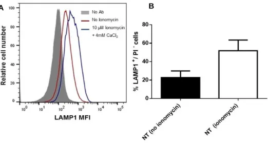
performed a lentiviral short-hairpin RNA (shRNA) screen, targeting the major families of membrane trafficking regulators (Encarnação et al., 2016). For this, the human monocytic cell line THP1 was transduced with lentiviral shRNA hairpins and then stimulated with the Ca2+ ionophore ionomycin, to
increase intracellular Ca2+ concentration. Lysosome exocytosis was analysed by measuring the cell
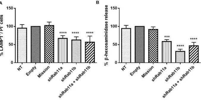
surface expression of the LE/lysosome marker LAMP1, by flow cytometry. Several trafficking regulators were found to impair lysosome exocytosis when silenced and interestingly, the majority were Rab GTPases. Surprisingly, the silencing of Rab11b, which has described roles in endocytic recycling traffic, was found to impair lysosome exocytosis (unpublished data, Encarnação et al., 2016) ). The involvement of Rab11b, and its isoform Rab11a in lysosome exocytosis was further confirmed in HeLa cells (human cervical cancer cells). Indeed, our group observed that the silencing of Rab11a or Rab11b impairs LAMP1 cell surface expression levels and decreases the release of the lysosomal hydrolytic enzyme β-hexosaminidase, upon ionomycin stimulation, when compared with non-transduced (NT) cells, cells treated with an empty vector (Empty), or cells treated with a non-targeting shRNA (Mission) (Figure I.9, A/B unpublished). Furthermore, HeLa cells transduced simultaneously with Rab11a and Rab11b shRNAs do not show a higher decrease in LAMP1 surface expression and β-hexosaminidase release, upon ionomycin stimulation, when compared with HeLa cells transduced with only Rab11a or Rab11b shRNAs (Figure I.9 A/B, unpublished). These results suggest a role for the endocytic recycling pathway in Ca2+-regulated lysosome exocytosis, and that Rab11a and Rab11b do not have redundant functions
and probably act in different steps of the pathway. In fact, Rab11 and the endocytic recycling pathway have been linked before with the exocytosis of LROs. Indeed, our group showed that Rab11b plays a role in melanosome exocytosis from melanocytes (Tarafder et al., 2014). Moreover, during lytic granule secretion by CTLs, it was proposed that Rab11-positive vesicles interact with immature vesicles, and deliver proteins required for the late stages of exocytosis (Sluijs et al., 2013). Nevertheless, the role of Rab11 in lysosome exocytosis remains elusive.
18
Figure I.9 - LAMP1 cell surface expression levels and β-hexosaminidase release in HeLa cells silenced for Rab11a, Rab11b or both. HeLa cells transduced with lentiviruses encoding shRNAs for Rab11a, Rab1b or both
were selected for 6 days in puromycin. Cells were the treated with 10 µM ionomycin and 4 mM CaCl2 for 10 minutes
at 37ºC, to trigger lysosome exocytosis. A. Cells were collected and stained with an anti-LAMP1 antibody. Non-transduced cells (NT), cells Non-transduced with an empty vector (empty) and cells Non-transduced with a non-targeting shRNA (Mission) were used as negative controls. The results are presented as percentage (%) of cells positive for LAMP1 and negative for propidium iodide (LAMP1+/PI-). All results were normalized to empty (100%) and are
represented as mean ± SD. B. Cellular extracts and supernatants containing released β-hexosaminidase were collected and analysed. Non-transduced cells (NT), cells transduced with an empty vector (Empty) and cells transduced with a non-targeting shRNA (Mission) were used as negative controls. The results are presented as percentage (%) of released β-hexosaminidase. All results were normalized to empty (100%) and are represented as mean ± SEM
19
4. Objectives
Since Rab11, similar to other Rab GTPases, exerts its function through the interaction with effector proteins, the role of Rab11 in the regulation of lysosome exocytosis is likely dependent on proteins that interact with Rab11.
Thus, the main objective of this thesis was to find proteins that interact with Rab11 and play a role in Ca2+-triggered lysosome exocytosis. Moreover, we want to characterize spatially and temporally the
21
II.
Materials and Methods
1. Cell culture
HeLa human cervical cancer cells were cultured in Dulbecco’s Modified Eagle’s Medium (DMEM, Gibco), supplemented with 10% heat-inactivated fetal bovine serum (FBS, Gibco), 100 U/mL penicillin/streptomycin (Gibco), 2 mM L-glutamine (Gibco) and 20 mM HEPES (Gibco), and kept at 37ºC in a 5% CO2.
2. Gene silencing
Rab11 effectors were silenced using siGENOME SMARTpool Oligos (Thermo Scientific Dharmacon) specific for human genes. The list of small interfering RNA (siRNA) sequences is described in Table 1. As a control, a non-targeting siRNA sequence (siControl) (Thermo Scientific Dharmacon) was used. HeLa cells (7x10⁴ cell/well) were seeded 24 hours before transfection, in 24 well-plates using
DMEM supplemented as described above, without antibiotics. Transfection was performed using 40 nM of siRNA and 2 µL of transfection reagent DharmaFECT 1 (Dharmacon) in 100 µL of Opti-MEM (Gibco), as indicated by the manufacturer. Twenty-four hours after the silencing, the transfection medium was replaced by complete DMEM. All assays were performed 72 hours after siRNA transfection.
Table II.1 - siRNAs used for silencing Rab11 effectors
Gene siRNA sequence siControl UAAGGCUAUGAAGAGAUAC siFIP1-C CAAACAGAAGGAAACGAUA; GCUAACUGCAGCUUGGGAA; AGUGAGAACUUGAACAAUG; UCCGCGAGCUGGAAGACUA. siFIP2 GGAUGAAGGUGAAUUGUGU; CGAGCUACCUGGAUUGCUA; GGAACAAUAUGACCGCAAG; GAUAAGAUGAAGGGUAGAA. siMyosin Va GAACAAAUGUGCACUCUUU; GAUCAUCUGCUCUGGAUUA; AAAGUAAGGUCGUUGCUAA; CGCAGGAGGUACAAGAUUA. siMyosin Vb GGACUUACCUCUUGGAGAA; CAAGUUGGCCUAACAGUGA; GCAGAUCUGGCCUACAAUA; ACAGUGGCCUUUAUACGAA. siSec8 GAAUUGAGCAUAAGCAUGU; UAACUGAGUACUUGGAUAU; GCCGAGUUGUGCAGCGUAA; ACUGAGUGACCUUCGACUA. siSec15 UAACUGAACUGCUGAAAGU; GUACUAGUCCGAAGUCUGA; CAAGUAAGCCACUAUCGAU; CGGGAAACAUUUGAGAAUU. siExo70 GGUUAAAGGUGACUGAUUA; GACCUUCGACUCCCUGAUA; CUAAGCACCUAUAUCUGUA; CGGAGAAGUACAUCAAGUA.
22
3. Cell transfection
HeLa cells were seeded, 24 hours before transfection in DMEM supplemented as described above, without antibiotics or FBS. The cells were transfected with 1 µg DNA and 2 µL Lipofectamine 2000 (Invitrogen) in 24-well plates, 10 µg DNA and 20 µL Lipofectamine 2000 in 10 cm dishes or 0.5 µg DNA and 1 µL Lipofectamin 2000 in Nunc™ Lab-Tek™ Chambered Coverglasses (ThermoFisher Scientific), in Opti-MEM, according to manufacturer’s instructions. DNA plasmids used are summarized Table 2. Five hours after transfection, medium was replaced by complete DMEM and cells were analysed 24 hours later.
Table II.2 - DNA plasmids used to transfect HeLa cells.
Gene Plasmids
GFP alone pENTR GFP vector.
Rab11a mRab11a GFP;
pCDNA-ENTR-BP-cherry mRab11a.
Rab11b pENTR GFP C2 mRab11b;
pCDNA-ENTR-BP-cherry mRab11b.
FIPs pEGFP-N1-FIP1C/RCP;
pEGFP-N3-FIP2-GFP; gift from R.Prekeris
Myosin V Myosin Va-GFP, gift from Dra. M. João Amorim;
Exocyst subunits
pGFP-N1 Sec8
pJ3 Myc-EGFP rat Sec15; pEGFP-C1 rat Exo70; pCDNA3.1 Exo70-cherry.
4. RNA extraction, cDNA production and real-time quantitative PCR
RNA from HeLa cell lysates was extracted using the RNeasy Mini Kit (Qiagen) according to the manufacturer’s instructions. RNA was reverse-transcribed into complementary DNA (cDNA), by incubating 1 µg of total RNA with 1 mM of dNTP mix (Thermo Scientific) and 0.3 µg/uL of random primers p(DN) (Roche) at 65ºC for 5 min. After placing samples for a few seconds on ice, they were incubated with 2x first strand buffer (Invitrogen), 20 mM DTT (Invitrogen) and 40 U/µL of Recombinant Ribonuclease Inhibitor RNaseOUT (Invitrogen) at 25ºC for 2 minutes. Finally, 50 U/µL of Superscript II
23 Reverse Transcriptase (Invitrogen) were added and samples were incubated at 25ºC for 10 minutes, 42ºC for 50 minutes and finally at 70ºC for 15 minutes.
To perform real-time quantitative PCR (qRT-PCR), Fast Start Essential DNA Green Master (Roche) kit was used according to the manufacturer’s instructions. Analysis was done in the qPCR Roche Light Cycler. Βeta-actin, a housekeeping gene, was used as an endogenous control to normalize the expression level of each gene analysed. Primers used are summarized in Table 3.
Table II.3 - Primers used in qRT-PCR assays.
5. Immunoprecipitation
Cells were lysed with ice-cold modified radioimmunoprecipitation lysis buffer (RIPA) (50 mM Tris- HCl pH 7.5; 1 mM EDTA; 1 mM EGTA; 150 mM NaCl; 2 mM MgCl2; 1 mM DTT; 1% IGEPAL) containing
protease (0.5 x) and phosphatase (0.5 µM) inhibithors, for 30 minutes at 4˚C, under constant agitation. After centrifugation at 18,800 x g for 30 minutes, at 4˚C, supernatants were collected and protein concentration was determined using DC protein assay kit (Bio-Rad), according to the manufacturer’s instructions.
Gene Primer sequence
Actin B Forward 5’-GCAAAGACCTGTACGCCAAC-3’ Reverse 5’-AGTACTTGCGCTCAGGAGGA-3’ FIP1-C Forward 5’-CAAGGAGCGAGGAGAAATTG-3’ Reverse 5’-GGTGTCTGACCCACTGTCCT-3’ FIP2 Forward 5’-TGGGGGATCTGATAGCCCTT-3’ Reverse 5’-ACTCATATGAAAACTTGAAGATGGC-3’ Sec8 Forward 5’-ATGGCCAGCAAGCACTATCT-3’ Reverse 5’-AGGTGCCGGTGTAGTTCATC-3’ Sec15 Forward 5’-CTGACCCTGCTTGAGAAGATGA3’ Reverse 5’-GCCACGGTGTCAATGAGTTTC-3’ Exo70 Forward 5’-CATGGGTTATCAGGGGATTTG-3’ Reverse 5’-GAGGTCCAGGTGTGGGTAGA-3’ Myosin Va Forward 5’-CAGTGGTCAGAACATGGGTG-3’ Reverse 5’-TCGCATGGCATACTTAGCTG-3’ Myosin Vb Forward 5’-AGAACTGGAGGAGGAGCGAT-3’ Reverse 5’-GGTTTGATGGGTTCCGCCTA-3’