Risk factors for decreased bone
density in premenopausal women
1Departamento de Ginecologia e Obstetrícia, Faculdade de Medicina,
Pontifícia Universidade Católica do Rio Grande do Sul, Porto Alegre, RS, Brasil
2Serviço de Endocrinologia, Departamento de Medicina Interna,
Hospital de Clínicas de Porto Alegre, Universidade Federal do Rio Grande do Sul, Porto Alegre, RS, Brasil
C. Krahe1,
R. Friedman2
and J.L. Gross2
Abstract
Osteoporosis is a major health problem. Little is known about the risk factors in premenopause. Sixty 40-50-year old patients with regular menses were studied cross-sectionally. None of the patients were on drugs known to interfere with bone mass. Patients answered a dietary inquiry and had their bone mineral density (BMD) measured. The Z scores were used for the comparisons. A blood sample was taken for the determination of FSH, SHBG, estradiol, testosterone, calcium and alkaline phosphatase. Calcium and creatinine were measured in 24-h urine. A Z score less than -1 was observed for the lumbar spine of 14 patients (23.3%), and for the femur of 24 patients (40%). Patients with a Z score less than -1 for the lumbar spine were older than patients with a Z score ≥-1 (45.7 vs 43.8 years) and presented higher values of alkaline phosphatase (71.1 ± 18.2 vs 57.1 ± 14.3 IU/l). Multiple regression analysis showed that a lower lumbar spine BMD was associated with higher values of alkaline phosphatase, lower calcium ingestion, a smaller body mass index (BMI), less frequent exercising, and older age. The patients with a Z score less than -1 for the femur were shorter than patients with a Z score ≥-1 (158.2 vs 161.3 cm). Multiple regression analysis showed that a lower femoral BMD was associated with lower BMI, higher alkaline phosphatase and caffeine intake, and less frequent exercising. A lower than expected BMD was observed in a significant proportion of premenopausal women and was associated with lower calcium intake, relatively lower physical activity and lower BMI. We conclude that the classical risk factors for osteoporosis may be present before ovarian failure, and their effect may be partly independent of estrogen levels.
Correspondence
J.L. Gross
Serviço de Endocrinologia Hospital de Clínicas de Porto Alegre Rua Ramiro Barcelos, 2350, sala 2030 90035-003 Porto Alegre, RS Brasil
Fax: 55 (051) 332-5188 E-mail: gross@hotnet.net
Received September 12, 1996 Accepted July 16, 1997
Key words
•Osteoporosis •Bone loss •Premenopause •Osteopenia •Densitometry
Introduction
Osteoporosis is a major health problem because it is associated with an increase in fracture rate (1,2). More than 1.5 million Americans are expected to have fractures every year, a cost of about 10 billion dollars (1,2). The disorder affects mainly women
after 50 years of age. From the onset of ovarian failure a woman loses 3 to 5% of her bone mass per year (3). Several other factors are associated with bone loss: low calcium intake, less frequent exercise, smoking and alcohol abuse, certain drugs, and low expo-sure to sunlight, among others (4,5).
therapeutic options are limited, costly and imply some risk. It is therefore important to detect risk factors for the development of osteoporosis as early as possible (6-8) so that preventive measures can be adopted. Most of the research in this field has been carried out on postmenopausal women and little is known about premenopause. Determinations of bone mineral density (BMD) have been suggested as a method to identify individu-als at risk (9). A decrease of 1 SD in BMD significantly increases the risk of fractures by 50%, irrespective of the site where the measurement was taken (9).
Thus, the aim of the present study was to measure BMD in 40-50-year old premeno-pausal women with normal ovarian function and to determine the factors associated with the disorder.
Patients and Methods
A cross-sectional study of 60 white women, 40 to 50 years old, with regular menses, was performed. The exclusion crite-ria were chronic illnesses and current use of estrogen, progesterone, androgen, adrenal steroids, thyroid hormones or any other drugs that might affect bone mass (tamoxifen, di-uretics, anticonvulsants, and barbiturates). Patients who had used hormonal contracep-tive pills in the past or who had received occasional treatment with estrogen and/or progesterone were not excluded. The proto-col was approved by the Ethics Committee of the Hospital de Clínicas de Porto Alegre. Patients were recruited from the private prac-tice of one of the authors (CK). A total of 220 patients aged 40 to 50 years were identified and contacted. Eighty-two agreed to attend a meeting where the protocol was presented, 12 were excluded on the basis of the above criteria, and an additional 10 patients were subsequently excluded for not having con-cluded the tests.
Patients answered a questionnaire inquir-ing about their age, smokinquir-ing habits, physical
activity, alcohol consumption over the last 5 years, and knowledge of family history of osteoporosis. Physical activity was analyzed as sports practice, and categorized as “posi-tive”, i.e., regularly practiced throughout the year (at least twice a week), or “negative”, i.e., all other situations. The following ac-tivities were considered to be sports: walk-ing, cyclwalk-ing, swimmwalk-ing, gymnastics, or court sports. A four-day complete dietary inquiry was obtained, and the energy, alcohol, caf-feine and calcium intakes were calculated (10,11). Patients were weighed and meas-ured and BMI was reported as kg/m2. A blood sample was taken between the 5th and 10thday of the cycle for the determination of FSH (immunoradiometric assay, reference values 5-20 mIU/ml, Irma-Count®), SHBG (immunoradiometric assay, 20-130 nmol/l, Irma-Count®), testosterone (radioimmunoas-say, 20-80 ng/dl, COAT-A-COUNT®), es-tradiol (radioimmunoassay, 25-150 pg/ml, COAT-A-COUNT®), calcium (cresolphtha-lein-complexone method, 8.5-11.0 mg/dl) and alkaline phosphatase (enzymatic method, 35-90 IU/l). Calcium and creatinine (Jaffé’s reaction) were measured in 24-h urine samples.
BMD was measured by densitometry (dual-energy X-ray absorptiometry, Lunar DPX-L) for the lumbar spine (L1, L2-L4) and femur (neck, Ward’s triangle and tro-chanter). BMD (g/cm2) was compared to reference standards for young adults, and the Z scores were calculated accordingly.
stud-ied by stepwise multivariate regression anal-ysis. Taking into account the sample size, the independent variables to be entered into the multivariate analysis were the seven ones more strongly correlated with Z scores in the univariate analysis, or those with a previ-ously known association with BMD. Results are reported as mean ± SD (range) or median (range), unless otherwise stated.
Results
The 60 patients were 44.3 ± 2.9 (40-50) years old, weighed 60.2 ± 10.5 (47-90) kg, and had a height of 160.1 ± 6.04 (142-170) cm, and BMI of 23.5 ± 3.34 (19-34) kg/m2. The results of the densitometric studies are listed in Table 1. For an expected fre-quency of about 15.9%, 14 patients (23.3%) had at least one Z score under -1 at the lumbar spine level (χ2 = 1.978, 0.10<P<0.25), and 24 patients (40%) had a Z score under -1 for the femur (χ2 = 24.4, P<0.0001).
The dietary data are summarized in Table 2. Fifty-two percent of the patients had a daily calcium intake of less than 800 mg, which is the minimum recommended daily allowance.
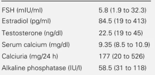
The results of the various hormonal and biochemical parameters are summarized in Table 3. No FSH higher than 40 mIU/ml or elevated testosterone values were observed. Hypercalcemia was not observed; 18 pa-tients had calciuria >4 mg kg-1 24 h-1. Three patients had alkaline phosphatase >90 IU/l (92, 104 and 118 IU/l, respectively).
Thirty-six (60%) patients had smoked in the past, but only 13 (21.7%) continued to do so. Forty percent began smoking during ado-lescence. None of the patients smoked more than 10 cigarettes a day.
Less than one quarter of the patients (21.6%) exercised regularly, 25% never prac-ticed any sport, and 53.4% exercised irregu-larly.
Twenty-five (41.6%) patients reported relatives with osteoporosis; 16 (26.7%) could
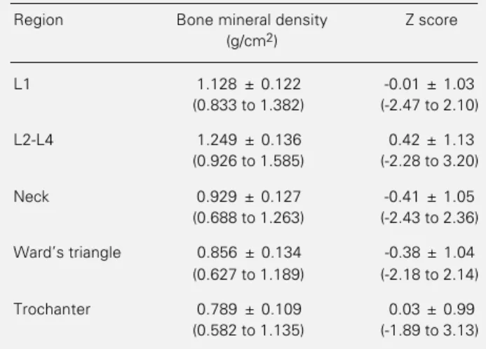
Table 1 - Bone mineral density and Z scores of premenopausal women.
Data are reported as means ± SD (range) for 60 subjects.
Region Bone mineral density Z score (g/cm2)
L1 1.128 ± 0.122 -0.01 ± 1.03 (0.833 to 1.382) (-2.47 to 2.10)
L2-L4 1.249 ± 0.136 0.42 ± 1.13 (0.926 to 1.585) (-2.28 to 3.20)
Neck 0.929 ± 0.127 -0.41 ± 1.05 (0.688 to 1.263) (-2.43 to 2.36)
Ward’s triangle 0.856 ± 0.134 -0.38 ± 1.04 (0.627 to 1.189) (-2.18 to 2.14)
Trochanter 0.789 ± 0.109 0.03 ± 0.99 (0.582 to 1.135) (-1.89 to 3.13)
Table 2 - Relevant data obtained from a 4-day dietary inquiry of premenopausal women who participated in the study.
Data are reported as means ± SD (range) or as median (range) for 60 subjects.
Total caloric intake (kcal/day) 1738.6 ± 398.9 (994 to 2967.3)
Fiber (g/day) 2.60 (0.54 to 10.82)
Alcohol (g/day) 4.61 (0 to 51.32)
Caffeine (mg/day) 104.4 (0.79 to 461.7)
Calcium (mg/day) 775.6 ± 255.2 (235.7 to 1392.3) not report if someone in the family had the
disease.
When lumbar spine Z score was used as the classification criterion, patients in group 1 had a mean age of 45.7 years and patients in group 2 had a mean age of 43.8 years (P = 0.029); in group 1, mean alkaline phos-phatase was 71.1 ± 18.2 (48.0-118.0) IU/l vs
57.1 ± 14.3 (31.0-104.0) IU/l in group 2 (P = 0.007). The other variables did not show significant differences.
= 0.0003) and sports practice (ß = 0.27, P = 0.019) were higher in patients with a higher BMD. Age presented a borderline negative association with BMD at L1 (ß = -0.23, P = 0.064). When the Z score for L2-L4 was entered as the dependent variable, the same negative correlation for alkaline phosphatase (ß = -0.30, P = 0.024), age (ß = -0.26, P = 0.044), and positive correlation for BMI (ß = 0.44, P = 0.0011) and sports practice (ß = 0.25, P = 0.033) became evident. Both cal-cium (ß = 0.24) and caffeine (ß = 0.25) ingestion had a borderline significant posi-tive association with BMD at this level (P = 0.071 and P = 0.066, respectively).
When the femoral BMD was used as the classification criterion, group 2 patients were taller (mean 161.3 cm) than group 1 patients (158.2 cm, P = 0.034). The other variables investigated were not significantly different. Stepwise multiple regression analysis, with the Z score for the femoral neck as the dependent variable, showed that BMI (ß = 0.51, P = 0.0003) and caffeine (ß = 0.32, P = 0.024) were positively correlated with Z score. Alkaline phosphatase showed a bor-derline negative correlation (ß = -0.20, P = 0.089), and sports practice showed a border-line positive correlation (ß = 0.20, P = 0.090) with Z score.
When the Z score for Ward’s triangle was entered as the dependent variable, caffeine ingestion (ß = 0.35, P = 0.022) and BMI (ß = 0.31, P = 0.034) were positively correlated.
Entering the Z score for the trochanter as the dependent variable resulted in a positive correlation for the BMI (ß = 0.39, P = 0.0086). Knowledge of osteoporosis in the family was not entered into the multivariate analysis because 16 patients did not know whether or not they had relatives with osteoporosis. Thus, the large number of missing values would have reduced the power of the analysis.
Discussion
In a sample of white middle class women in the fifth decade of life with normal menses, 14 (23.3%) presented at least one Z score lower than -1 at the lumbar spine level, and 24 (40%) of them had a Z score under -1 at the femoral level. The hormonal and bio-chemical profiles of the patients were within the normal range. The role of oral contracep-tives in BMD could not be evaluated be-cause the majority of patients in this sample had used these preparations in intermittent or not recollectable patterns.
BMI was higher in patients with a higher BMD; alkaline phosphatase, although in the normal range, was higher in patients with greater bone loss. A higher BMI is normally associated with a higher BMD (4,5,12), and this is observed in premenopausal and even in younger women (13). In adults, alkaline phosphatase is a well-established indirect marker of bone resorption, and higher values are associated with lower BMD (5,14).
The practice of sports was associated with a higher BMD at the lumbar spine level and tended to be associated with a higher BMD at the femoral neck level, in agreement with previous reports (15).
Smoking and alcohol consumption showed no association with a lower BMD, although both were only moderate in the sample.
Patients with higher femoral BMD had a higher caffeine intake. It is known that high intakes of caffeine are associated with bone loss (16). In this sample, the overall caffeine Table 3 - Hormonal and biochemical parameters
of premenopausal women who participated in the study.
Data are reported as median (range) for 60 sub-jects.
FSH (mIU/ml) 5.8 (1.9 to 32.3) Estradiol (pg/ml) 84.5 (19 to 413)
Testosterone (ng/dl) 22.5 (19 to 45)
Serum calcium (mg/dl) 9.35 (8.5 to 10.9)
Calciuria (mg/24 h) 177 (20 to 526)
intake was in fact low, and the patients who drank more coffee were those who ate more and were heavier, thus explaining this appar-ent contradiction. Age is a well-established risk factor for osteoporosis (4), but its effect has mostly been observed in postmenopausal subjects. The finding of a borderline asso-ciation with lower bone mass in this sample is further evidence that the process of bone loss starts long before menopause. Further-more, our data show that age per se is not the
main determinant of BMD in this subset of patients, and that the other variables hold independent associations with bone loss.
Daily calcium ingestion was less than 800 mg in 52% of the patients; this is a well-established risk factor for a low BMD (17). In fact, stepwise multivariate analysis showed that a lower calcium ingestion was associ-ated with lower BMD at L1 and L2-L4. A higher calcium intake has been recommended starting during adolescence as a preventive measure (6-8,17). In the present patient sample, all drugs and systemic conditions known to be associated with bone loss were excluded. Since all of these women had regu-lar menses and normal estrogen levels, the highly significant association of estrogen de-ficiency with osteoporosis was ruled out. Therefore, the only known major risk factor remaining in this study population was low calcium ingestion (4,8). A reduced BMD observed cross-sectionally is either the re-sult of bone mass loss, or reflects a lower than expected peak of bone mass achieved
during adulthood (1,4). Since our patients did not present the factors known to be asso-ciated with bone loss, the lower BMD ob-served may indicate a lower peak of bone mass, and this in turn may be related to deficient calcium intake.
Finally, it should be pointed out that a selection bias could not be excluded in this study. Socioeconomic and educational fac-tors may have contributed to some of the findings. This study should be extended to other social strata in order to determine whether these findings are confined to a certain class or represent a generalized phe-nomenon in our population.
Our data show that in 40-50-year old premenopausal women a higher than ex-pected frequency of low BMD was associ-ated with lower calcium intake, relatively less physical activity, and lower BMI. This clearly indicates that the classical risk fac-tors are operative before the cessation of menses occurs, and that their effect may be independent of estrogen levels, most likely leading to a lower bone mass peak during early adult life.
Acknowledgments
Special thanks are due to Prof. Sídia Jaques for statistical advice, to Izabel B. Streit for the dietary analysis, to Laboratório Weinmann for hormonal and biochemical determinations, and to Radimagem for the densitometric studies.
References
1. Riggs BL & Melton III LJ (1986). Involu-tional osteoporosis. New England Journal of Medicine,314: 1676-1686.
2. Riggs BL & Melton III LJ (1992). The pre-vention and treatment of osteoporosis.
New England Journal of Medicine, 327: 620-627.
3. Prior JC, Vigna YM, Schechter MT & Bur-gess AE (1990). Spinal bone loss and ovu-latory disturbances. New England Journal of Medicine, 323: 1222-1227.
4. Cummings SR, Nevitt MC, Browner WS, Stone K, Fox KM, Ensrud KE, Cauley J, Black D & Vogt TM (1995). Risk factors for hip fractures in white women. New Eng-land Journal of Medicine, 332: 767-773. 5. Christiansen C & Riis BJ (1989). New
methods for identifying “at risk” patients for osteoporosis. Clinical Rheumatology,
8: 52-55.
6. Holbrook TL, Barret-Connor E & Wingard DL (1988). Dietary calcium and risk of hip fracture: 14-year prospective population study. Lancet, II: 1046-1049.
8. Baran D, Sorensen A, Grimes J, Lew R, Karellas A, Johnson B & Roche J (1990). Dietary modification with dairy products for preventing vertebral bone loss in pre-menopausal women: A three-year pro-spective study. Journal of Clinical Endo-crinology and Metabolism, 70: 264-270. 9. Marshall D, Johnell O & Wedwl H (1996).
Meta-analysis of how well measures of bone mineral density predict occurrence of osteoporotic fractures. British Medical Journal, 312: 1254-1259.
10. Burke B (1947). The dietary history as a tool in research. Journal of the American Dietary Association, 23: 1041-1046. 11. Williet WC (1994). Future directions in
the development of food-frequency ques-tionnaires. American Journal of Clinical Nutrition, 59 (Suppl): 1715-1745.
12. Reid IR, Ames R, Evans MC, Sharpe S, Gamble G, France JT, Lim TMT & Cundy TF (1992). Determinants of total body and regional bone mineral density in normal post menopausal women: a key role for fat mass. Journal of Clinical Endocrinol-ogy and Metabolism, 75: 45-51. 13. Drinkwater BL, Bruemner BS & Chesnut
CH (1990). Menstrual history as a deter-minant of current bone density in young athletes. Journal of the American Medical Association, 263: 545-548.
14. Christiansen C, Riis BJ & Rodbro P (1987). Prediction of rapid bone loss in postmeno-pausal women. Lancet, 16: 1105-1108.
15. Prince RJ, Smith M, Dick IM, Price RI, Webb PG, Henderson NK & Harris MM (1991). Prevention of postmenopausal os-teoporosis: a comparative study of exer-cise, calcium supplementation, and hor-mone replacement therapy. New England Journal of Medicine, 325: 1189-1195. 16. Kiel DP, Felson DT, Hannan MT,
Ander-son JJ & WilAnder-son PWF (1990). Caffeine and the risk of hip fracture: The Framing-ham study. American Journal of Epidemi-ology, 132: 675-684.
17. Picard D, Ste-Marie LG, Coutu D, Carrier L, Chartrand R, Lepage R, Fugère P & D’Amour P (1988). Premenopausal bone mineral content relates to height, weight and calcium intake during early adulthood.