Risk factors for decreased bone density in premenopausal women
Texto
Imagem
Documentos relacionados
A stepwise multiple regression model was used with lumbar spine, femoral neck bone and total BMD as independent variables and body weight, height puberty stage, age,
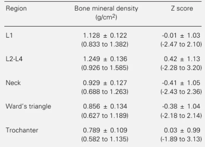
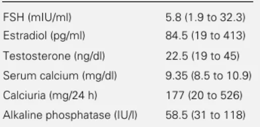
In the present study, women with primary ovarian insuf- ficiency had lower BMD than premenopausal women of similar age and a higher prevalence of osteopenia/osteo- porosis than
When considering renal replacement therapy (RRT) for obese patients, several studies show that in hemodialysis (HD), body mass index (BMI) is as- sociated solidly with a lower risk
Results: The automated analysis showed that, in comparison with the controls, the patients with PH showed lower 10th percentile values for lung density, higher vascular volumes in
The association of serum uric acid (SUA) with cardiovascular risk factors was gender-specific: in women, higher SUA was associated with increasing BMI, even after
tive factors independently associated with bone mass in lumbar spine were BMI, FEV1 (% predicted), fat mass, body weight, age of puberty, body cell mass in children and
O objetivo dessa pesquisa foi a realização de uma investigação acerca da evolução da comunicação e da tecnologia da informação sobre ela, e os impactos ambientais que
Bone mineral density was not influenced by anticonvulsant therapy, period of treatment, body mass index, serum levels of calcium, alkaline phosphatase, albumin and ionized
