Mariana Pracucio Gigliotti*, Guilherme Janson**, Sérgio Estelita Cavalcante Barros***, Kelly Chiqueto****, Marcos Roberto de Freitas*****
Influence of inter-root septum width on
mini-implant stability
Objective: The purpose of this study was to evaluate the influence of the inter-radicular septum width in the insertion site of self-drilling mini-implants on the stability degree of these anchorage devices. Methods: The sample consisted of 40 mini-implants insert-ed in the inter-radicular septum between maxillary second premolars and first molars in 21 patients to provide skeletal anchorage for anterior retraction. The post-surgical radiographs were used to measure the septum width in the insertion site (ISW). In this regard, the mini-implants were divided in two groups: group 1 (critical areas, ISW≤3 mm) and group 2 (non-critical areas, ISW>3 mm). The degree of mobility (DM) was monthly quantified to determine mini-implant stability, and the success rate of these devices was calculated. This study also evaluated the sensitivity degree during minis-crew load, amount of plaque around the minisminis-crew, insertion height, and total evalu-ation period. Results: The results showed no significant difference in mobility degree and success rate between groups 1 and 2. The total success rate found was 90% and no variable was associated with the miniscrew failure. Nevertheless, the results showed that greater patient sensitivity degree was associated to the mini-implant mobility and the failure of these anchorage devices happened in a short time after their insertion.
Conclusion: Septum width in the insertion site did not influence the self-drilling mini-implant stability evaluated in this study.
Abstract
Keywords: Orthodontic anchorage procedures. Dental implants. Dental radiography. Tooth root.
* MSc in Orthodontics, Bauru Dental School (FOB) - University of São Paulo (USP).
** Professor and Head, Department of Pediatric Dentistry, Orthodontics and Public Health, FOB-USP. Coordinator of the Applied Dental Sciences Pro-gram, FOB-USP. Member of the “Royal College of Dentists of Canada”.
*** Master, PhD and Postdoctoral in Orthodontics, FOB-USP. **** MSc and PhD in Orthodontics, FOB-USP.
intROduCtiOn
Mini-implants have become a routine anchor-age method in orthodontic practice given their high predictability and scientifically proven ben-efits.1 The success rate of these devices ranges
from 70% to 95%.2-7 On the other hand,
consid-erable failure rates have prompted several stud-ies that seek to determine the risk factors which may compromise mini-implant success.
According to the literature, location, angle of insertion, cortical bone thickness and bone tissue quality, presence of attached gingiva, mini-implant features, degree of primary stabil-ity and load intensstabil-ity,10,14 hygiene and degree
of peri-implant tissue inflammation3,5,11 are all
factors associated with mini-implant stability. Moreover, some recent studies have found that proximity and contact between mini-implant and tooth root are significant risk factors in the failure of this anchorage system.2,15,16,17
How-ever, contradictory results regarding the degree of influence of these various factors on the suc-cess rate of mini-implants are often found in the literature, given sample heterogeneity and the wide range of variables.
It is known that self-drilling mini-implants are the state-of-the-art device for orthodontic anchorage. Furthermore, studies have shown a larger contact area between the surface of mini-implants and bone tissue, thereby en-hancing stability.19
One should also consider that the surgical risk of injury to tooth roots adjacent to the mini-implant has been significantly reduced by ad-vances in this anchorage system, especially when three-dimensional surgical orientation guides are employed.19,20,21 Although risks inherent in the
insertion procedure have been reduced, thereby facilitating the placement of these mini-implants in areas with critical dimensions, few studies have evaluated whether mini-implants placed in close proximity to the periodontal ligament may have their stability compromised.2,15,16,17
This study, therefore, aimed to compare the stability and success rate of self-drilling mini-im-plants placed in interradicular septa with criti-cal and non-criticriti-cal mesiodistal dimensions, i.e., septa with width equal to 3 mm and greater than 3 mm, respectively.
MAtERiAL And MEtHOdS Material
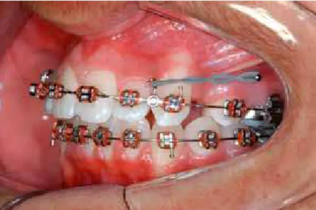
Twenty-one patients were selected (9 males, 12 females, mean age 16.99 ± 5.08 years) from the Clinic of Orthodontics, School of Dentist-ry of Bauru (FOB-USP) who were undergoing orthodontic treatment involving premolar ex-tractions and requiring maximum anchorage for anterior retraction. The selection criteria used in this study were: Mini-implants located in the in-terradicular septum, between second premolars and maxillary first molars, self-drilling mini-im-plants (length = 7 mm and diameter = 1.5 mm, Absoanchor, Dentos®), inserted by the same
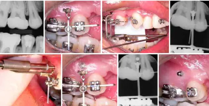
den-tist (Fig 1). The surgical protocol recommended and described by Barros20,21,22 was employed in
this study as it makes use of a three-dimensional graded radiographic-surgical guide (GRSG), al-lowing a perpendicular insertion path with rea-sonable predictability of final mini-implant posi-tioning (Fig 2).
However, some exclusion criteria were used: Absence of any local or systemic condition that could influence mini-implant stability, such as Active periodontal disease, smoking and diabetes, mini-implants placed in the mandible and with indications other than to provide anchorage for anterior retraction.
Thus, this study utilized 40 mini-implants, which were divided according to the width of the interradicular septum where they were inserted:
≤3.0 mm (group 1, critical areas) and >3.0 mm (group 2, non-critical areas).
Methods
Measurement of postoperative radiographs
Postoperative radiographs were obtained with a surgical radiographic positioner (SRP) in con-junction with GRSG and followed the technique of parallelism/bitewing radiographs (Fig 3). In these radiographs, the following variables were as-sessed: Width of the interradicular septum at the insertion site (ISW; Fig 4) and mini-implant inser-tion height (IH; Fig 5).
A Spectro II X-ray machine (Dabi Atlante, Ri-beirão Preto, Brazil) was used with 50 kVp volt-age, 10 mA current, and exposure time from 0.5 to 0.7 seconds. After processing, the radiographs were scanned in a 35 mm slide scanner (Sprint Scan 35 Plus, Version 2.7.2, Polaroid Corpora-tion) with 675 dpi resolution and 1:1 ratio. Sub-sequently, the images were measured using Adobe Photoshop software (version 7.0, Adobe Corpora-tion) and manipulated by the same examiner with an accuracy of 0.1 mm.21 High resolution
scan-ning allowed up to 300% magnification without any loss in quality.
Assessment of mini-implant stability
A
a
c b
d
B C
FIGURE 3 - Use of the SRP attached to the GRSG.
FIGURE 5 - Measurement of the insertion height of the mini-implant (IH): smallest distance between the alveolar bone crest and the center of the mini-implant.
FIGURE 4 - Measurement of the septum width on the insertion site (ISW): distance between the internal limits of the lamina dura of the dental roots adjacents to the mini-implant.
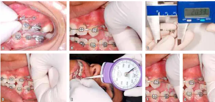
FIGURE 6 - A) Parts of the ATR: (a) winding lock for rod stabilization; (b) movable rod to quantify mobility; (c) concavity for mini-implant head attachment; (d) loop for force application with gauge. B) Open-ing on the windOpen-ing latch. C) ATR length reducal.
orthodontic tension gauge (Correx series 040-712-00, Dentaurum Orthodontics), with the aim of assigning numerical values to the degree of mo-bility of mini-implants.
Figure 7 illustrates the method used to evalu-ate the horizontal mobility of mini-implants, which will be described in the following steps:
» Step 1: First, it was necessary to define a random reference point such as the distal tie-wing of the canine bracket. ATR length was then adjusted according to the distance between the head of the mini-implant and the desired point.
To this end, part “c” of the ATR was then con-nected to the head of the mini-implant (Fig 7A), while the tip of part “b” touched the reference point (Fig 7B). The device was then locked with-in this dimension (distance from the mwith-ini-im- mini-im-plant to the distal tie-wing of the canine bracket) through the self-threading lock (a). This length was defined as the initial measurement and was measured with a digital caliper (Fig 7C).
A
D
B
E
C
F G
part “d” and pulled mesially to apply 400 g of force (Fig 7E). At this point, it was checked whether any movement occurred at the end of the device’s removable rod (part b) relative to the reference point.
» Step 3: To define the degree of mobility the amount of movement experienced by the end of the removable rod relative to the refer-ence point was evaluated (Figs 7F and 7G). If no movement occurred the mini-implant was considered stable, i.e., the difference between initial and final ATR measurements was zero. However, if mesial movement of the removable rod end (b) was observed during evaluation, the mini-implant was deemed to have mobility. Thus, the device had its length reduced (final measurement) to make sure that the end of the removable rod (b) would once again coincide with the reference point during force
applica-tion. After this procedure, the final measure-ment was taken with a digital caliper. Measure-ment of the degree of mobility was therefore considered representative of mini-implant sta-bility, and equal to the difference between the final and initial measurements of ATR length.
The average degree of mobility of each mini-implant was obtained by calculating the means of monthly measurements, and the success rate was defined by the number of mini-implants that re-mained clinically stable divided by the total num-ber of mini-implants evaluated in the study.
Evaluation of stability-related factors
Some factors that could affect mini-implant stability in this study were clinically evaluated: • Insertion site (IS): Divided between (1)
re-gion of attached gingiva, (2) rere-gion of alveo-lar mucosa or (3) mucogingival line.
• Degree of sensitivity (DS) measured month-ly during force application and anamonth-lyzed by means of scores: (0) When the patient re-ported no discomfort during force applica-tion, (1) when the patient reported slight dis-comfort, (2) when the patient reported pain, but it was bearable, and (3) when sensitivity was considered unbearable by the patient. • Evaluation of peri-implant biofilm using the
Modified Plaque Index (MPI) for dental im-plants since no specific index for mini-im-plants was found in the literature. This index uses a zero (0) score when there is no detect-able plaque, (1) when plaque is detected by sliding a probe, (2) when plaque is visible to the naked eye, and (3) when soft matter is abundant.
Statistical Analysis
Method error and compatibility between groups
Calculation of method error was performed for variables ISW and IH on 15 mini-implants distributed in both groups of this study. The formula proposed by Dahlberg24 (Se2=Σd2/2n)
and the paired t-test were used to perform the calculation of random and systematic errors, re-spectively.
As for the monthly assessment of degree of mobility (DM) using ATR, parameters were used to ensure that measurements were repro-ducible: ATR’s position relative to the reference point was checked by removing the device and putting it back in position. ATR positioning on the digital caliper had to be parallel to the cali-per ruler. The value obtained for ATR length was checked by repositioning the device on the caliper. ATR locking was checked prior to application of force by the tension gauge. The visual analysis of presence or absence of move-ment at the ATR’s removable rod end relative to the reference point was performed with utmost thoroughness.
T-test was used between groups 1 and 2 to check sample homogeneity for variables IH, MPI and OP.
Comparison between groups and variables
The Kolmogorov-Smirnov test was applied and showed absence of normal distribution for variables DM and DS, indicating the application of nonparametric tests for these variables and parametric tests for the others.
For the comparative analyses the following statistical tests were conducted:
• Descriptive statistics: Means, standard devia-tions, maximum and minimum value of vari-ables ISW, IH, DM, DS, MPI and OP.
• Nonparametric Mann-Whitney test: To com-pare groups 1 and 2 for differences in the de-gree of mobility of mini-implants.
• Fisher’s exact test: To check for any associa-tion between the success rates of mini-im-plants in groups 1 and 2.
• T-test and chi-square test: To compare all variables between the success and failure groups, and determine the risk factors associ-ated with mini-implant failures.
All statistical tests were performed with Sta-tistica software (Version 7.0, StatSoft Inc., Tulsa, OK, USA), adopting a significance level of p<0.05.
RESuLtS
Method error results yielded very small val-ues for random errors (0.0577 to 0.0912) and no significant systematic errors (Table 1). In addi-tion, both groups showed compatibility regard-ing insertion height, amount of plaque and mini-implant observation period (Table 2).
Table 3 shows the results yielded by descrip-tive statistics of the 40 mini-implants for vari-ables ISW, IH, DM, MPI, DS and OP. Table 4 shows a significant difference between groups 1 and 2 in terms of variable ISW.
similar (Tables 5 and 6).
An analysis of the risk factors involved in mini-implant stability in this investigation re-vealed that none of the variables could be
associ-ated with mini-implant failure. However, differ-ences were observed in degree of sensitivity and mini-implant observation period in the success and failure groups (Table 7).
Variables
1st measurement (n=15) 2nd measurement (n=15)
gl p Dahlberg
Mean SD Mean SD
Interradicular septum
width (ISW) 2.90 0.68 2.88 0.67 28 0.957 0.0632
Insertion height (IH) 3.05 0.69 3.02 0.75 28 0.901 0.0912
TABLE 1 - Results of paired t-test and Dahlberg’s formula24 as applied to variables ISW and IH for assessment of systematic and random errors,
respec-tively.
TABLE 2 - Compatibility between groups 1 and 2 concerning variables IH, MPI and OP (t-test).
Variables
Group 1 (≤3 mm)
Group 2
(>3 mm) p
Mean (SD) Mean (SD)
Insertion
height (mm) 3.13 (0.82) 3.08 (0.62) 0.83
Modified plaque
index (MPI) 1.52 (0.79) 1.52 (0.61) 0.25
Observation period (months)
8.55 (3.64) 9.90 (2.40) 0.17
TABLE 3 - Results yielded by descriptive statistics of the 40 mini-implants for variables ISW, IH, DM, MPI, DS and OP.
Variables N=40
Mean SD Min. Max.
Interradicular septum
width (ISW) 3.05 0.82 1.60 4.50
Insertion height (IH) 3.11 0.72 1.70 5.40
Degree of mobility (DM) 0.07 0.23 0.00 0.88
Modified plaque index (MPI) 1.52 0.70 0.00 2.62
Degree of sensitivity (DS) 0.23 0.73 0.00 3.00
Observation period (months) 9.22 3.12 1.00 12.00
TABLE 4 - T-test results between groups 1 and 2 for variable ISW.
*Statistically significant p<0.05.
Group N ISW p
Mean SD
Group 1 (ISW ≤ 3 mm) 20 2.38 0.44 0.00* Group 2 (ISW > 3 mm) 20 3.71 0.50 0.00*
TABLE 5 - Results of statistical analysis and Mann-Whitney test for de-gree of mobility between groups 1 and 2.
Group
Degree of mobility (DM)
p
n Mean SD Minimum Maximum
Group 1
(≤ 3 mm) 20 0.11 0.28 0.00 0.88 0.59
Group 2
(> 3 mm) 20 0.04 0.18 0.00 0.82 G1=G2
TABLE 6 - Results of Fisher’s exact test to assess success rate associa-tion between groups 1 and 2.
Group 1
(≤ 3 mm) (> 3 mm)Group 2 Total p Success 17 (42.5%) 19 (47.5%) 36 (90%) 0.30
Failure 3 (7.5%) 1 (2.5%) 4 (10%) 0.30
Total 20 20 40 0.30
TABLE 7 - Analysis of factors associated with mini-implant failure.
¤Student’s t-test, ¥Chi-square. *Statistically significant: p<0.05.
Variables Success
Mean (SD)
Failure
Mean (SD) p
Total 36 (90%) 4 (10%)
Insertion site (IS) 0.94¥
Attached Gingiva 16 (40%) 2 (5%)
Mucogingival line 12 (30%) 1 (2.5%)
Alveolar mucosa 8 (20%) 1 (2.5%)
Modified plaque index (MPI) 1.47 (0.72) 2.00 (0.00) 0.15¤
Degree of sensitivity (DS) 0.00 (0.00) 2.37 (0.47) 0.00*¤
Observation period (OP) 10.05 (1.94) 1.75 (0.50) 0.00*¤
Septum width at
diSCuSSiOn Sample
The sample utilized in this study (40 mini-implants) may be relatively small compared to previous studies that sought to determine risk factors for mini-implant success.1,3,5-8,11
In fact, sample size is a very important factor, but no less important are the selection criteria.26
Since this study followed strict selection criteria in determining its sample, the number of mini-implants was understandably reduced. Moreover, the results were influenced by a smaller number of uncontrolled variables, thereby contributing to the veracity of the inferences, which might not occur if mini-implants with different characteris-tics were compared, inserted and used under un-controlled conditions, which would excessively increase the number of variables involved in the stability of these anchorage devices.1,7
Sample selection included only mini-im-plants inserted with the aid of GRSG, since the use of surgical guides for mini-implant installa-tion in areas of interradicular septum is manda-tory to prevent injury to adjacent structures,20,27
and when guides are not used the success of this procedure depends almost exclusively on the skill and experience of the surgeon.
Factors age and gender were not standard-ized since several studies have shown that these characteristics are not directly associated with de-creased stability or mini-implant success rate.3-8,11
Results
Periapical/bitewing radiographs are often employed to evaluate the final positioning of mini-implants.2,7 Their accuracy, however,
de-pends on standardized implementation and/or the aid of radiographic and surgical guides.
An examination of postoperative radiographs showed that septum width at mini-implant in-sertion exhibited a mean value of 3.05±0.82 mm. It is noteworthy that the mini-implants were inserted in a more coronal region of the
septum (3.11 ± 0.72 mm, Table 3), which usu-ally has a shorter interradicular distance com-pared to a more apical region.28 The results of
this study were similar to those reported by Hernández et al28 and Hu et al29 but slightly
higher than those achieved by Deguchi et al8
and Poggio et al.9 It should be emphasized that,
unlike the methodology used in this study, the measuring method employed in these investiga-tions made use of computed tomography and cross-sections of human maxillas, which may explain the different findings.
In this study, septum width was defined as the distance between the inner boundaries of the lamina dura of the roots adjacent to the mini-implants, not including the periodontal ligament space, as it is not advisable to encroach upon it during mini-implant insertion. Howev-er, some studies use the cementum as bound-ary6,9,28 and include the periodontal ligament
space in their measurements, which inevitably yields values that are different from those at-tained in this study.
As yet, the literature has not reached a con-sensus on the minimum distance required be-tween mini-implants and tooth roots. Most stud-ies merely speculate on the ideal “safety margin,” but fail to show accurate values for such dis-tance. Only a small number of studies have ex-amined the proximity of mini-implants to tooth roots and the influence of such proximity on the stability of this anchorage system.2,6,16
Poggio et al9 and Schnelle et al30 recommend
Accurate assessment of mini-implant stability is usually reported only in animal experiments.19
Few authors report a more categorical assess-ment of mini-implant mobility in humans,14
and most of them only check for the presence or absence of mobility with clinical tweezers.5,11
Furthermore, this assessment is never evaluated monthly but only a few months after mini-im-plant installation.
The results of this study demonstrate that mini-implants in groups 1 and 2 had a similar degree of mobility (Table 5) and no association was observed between the success rate of mini-implants and septum width at the insertion site (Table 6). Corroborating these results, Motoyo-shi et al6 also found no differences when
evalu-ating a sample of standardized mini-implants. The accuracy of mini-implant insertion found in the present study—even in septa with criti-cal dimensions—was due to the use of GRSG. In the study by Motoyoshi et al6 no guide was used,
however, the authors achieved a certain degree of precision with their surgical technique using CT scans to define the ideal insertion height.
The total success rate found in the sample was 90% (Table 7), similar to the work of Mo-toyoshi et al,6 who also used Absoanchor®
mini-implants. An analysis of the factors associated with mini-implant failure showed that none of the variables assessed in this study has any bear-ing on the success rate of mini-implants.
The similarity found between the success rate of mini-implants placed in different inser-tion sites had no precedent in previous studies, in which the absence of keratinized mucosa signifi-cantly increased the risk of infection and mini-implant failure.11 However, all mini-implants in
this study were included in a more coronal re-gion of the septum, i.e., in a rere-gion of alveolar mucosa near the mucogingival junction. Since in the studies cited above the authors provide no information regarding mini-implant insertion height, one can speculate that in these samples
some mini-implants may have been placed in apical regions where the cortical bone is thin-ner and proximity to frenum areas facilitate the process of inflammation and tissue hyperplasia around the mini-implant.3 Additionally, since
few mini-implants were lost (4 mini-implants in the failure group), the odds of finding signifi-cant values to support comparison analyses and thereby determine risk factors with reliability are reduced.
Generally, studies only assess the quality of patient toothbrushing5,11 and/or the presence of
inflammation around the implant.3,5,11 The
re-sults showed no significant difference between the plaque index found in successful and failed mini-implants (Table 7). Inflammation around the mini-implant was not evaluated since it was hardly noticed during treatment given that patients were constantly instructed on oral hy-giene. This fact is reflected in the mean value of MPI (1.52±0.70, Table 7), which demonstrates that most of the mini-implants did not show abundant plaque.
During the monthly evaluation of the mini-implants it was noted that sensitivity was non-existent in devices with no mobility. However, degree of sensitivity increased significantly as mini-implants lost stability (Table 7). This sensi-tivity is probably related to compression of the surrounding soft tissues caused by the move-ment of mini-implants with a high degree of mobility since in general there is no such thing as spontaneous pain, but only pain caused by movement. Moreover, this traumatic condition favors peri-implant tissue inflammation, pro-gressively increasing sensitivity in the region.
Table 7 shows that evaluation of failed mini-implants lasted approximately 1.75 months, and successful mini-implants were assessed for al-most 10 months, which is consistent with the findings of Moon et al7 and Cheng et al.11 A
in most studies, mini-implant failure occurs al-most immediately after installation. This short-term failure is therefore directly related to the primary stability of mini-implants.10
Even when measured in isolation, ISW ap-peared not to be associated with failures in this an-chorage system (Table 7). It was observed that the widths of interradicular septa with critical dimen-sions are not considered a risk factor for the stabil-ity and success of self-drilling mini-implants. It is speculated that this lack of correlation between septum width and mini-implant success rate is directly linked to the use of three-dimensional ra-diographic-surgical guides, which enabled highly accurate and safe mini-implant insertion.21,22
Given the small number of studies using simi-lar methodologies—since it was only recently that a surge of interest in these risk factors began to
oc-cur—further studies are warranted if the relevant literature is to reach any lasting agreement.
COnCLuSiOnS
Based on the results attained for the study sample and in accordance with the methodology applied, it was concluded that:
1. No statistically significant difference was found in the degree of mobility and success rate of self-drilling mini-implants placed in septa with critical (≤3 mm) and noncritical (>3 mm) mesiodistal width.
Contact address Mariana Pracucio Gigliotti
Rua José Lúcio de Carvalho, 558 Centro CEP: 17.201-150 - Jaú / SP, Brazil E-mail: [email protected]
1. Janssen KI, Raghoebar GM, Vissink A, Sandham A. Skeletal anchorage in orthodontics: a review of various systems in animal and human studies. Int J Oral Maxillofac Implants. 2008 Jan-Feb;23(1):75-88.
2. Kuroda S, Yamada K, Deguchi T, Hashimoto T, Kyung HM, Takano-Yamamoto T. Root proximity is a major factor for screw failure in orthodontic anchorage. Am J Orthod Dentofacial Orthop. 2007 Apr;131(4 Suppl):S68-73.
3. Miyawaki S, Koyama I, Inoue M, Mishima K, Sugahara T, Takano-Yamamoto T. Factors associated with the stability of titanium screws placed in the posterior region for orthodontic anchorage. Am J Orthod Dentofacial Orthop. 2003 Oct;124(4):373-8.
4. Motoyoshi M, Hirabayashi M, Uemura M, Shimizu N. Recommended placement torque when tightening an orthodontic mini-implant. Clin Oral Implants Res. 2006 Feb;17(1):109-14.
5. Park HS, Jeong SH, Kwon OW. Factors affecting the clinical success of screw implants used as orthodontic anchorage. Am J Orthod Dentofacial Orthop. 2006 Jul;130(1):18-25. 6. Motoyoshi M, Yoshida T, Ono A, Shimizu N. Effect of cortical
bone thickness and implant placement torque on stability of orthodontic mini-implants. Int J Oral Maxillofac Implants. 2007 Sep-Oct;22(5):779-84.
7. Moon CH, Lee DG, Lee HS, Im JS, Baek SH. Factors associated with the success rate of orthodontic miniscrews placed in the upper and lower posterior buccal region. Angle Orthod. 2008 Jan;78(1):101-6.
8. Deguchi T, Nasu M, Murakami K, Yabuuchi T, Kamioka H, Takano-Yamamoto T. Quantitative evaluation of cortical bone thickness with computed tomographic scanning for orthodontic implants. Am J Orthod Dentofacial Orthop. 2006 Jun;129(6):721.e7-12.
9. Poggio PM, Incorvati C, Velo S, Carano A. “Safe zones”: a guide for miniscrew positioning in the maxillary and mandibular arch. Angle Orthod. 2006 Mar;76(2):191-7.
10. Wilmes B, Rademacher C, Olthoff G, Drescher D. Parameters affecting primary stability of orthodontic mini-implants. J Orofac Orthop. 2006 May;67(3):162-74.
11. Cheng SJ, Tseng IY, Lee JJ, Kok SH. A prospective study of the risk factors associated with failure of mini-implants used for orthodontic anchorage. Int J Oral Maxillofac Implants. 2004 Jan-Feb;19(1):100-6.
12. Wilmes B, Su YY, Drescher D. Insertion angle impact on primary stability of orthodontic mini-implants. Angle Orthod. 2008 Nov;78(6):1065-70.
13. Chun YS, Lim WH. Bone density at interradicular sites: implications for orthodontic mini-implant placement. Orthod Craniofac Res. 2009 Feb;12(1):25-32.
14. Liou EJ, Pai BC, Lin JC. Do miniscrews remain stationary under orthodontic forces? Am J Orthod Dentofacial Orthop. 2004 Jul;126(1):42-7.
15. Chen YH, Chang HH, Chen YJ, Lee D, Chiang HH, Yao CC. Root contact during insertion of miniscrews for orthodontic anchorage increases the failure rate: an animal study. Clin Oral Implants Res. 2008 Jan;19(1):99-106.
16. Asscherickx K, Van de Vannet B, Wehrbein H, Sabzevar MM. Success rate of miniscrews relative to their position to adjacent roots. Eur J Orthod. 2008 Aug;30(4):330-5.
17. Kang YG, Kim JY, Lee YJ, Chung KR, Park YG. Stability of mini-screws invading dental roots and their impact on the paradental tissues in Beagles. Angle Orthod. 2009 Mar;79(2):248-55.
18. Heidemann W, Terheyden H, Louis GK. Analysis of the osseous/ metal interface of drill free screws and self-tapping screws. J Maxillofac Surg. 2001 Apr;29(2):69-74.
19. Kim JW, Ahn SJ, Chang YI. Histomorphometric and mechanical analyses of the drill-free screw as orthodontic anchorage. Am J Orthod Dentofacial Orthop. 2005 Aug;128(2):190-4.
REfEREnCES
20. Barros SEC, Janson G, Chiqueto K, Freitas MR, Henriques JF, Pinzan A. A three-dimensional radiographic-surgical guide for mini-implant placement. J Clin Orthod. 2006 Sep;40(9):548-54.
21. Barros SEC, Janson G, Chiqueto K, Janson M, Freitas MR. Predictable drill-free screw positioning with a graduated 3-D radiographic-surgical guide: a preliminary report. Am J Orthod Dentofacial Orthop. 2009 Nov;136(5):722-35.
22. Barros SEC. Avaliação da precisão de um guia
radiográico-cirúrgico para inserção de mini-implantes [dissertação]. Bauru (SP): Universidade de São Paulo; 2008.
23. Mombelli A, Van Oosten MA, Schurch E Jr, Land NP. The microbiota associated with successful or failing osseointegrated titanium implants. Oral Microbiol Immunol. 1987 Dec;2(4):145-51.
24. Dahlberg G. Statistical methods for medical and biological students. New York: Interscience; 1940.
25. Houston WJ. The analysis of errors in orthodontic measurements. Am J Orthod. 1983 May;83(5):382-90. 26. Padovani CR. Estatística na metodologia da investigação
cientíica. Botucatu (SP): UNESP; 1995.
27. Kravitz ND, Kusnoto B. Risks and complications of orthodontic miniscrews. Am J Orthod Dentofacial Orthop. 2007 Apr;131(4 Suppl):S43-51.
28. Hernandez LC, Montoto G, Puente Rodriguez M, Galban L, Martinez V. Bone map for a safe placement of miniscrews generated by computed tomography. Clin Oral Implants Res. 2008 Jun;19(6):576-81.
29. Hu KS, Kang MK, Kim TW, Kim KH, Kim HJ. Relationships between dental roots and surrounding tissues for orthodontic miniscrew installation. Angle Orthod. 2009 Jan;79(1):37-45.
30. Schnelle MA, Beck FM, Jaynes RM, Huja SS. A radiographic evaluation of the availability of bone for placement of miniscrews. Angle Orthod. 2004 Dec;74(6):832-7.
Submitted: July 2009
* Access www.dentalpress.com.br/journal to read the full article.
Influence of inter-root septum width on
mini-implant stability
Mariana Pracucio Gigliotti**, Guilherme Janson***, Sérgio Estelita Cavalcante Barros****, Kelly Chiqueto*****, Marcos Roberto de Freitas******
Objective: The purpose of this study was to evaluate the influence of the inter-radicular septum width in the insertion site of self-drilling mini-implants on the stability degree of these anchorage devices. Methods: The sample consisted of 40 mini-implants insert-ed in the inter-radicular septum between maxillary second premolars and first molars in 21 patients to provide skeletal anchorage for anterior retraction. The post-surgical radiographs were used to measure the septum width in the insertion site (ISW). In this regard, the mini-implants were divided in two groups: group 1 (critical areas, ISW≤3 mm) and group 2 (non-critical areas, ISW>3 mm). The degree of mobility (DM) was monthly quantified to determine mini-implant stability, and the success rate of these devices was calculated. This study also evaluated the sensitivity degree during minis-crew load, amount of plaque around the minisminis-crew, insertion height, and total evalu-ation period. Results: The results showed no significant difference in mobility degree and success rate between groups 1 and 2. The total success rate found was 90% and no variable was associated with the miniscrew failure. Nevertheless, the results showed that greater patient sensitivity degree was associated to the mini-implant mobility and the failure of these anchorage devices happened in a short time after their insertion.
Conclusion: Septum width in the insertion site did not influence the self-drilling mini-implant stability evaluated in this study.
Abstract
Keywords: Orthodontic anchorage procedures. Dental implants. Dental radiography. Tooth root.
** MSc in Orthodontics, Bauru Dental School (FOB) - University of São Paulo (USP).
*** Professor and Head, Department of Pediatric Dentistry, Orthodontics and Public Health, FOB-USP. Coordinator of the Applied Dental Sciences Pro-gram, FOB-USP. Member of the “Royal College of Dentists of Canada”.
**** Master, PhD and Postdoctoral in Orthodontics, FOB-USP. ***** MSc and PhD in Orthodontics, FOB-USP.
Questions to the authors
1) How can orthodontists ensure that a mini-implant is successfully inserted in a region of narrow interdental bone septum?
Despite the high success rate of mini-implants, even when installed in narrow septa, and although the installation procedure is apparently simple, or-thodontists should strive to be as thorough as pos-sible since this procedure is extremely technique-sensitive. The keys to success when inserting mini-implants in critical areas are: Accurate diagnosis by means of standardized bitewing radiographs or CT scans so that selection of insertion site and mini-implant diameter are carefully defined, use of a three-dimensional surgical guide, particularly for orthodontists who are new to mini-implants and, finally, professionals should not underesti-mate any surgical technique detail as these are essential for success in the use of mini-implants.
2) Are the rates of accidents and complica-tions higher in regions of narrow bone sep-tum?
Yes. These insertion areas are considered criti-cal due to a higher rate of accidents and compli-cations since the chance of tooth root contact or perforation increases considerably. Damage to tooth roots is mainly due to incorrect determina-tion of the site and/or angle of inserdetermina-tion of the mini-implant in the bone tissue, and when faced with a narrow bone septum any deviation from this insertion angle, however small, can lead to contact between mini-implant and tooth root, and even to tooth loss. Besides, one must con-sider that close proximity of the mini-implant to the tooth root in narrow septa also renders more frequent the encroachment of periodontal liga-ment space during the insertion procedure, which may affect the stability of this anchorage device. Therefore, the use of surgical guides is mandatory
Editor´s summary
Mini-implants feature a considerable clinical failure rate due to early or late instability. Thus, research has been searching for the risk factors associated with failure in the stability of skeletal anchorage devices. This study aimed to compare the stability and success rate of self-tapping mini-implants placed in inter-radicular septa with criti-cal and non-criticriti-cal mesiodistal dimensions, i.e., septa with width equal to or smaller than 3 mm and greater than 3 mm, respectively.
Twenty-one patients were selected who were undergoing orthodontic treatment and needed anchorage for anterior retraction, totaling 40 mini-implants. The devices were inserted in the inter-radicular septum between maxillary second premolars and first molars. The sample was di-vided into two groups: Group 1 (critical areas) and group 2 (non-critical areas), and septum
width at the insertion site was measured on post-operative radiographs. Mini-implant stability was evaluated monthly by assessing the degree of mobility by means of a very specific and sensitive methodology.
for accurate insertion of mini-implants in criti-cal areas. Moreover, selection of mini-implant di-ameter in narrow septa should be thorough and take into account, when measuring septum width on bitewing radiographs or CT scan sections, the periodontal ligament space of adjacent tooth roots (approximately 0.25 mm each). As a result, the rates of accidents and complications in septa with critical width can be reduced.
3) Research in the area of mini-implants has intensified in recent years. What issues still need further clarification as regards mini-im-plant stability?
The number of scientific works involving orthodontic mini-implants is indeed experi-encing continuous growth. However, there are important methodological difficulties to be overcome by scientific studies that focus on this topic. Actually, the variables that influence mini-implant stability are numerous, and there-fore difficult to study in isolation because they involve issues related to the patient, the clini-cian and the mini-implant features. To further complicate matters, most of these studies are not prospective, and as a consequence samples are poorly standardized, with strict selection criteria,
and the fact that a large number of variables are included yields sharply conflicting results in the literature. Thus, studies are inconclusive or show widely divergent conclusions regarding the defi-nition of variables that determine the stability or loss of these anchorage devices. The number of histological studies in animals has been growing and as a result some important factors have been brought to light concerning the understanding of peri-implant bone remodeling, the presence of osseointegration and extension of the bone/metal contact surface, but small sample sizes preclude the extrapolation of results. Many findings, there-fore, are still mere speculation. It should also be noted that the results achieved in these animal studies cannot be fully extrapolated to humans because differences between these organisms do not reproduce the same biological events. In summary, the theme of “mini-implant stability” still comprises an untold number of issues to be addressed and explained. It is essential that further studies be conducted with well defined methodologies and purposes to progressively en-hance the understanding of variables that need to be controlled by clinicians if these devices are to provide excellent stability and success in orth-odontic treatment.
Contact address Mariana Pracucio Gigliotti