Post-thoracotomy pain relief with subpleural analgesia or
thoracic epidural analgesia: randomized clinical trial
Alívio da dor pós-toracotomia com analgesia subpleural ou analgesia epidural
torácica: ensaio clínico randomizado
Aysu Hayriye Tezcan
I, Özgür Karakurt
II, Mehmet Ali Eryazgan
III, Semih Başkan
I, Dilşen Hatice Örnek
IV, Ramazan Baldemir
V,
Bülent Koçer
VI, Mustafa Baydar
VIIAnesthesiology and Reanimation Department, Ankara Numune Education and Research Hospital, Ankara, Turkey
ABSTRACT
CONTEXT AND OBJECTIVE: Post-thoracotomy pain is a severe and intense pain caused by trauma to ribs, muscles and peripheral nerves. The current study aimed to compare subpleural analgesia (SPA) with thoracic epidural analgesia (TEA) in patients undergoing thoracotomy.
DESIGN AND SETTING: Randomized study at Ankara Numune Education and Research Hospital, in Turkey. METHODS: Thirty patients presenting American Society of Anesthesiologists physical status I-III were scheduled for elective diagnostic thoracotomy. The patients were randomized to receive either patient-controlled SPA or patient-patient-controlled TEA for post-thoracotomy pain control over a 24-hour period. The two groups received a mixture of 3 µg/ml fentanyl along with 0.05% bupivacaine solution through a patient-controlled analgesia pump. Rescue analgesia was administered intravenously, consisting of 100 mg tramadol in both groups. A visual analogue scale was used to assess pain at rest and during coughing over the course of 24 hours postoperatively.
RESULTS: In the SPA group, all the patients required rescue analgesia, and ive patients (33%) required rescue analgesia in the TEA group (P < 0.05). Patients who received subpleural analgesia exhibited higher visual analogue scores at rest and on coughing than patients who received thoracic epidural analgesia. None of the patients had any side-efects postoperatively, such as hypotension or respiratory depression. CONCLUSION: Thoracic epidural analgesia is superior to subpleural analgesia for relieving post-thoracotomy pain. We suggest that studies on efective drug dosages for providing subpleural analgesia are necessary.
RESUMO
CONTEXTO E OBJETIVO: Dor pós-toracotomia é severa e intensa, causada por trauma de nervos peri-féricos, músculos e costelas. O objetivo foi comparar analgesia subpleural (SPA) com analgesia epidural torácica (TEA) em pacientes submetidos à toracotomia.
TIPO DE ESTUDO E LOCAL: Estudo randomizado no Hospital Educação e Pesquisa de Numune, em An-cara, Turquia.
MÉTODOS: Trinta pacientes com o estado físico I-III da Sociedade Americana de Anestesiologistas foram agendados para toracotomia diagnóstica eletiva e escolhidos aleatoriamente para receber, num período de 24 horas, SPA ou TEA, ambas controladas pelo próprio paciente, para controle da dor pós-toracotomia. Os dois grupos receberam mistura de 3 ug/ml de fentanil com solução de 0,05% de bupivacaína por meio de bomba de analgesia controlada pelo paciente. Foi administrada analgesia de resgate por via intrave-nosa, com 100 mg de tramadol, nos dois grupos. No pós-operatório, a escala visual analógica foi utilizada para medir presença de dor durante a tosse ou em repouso, ao longo de 24 horas.
RESULTADOS: No grupo SPA, todos os pacientes necessitaram de analgesia de resgate. Cinco pacientes (33%) necessitaram de analgesia de resgate no grupo TEA (P < 0,05). Os pacientes com SPA exibiram pon-tuações superiores na escala visual analógica, em repouso e ao tossir, em comparação aos que receberam TEA. Nenhum dos pacientes teve quaisquer efeitos secundários no pós-operatório, como hipotensão ou depressão respiratória.
CONCLUSÃO: A analgesia peridural torácica é superior à analgesia subpleural no alívio da dor pós-tora-cotomia. Consideramos que estudos sobre a dosagem de drogas eicazes para proporcionar analgesia subpleural são necessários.
IMD. Attending Physician, Anesthesiology and
Reanimation Department, Ankara Numune Education and Research Hospital, Ankara, Turkey.
IIMD. Attending Physician, Thoracic Surgery
Department, Ankara Numune Education and Research Hospital, Ankara, Turkey.
IIIMD. Resident, Thoracic Surgery Department,
Ankara Numune Education and Research Hospital, Ankara, Turkey.
IVMD. Associate Professor, Anesthesiology and
Reanimation Department, Ankara Numune Education and Research Hospital, Ankara, Turkey.
VMD. Resident, Anesthesiology and Reanimation
Department, Ankara Numune Education and Research Hospital, Ankara, Turkey.
VIMD. Associate Professor, Thoracic Surgery
Department, Ankara Numune Education and Research Hospital, Ankara, Turkey.
VIIMD. Attending Physician, Head of Department,
Anesthesiology and Reanimation Department, Ankara Numune Education and Research Hospital, Ankara, Turkey.
KEY WORDS:
Pain, postoperative. Analgesia, epidural. Pleura.
Thoracotomy.
Analgesia, patient-controlled.
PALAVRAS-CHAVE:
Dor pós-operatória. Analgesia epidural. Pleura.
Toracotomia.
INTRODUCTION
Post-thoracotomy pain is a severe and intense pain caused by trauma to ribs, muscles and peripheral nerves. Efective postop-erative analgesia helps to reduce postoppostop-erative morbidity through early mobilization and rehabilitation and also reduces the devel-opment of chronic post-thoracotomy syndrome.1,2 Various
analge-sic techniques have been developed to treat postoperative thora-cotomy pain.1-6 horacic epidural analgesia is the gold standard not
only for pain relief ater thoracotomy, but also because of its many beneicial efects, such as reduction of intraoperative opioid require-ments, improvement of postoperative cardiopulmonary function and suppression of stress response.1-6 horacic epidural block is
usually performed percutaneously, with considerable failure rates. Unfortunately, it is contraindicated in patients who are using anti-coagulant or antiplatelet drugs.7 Intercostal nerve block, intrathecal
administration of opioids and interpleural analgesia have also been developed as alternative regional techniques for post-thoracotomy pain management.5 Many of these techniques are claimed to
pro-vide good pain control, but studies to ascertain the ideal technique are still ongoing.5 here have not been enough studies on the subject
of subpleural catheters for patient-controlled subpleural analgesia.
OBJECTIVE
he current study aimed to compare subpleural analgesia (SPA) with thoracic epidural analgesia (TEA) in patient-controlled analgesia devices for patients undergoing thoracotomy.
METHODS
his randomized clinical study included 30 patients with American Society of Anesthesiologists (ASA) physical status I-III, ranging in age from 20 to 70 years, for whom thoracotomy was planned. Approval for the study was granted by the institutional ethics board and writ-ten informed consent was obtained from all patients. Any patients with ASA status IV or greater, previous history of thoracotomy, use of chronic pain medication or contraindication against receiving local anesthetics or thoracic epidural block were excluded from the study. All the surgical procedures were performed by the same surgeons.
he patients were instructed how to use a patient-controlled analgesia (PCA) pump (Abbot Pain Management Provider, Abbott Laboratories, North Chicago, IL 60064, USA) and how to assess pain on a visual analogue scale (VAS), before their surgery. All the patients were pre-medicated with 1-2 mg of midazolam intravenously before surgery. he intraoperative monitoring included ECG, invasive arterial blood pressure, pulse oximetry, end-tidal carbon dioxide (EtCO2), end-tidal sevolurane concentration and serial arterial blood gas (ABG) analysis.
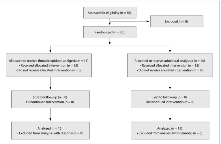
Patients were randomly assigned by means of the sealed envelope technique to either the thoracic epidural group (TEA group; n = 15) or the subpleural group (SPA group; n = 15). he patient inclusion and exclusion lowchart is described in
Figure 1. Allocation was organized by a member of the medical staf who was not included in the study.
Anesthesia comprising fentanyl (2 µg/kg) and propofol (2-2.5 mg/kg) was induced and tracheal intubation was facilitated using 0.6 mg/kg of rocuronium. To maintain anesthesia, the patients received sevolurane at 2% to 4% end-tidal concentration and 1 mcg/ kg/h of remifentanil infusion, intravenously. All the patients were ven-tilated with a 50% oxygen and 50% air mixture. Muscle relaxation was obtained by means of a 0.1 mg/kg rocuronium bolus. Ater surgery, the neuromuscular blockade was reversed and the trachea was extu-bated in the operating room. All the patients were then transferred to the post-anesthesia care unit, where they were observed for 24 hours. All patients in this unit received O2 via a face mask at 0.4 FiO2 and were nursed in a 30° head-up position.
In the thoracic epidural anesthesia group (TEA group; n = 15), before induction of anesthesia, an epidural catheter was inserted in the thoracic region between T4 and T6 by an anesthesiologist, to a depth of 3-5 cm into the epidural space. he catheter placement was conirmed using 3 ml of 2% lidocaine with 1:200,000 adrenaline. Heparin and low molecular weight heparin therapies were stopped at least 6 or 12 hours, respectively, before the catheter insertion.
In the subpleural analgesia group (SPA group; n = 15), before the surgical wound was closed, the parietal pleura was removed bluntly from the posterior chest wall towards the vertebral body through three intercostal spaces above the thoracotomy incision. An 18-gauge epi-dural catheter was advanced into the space at the level of the neck of the ribs and laid on the endothoracic fascia under direct viewing. he catheter was secured with 4-0 prolene sutures to maintain its position during lung expansion and it extruded through the chest wall.
Preoperative baseline variables (heart rate, mean arterial blood pressure (MAP), PaO2, PaCO2 and respiratory rate) were recorded for each patient. hese parameters together with anal-gesia and side efects (nausea/vomiting, pruritus, hypotension, respiratory depression and desaturation) were recorded in the post-anesthesia care unit at 0, 2, 8, 12 and 24 hours. Hypotension was deined as a drop in blood pressure of more than 25% of the baseline value. Respiratory depression was deined as respiratory rate of < 10/min. All the postoperative clinical outcome assessors and statistical analysis assessors were blinded. he member of the medical staf who monitored the PCA consumption, VAS scores and rescue analgesia requirements was blinded to the study.
Statistics
To detect a diference from 80% to 30% in the incidence of analgesic failure, with a one-tailed signiicance level of 5% (α = 0.05) and β of 0.2 (power 80%), a sample size of 15 patients was required in each group.
Demographic variables (age, weight and height) and duration of surgery were compared using Student’s t test. Categorical vari-ables were compared using the χ2 test. Pain scores, heart rate, mean
arterial blood pressure (MAP), PaO2 and PaCO2 at diferent time
intervals were compared using the Mann-Whitney U test. SPSS ver-sion 11.0 (SPSS Inc., Chicago, IL, USA) was used for all statistical analyses. P values < 0.05 were considered statistically signiicant.
RESULTS
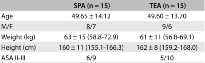
he demographic data from the two groups were similar with regard to age, height, weight, sex ratio and duration of surgery (Tables 1 and 2). he patients with patient-controlled subpleu-ral analgesia (SPA group) had higher visual analogue scale scores (VAS) at rest (Table 3) and on coughing scores (Table 4) at all measurement times than the patients with patient-controlled thoracic epidural analgesia (TEA group).
In the SPA group, all the patients required rescue analgesia using tramadol (100%). Five patients (33%) required rescue anal-gesia in the TEA group (P < 0.05). he mean dose of tramadol consumed as rescue analgesia postoperatively in the SPA group was 380 mg, compared with 120 mg in the TEA group (P = 0.002; Mann-Whitney U test).
he mean number of PCA boluses used was signiicantly lower in the TEA group: 7 in the TEA group versus 28 in the SPA group (P < 0.002; Mann-Whitney U test). he respiratory rate, heart rate, MAP, PaO2 and PaCO2 valueswere comparable
Figure 1. Patient inclusion and exclusion lowchart. Allocated to receive thoracic epidural analgesia (n = 15)
• Received allocated intervention (n = 15) • Did not receive allocated intervention (n = 0)
Lost to follow-up (n = 0) Discontinued intervention (n = 0)
Assessed for eligibility (n = 30)
Randomized (n = 30)
Analyzed (n = 15)
• Excluded from analysis (with reasons) (n = 0)
Allocated to receive subpleural analgesia (n = 15) • Received allocated intervention (n = 15) • Did not receive allocated intervention (n = 0)
Lost to follow-up (n = 0) Discontinued intervention (n = 0)
Analyzed (n = 15)
he mechanism of subpleural analgesia might be explained by the spread of local anesthetic to the posterior wall of the tho-rax, i.e. towards the vertebral column, and its difusion to the paravertebral space, which contains the thoracic spinal nerves.4,8
Kanai et al. reported that subpleural analgesia provided suc-cessfully adequate pain control in two-thirds of their patients, through continuous infusion of 0.125% bupivacaine at 8 ml/h. However, in the current study, in the SPA group, all the patients required rescue analgesia. he failure to provide adequate post-thoracotomy pain relief could be attributed to dislodgement of the subpleural catheter or inadequate and limited difusion of local anesthetic to the paravertebral space. he subpleural space is sep-arated from the paravertebral space by the endothoracic or extra-pleural fascia,4,9 and this barrier may prevent adequate difusion
of local anesthetic to the nerve endings.4,10 he deiciency of this
study might be the low doses of local anesthetic usage, but there were not enough data about the ideal local anesthetic dosage for this kind of subpleural block, especially using bupivacaine, which has a long elimination time and cardiac side efects. Because of the unpredictable cardiac side efects, the concentration of bupiva-caine used in this study was limited and fentanyl was added to the infusion solution to improve the analgesic quality.11-13
he most important advantages of patient-controlled epi-dural analgesia were the reduction of prolonged ventilation, reduction of re-intubation, improvements of pulmonary func-tions and early mobilization of the patient. he disadvantages of this technique were hypotension, urinary retention, pruritus and possible technical failure.2 Local anesthetic plus an opioid
combination in PCA is believed to provide synergistic analgesia, thus requiring smaller doses and fewer side efects.5 Epidurally
administered opioids produce segmental analgesia and improve the quality and duration of the sensory block produced by local anesthetics,14,15 which may explain the better pain relief in the
TEA group. In our clinical practice, we usually use an opioid and local anesthetic mixture for TEA solutions and, even at low con-centrations, adequate analgesia levels are obtained. As mentioned in relation to this current study, in clinical practice low doses of bupivacaine with TEA are suiciently eicient to deal with post-thoracotomy pain.
Kanazi et al.4 determined that the pain scores when
cough-ing were higher than at rest in all their patients and at all times, whether using TEA or SPA, and that VAS scores when cough-ing were always lower in the TEA group than in the SPA group. In that study, VAS scores at rest in the presence of thoracic epi-dural analgesia ranged from 1 cm to 6 cm. hose indings are similar to the indings of the current study (Tables 3 and 4).
here was no diference between the two groups of the cur-rent study in relation to the incidence of hypotension. However, Kanazi et al.4 reported that the incidence of hypotension was
Table 1. Demographic data and preoperative variables
Values are mean ± standard deviation (95% conidence interval) or number (P > 0.05). SPA = subpleural analgesia; TEA = thoracic epidural analgesia; M = male; F = female; ASA = American Society of Anesthesiologists.
SPA (n = 15) TEA (n = 15)
Age 49.65 ± 14.12 49.60 ± 13.70
M/F 8/7 9/6
Weight (kg) 63 ± 15 (58.8-72.9) 61 ± 11 (56.8-69.1) Height (cm) 160 ± 11 (155.1-166.3) 162 ± 8 (159.2-168.0)
ASA II-III 6/9 5/10
Table 2. Postoperative variables
Values are mean ± standard deviation (95% conidence interval) or number. SPA = subpleural analgesia; TEA = thoracic epidural analgesia; MAP = mean arterial pressure, H = heart rate, RR = respiratory rate; P > 0.05.
SPA (n = 15) TEA (n = 15)
HR (per minute) 91 ± 7 (89.8-96.2) 88 ± 8 (82.5-93.7) MAP (mmHg) 95.3 ± 10 (85.3-98.5) 90.1 ± 7(83.2-94.3) Duration of
surgery (h) 3.55 ± 0.93 (3.30-4.10) 3.95 ± 0.96 (3.28-4.15) PaO2 (mmHg) 105 ± 10 (98.6-105.2) 108 ± 15 (98.7-104.8)
PaCO2 (mmHg) 37 ± 4 (35.9-40) 37 ± 4 (36.8-39.4) RR (per minute) 18 ± 2 (16.5-19.5) 18 ± 2 (16.3-19.5)
Table 3. Visual analogue scale for pain at rest
Values are medians. SPA = subpleural analgesia; TEA = thoracic epidural analgesia.
Time (hour) SPA TEA P
0 7 (9-4) 3 (4-1) 0.001
2 6 ( 8-4) 3 (4-1) 0.001
8 4 (7-3) 2 (3-1) 0.001
12 4 (7-3) 2 (3-1) 0.001
24 4 (6-3) 2 (3-1) 0.001
Values are medians. SPA = subpleural analgesia; TEA = thoracic epidural analgesia.
Table 4. Visual analogue scale for pain during coughing
Time (hour) SPA TEA P
0 8 (10-5) 6 (7-3) 0.001
2 7 (10-5) 5 (6-2) 0.001
8 6 (9-4) 4 (6-2) 0.001
12 6 (9-4) 4 (6-2) 0.001
24 6 (9-4) 4 (6-2) 0.001
between the groups during the study period. None of the patients had hypotension or side efects. Oxygenation was satisfactory (PaO2 > 90 mmHg) in all the patients. None of the patients in either group showed respiratory depression (Table 2).
DISCUSSION
higher with thoracic epidural analgesia than with subpleural analgesia. his diference might be attributable to low concentra-tions of local anesthetic.
he strong point of the present study was that it showed that minimal bupivacaine doses were needed for efective thoracic epidural analgesia. he most important limitation of the study was the small sample size.
here are no published data identifying equipotent doses of bupivacaine for use in thoracic epidural and subpleural analge-sia. Previous studies have suggested that local anesthetic doses for TEA should be half those of subpleural analgesia.4,5 In the
current study, the starting bolus doses were given at a ratio of 1:2, but the PCA doses were lower than the doses used in the study by Kanazi et al. For supplemental therapy, paracetamol and diclofenac sodium were used in the current study. hese bupiva-caine doses in the TEA group were suicient for analgesia, and reduced the rate of complications. However, they were not sui-cient in the SPA group.
CONCLUSION
In conclusion, TEA is better than SPA for providing post-thora-cotomy pain relief. In order to avoid cardiac side efects of bupi-vacaine; the doses of the drug in the SPA group were limited and set as twice those of the TEA group. he local anesthetics and opioid doses in the TEA group in this study were safe and efec-tive, but were insuicient in the SPA group even with parenteral supportive analgesic therapy. Subpleural analgesia with this dose regimen is not recommended for clinical practice. Further stud-ies to determine local anesthetic doses and concentrations for this kind of subpleural analgesia are needed in order to achieve better analgesia for thoracic surgery.
REFERENCES
1. Gulbahar G, Kocer B, Muratli SN, et al. A comparison of epidural and
paravertebral catheterisation techniques in post-thoracotomy pain
management. Eur J Cardiothorac Surg. 2010;37(2):467-72.
2. Behera BK, Puri GD, Ghai B. Patient-controlled epidural analgesia with
fentanyl and bupivacaine provides better analgesia than intravenous
morphine patient-controlled analgesia for early thoracotomy pain.
J Postgrad Med. 2008;54(2):86-90.
3. Sentürk M, Ozcan PE, Talu GK, et al. The efects of three diferent
analgesia techniques on long-term postthoracotomy pain. Anesth
Analg. 2002;94(1):11-5.
4. Kanazi GE, Ayoub CM, Aouad M, et al. Subpleural block is less
efective than thoracic epidural analgesia for post-thoracotomy pain:
a randomised controlled study. Eur J Anaesthesiol. 2012;29(4):186-91.
5. Joshi GP, Bonnet F, Shah R, et al. A systematic review of randomized
trials evaluating regional techniques for postthoracotomy analgesia.
Anesth Analg. 2008;107(3):1026-40.
6. Bachmann-Mennenga B, Biscoping J, Kuhn DF, et al. Intercostal
nerve block, interpleural analgesia, thoracic epidural block or
systemic opioid application for pain relief after thoracotomy? Eur J
Cardiothorac Surg. 1993;7(1):12-8.
7. Helms O, Mariano J, Hentz JG, et al. Intra-operative paravertebral block
for postoperative analgesia in thoracotomy patients: a randomized,
double-blind, placebo-controlled study. Eur J Cardiothorac Surg.
2011;40(4):902-6.
8. Richardson J, Lönnqvist PA, Naja Z. Bilateral thoracic paravertebral
block: potential and practice. Br J Anaesth. 2011;106(2):164-71.
9. Karmakar MK. Thoracic paravertebral block. Anesthesiology.
2001;95(3):771-80.
10. McKenzie AG, Mathe S. Interpleural local anaesthesia: anatomical
basis for mechanism of action. Br J Anaesth. 1996;76(2):297-9.
11. Macias A, Monedero P, Adame M, et al. A randomized, double-blinded
comparison of thoracic epidural ropivacaine, ropivacaine/fentanyl, or
bupivacaine/fentanyl for postthoracotomy analgesia. Anesth Analg.
2002;95(5):1344-50.
12. Concha M, Dagnino J, Cariaga M, et al. Analgesia after thoracotomy:
epidural fentanyl/bupivacaine compared with intercostal nerve
block plus intravenous morphine. J Cardiothorac Vasc Anesth.
2004;18(3):322-6.
13. Baidya DK, Khanna P, Maitra S. Analgesic eicacy and safety of
thoracic paravertebral and epidural analgesia for thoracic surgery:
a systematic review and meta-analysis. Interact Cardiovasc Thorac
Surg. 2014;18(5):626-35.
14. Ginosar Y, Riley ET, Angst MS. The site of action of epidural fentanyl in
humans: the diference between infusion and bolus administration.
Anesth Analg. 2003;97(5):1428-38.
15. Kanai A, Osawa S, Suzuki A, et al. Regression of sensory and motor
blockade, and analgesia during continuous epidural infusion of
ropivacaine and fentanyl in comparison with other local anesthetics.
Pain Med. 2007;8(7):546-53.
Sources of funding: None Conlict of interest: None
Date of irst submission: March 2, 2015 Last received: May 12, 2015
Accepted: May 24, 2015
Address for correspondence: Dr. Aysu Hayriye Tezcan
Anesthesiology and Reanimation Department
Ankara Numune Education and Research Hospital,
Ulku District, Talatpasa Street no. 5
Altindag, Ankara 06110, Turkey
Tel. 090 05326735711