Improved model for the establishment and evaluation of
detrusor overactivity in female Wistar rats
_______________________________________________
Nian-zhao Zhang
1, Lin Ma
1, Jian-bo Zhang
2, Jun Chen
11Department of Urology, Qilu Hospital, Shandong University and 2Department of Pathology, Shandong
Tumor Hospital, Jinan, P.R. China
ABSTRACT
ARTICLE
INFO
______________________________________________________________ ______________________
Objective: To improve the model for establishment and evaluation of detrusor overac-tivity in female Wistar rats.
Materials and Methods: We ligated the perineal urethra of female Wistar rats and then performed filling cystometry. The probability of detrusor overactivity, bladder capacity, peak voiding pressure and histological changes were investigated.
Results: Detrusor overactivity ratio of the obstruction group was 32.4%. Bladder ca-pacity increased from 0.273 ± 0.036mL in control group to 0.89 ± 0.19mL in detrusor overactivity group (P < 0.001), and peak voiding pressure increased from 45.9 ± 4.1 cm.H2O to 63.5 ± 17.4cm.H2O (P = 0.007). For obstruction group, compared to no de-trusor overactivity rats, dede-trusor overactivity rats had higher bladder capacity (0.89 ± 0.19mL versus 0.43 ± 0.09mL, P < 0.001) and higher peak voiding pressure (63.5 ± 17.4cm.H2O versus 44.8 ± 6.2cm.H2O, P = 0.005). Detrusor overactivity rats were clas-sified according to peak voiding pressure (49.2 ± 4.2cm.H2O versus 80.8 ± 7.1cm.H2O, P < 0.001). Moreover, bladder weight increased significantly in detrusor overactivity rats (P = 0.003, P = 0.028) and detrusor histological hypertrophy was observed.
Conclusions: Ligating perineal urethra and filling cystometry with intra-urethral can-nula approach is a simple and easily reproducible method to establish and evaluate the model of detrusor overactivity in rats.
Key words:
Lower Urinary Tract Symptoms; Urinary Bladder; Urodynamics
Int Braz J Urol. 2014; 40: 414-22
_____________________
Submitted for publication: November 12, 2012
_____________________
Accepted after revision: September 18, 2013
INTRODUCTION
Detrusor overactivity (DO) is an urody-namic observation characterized by involunta-ry detrusor contractions during the filling phase (non-voiding contractions). These contractions, which may be spontaneous or provoked, produ-ce a wave form on the cystometrogram, of va-riable duration and amplitude. The contractions may be phasic or terminal (1,2). DO may occur in patients with bladder outlet obstruction (BOO), which can be caused by urethral strictures, con-genital malformations such as posterior urethral
The most common method to create a mo-del of DO rats involved reduction of the urethral diameter by placing a suture around the urethra (7), and significant increases were demonstrated in voiding frequency, voiding pressure and bladder capacity. Moreover, DO rats showed a pronounced “non-voiding contractions” during cystometry, and the categorization of different types of DO in conscious rats has been described (3). However, many disadvantages presented in the model of DO rats established in the previous studies. For exam-ple, DO rats were established by partial obstruc-tion with a method of transabdominal pathway, and the abdomen was opened through a midline incision (7). The architecture of cavitas pelvis was dissected, and the trauma might induce impair-ment of nerves and deformity of bladder which might influence the function of bladder. Therefo-re, in the study, we improved the method of esta-blishment and evaluation of DO model in female Wistar rats.
MATERIALS AND METHODS
Animals
A total of 50 female Wistar rats (Labora-tory Animal Center of Shandong University, Chi-na) with an initial weight of 192-204g were used in our study. The rats were housed individually on a 12h light/12h dark cycle at 22-24ºC in mesh--bottom cages with free access to food and wa-ter ad libitum. The rats were acclimatized to the facilities for 7 days, and all experimental proto-cols were approved by the Animal Research Ethics Committee of Shandong University.
Before the study, initial cystometric inves-tigation of the 50 rats was performed according to the method of section 2.4-cystometric investiga-tion below, and no DO rat was found (Figure-1a). Then, forty rats were used in an obstruction group (underwent obstruction surgical procedures) and 10 rats were used in a control group (without obs-truction surgical procedures), randomly.
Surgical procedures
In the study, the 40 rats of the obstruction group were anaesthetized with chloral hydrate (350mg/kg i.p.). The urethra was exposed and the
urinary bladder was catheterized with a human epidural catheter (F3, 1.0mm outer diameter) via the urethral orifice (Figures-2a and b). Care was taken to identify the urethra and vagina, and a needle with 4/0 silk suture was pierced around the perineal urethra (Figure-2c). A constant degree of partial obstruction was created by tying the silk ligature loosely around the urethra in the presence of the intraluminal indwelling catheter (Figure--2d), and then the epidural catheter was removed (Figure-2e). The rats were observed overnight in the recovery room, and then returned to normal conditions with food and water ad libitum. Six weeks later, the ligature around urethra was re-moved in the obstruction rats.
All of the obstruction rats were performed by one investigator. No prophylactic antibiotic was used after the surgical procedures. Moreover, in our preliminary experiment, we dissected an obstruction rat after surgical procedures for the demonstration to identify the location of ligature (Figure-2a,b and c).
Ligature removal
Two rats died within one week after the surgical procedures in the obstruction group, and acute retention was probably the cause of death. Moreover, the rats tried to remove the ligature, and the ligature ablated in 4 rats, which were excluded from the study. Therefore, the ligature was remo-ved in only 34 rats after 6 weeks of obstruction.
The rats were anesthetized with chloral hydrate (350mg/kg i.p.). The ligature around ure-thra (Figure-3a) was cut and carefully removed to make the stenosis disappeared and enable the transurethral cystometric investigation (Figure--2b). After the ligature around urethra was remo-ved, the rats were allowed to recover for two days. Then, cystometric investigation was performed in the 34 obstruction rats and the 10 control rats, and the probability of DO was analyzed.
Cystometric investigation
Figure 1 - a) the initial cystometric investigation of the 50 rats before our study (two voiding contractions were observed and the number indicated peak voiding pressure). b) the cystometric investigation of the 10 control rats 6 weeks later (one voiding contraction was observed and the number indicated peak voiding pressure). c) the cystometric investigation of the 6 detrusor overactivity rats 6 weeks later (one voiding contraction was observed and the number indicated peak voiding pressure, the low peak voiding pressure group). d) the cystometric investigation of the 5 detrusor overactivity rats 6 weeks later (one voiding contraction was observed and the number indicated peak voiding pressure, the high peak voiding pressure group).
A
B
C
D
human epidural catheter (F2, 0.7mm outer diame-ter, 0.4mm internal diameter), which was connec-ted via a T-tube to urodynamic testing machine (Laborie medical technologies, Corp) and infusion pump (LION WZ-50C6 microinfusion pump, Zhe-jiang University, China).
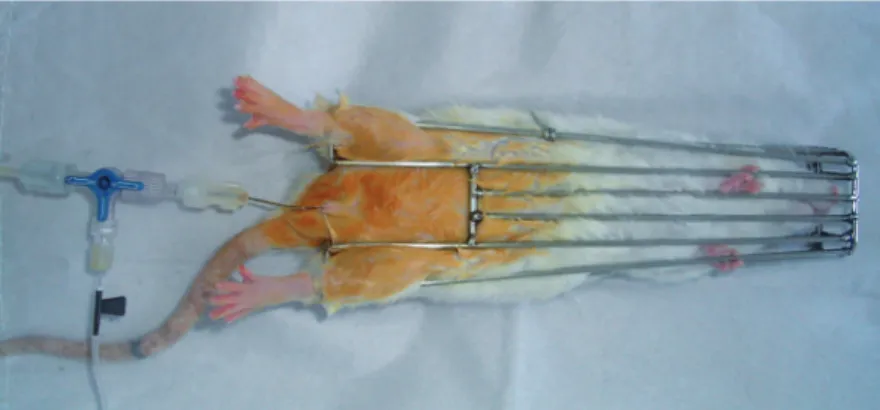
The rat was placed supine and the ure-thral orifice could be observed clearly. Data were collected and analyzed after the animals were seen to be resting quietly in the restraining devi-ce (Figure-4). The cystometric investigation was performed by infusing warm saline (37-38ºC) at a rate of 9mL/h, and the infusion was stopped
Figure 2 - a, b, c, d and e: the surgical procedures for obstruction rats. A, B and C: the dissection of an obstruction rat to identify the location of ligature.
Figure 3 - a) the ligature of obstruction rats 6 weeks later. b) the intra-urethral cannula introduction was performed after the ligature removal.
Figure 4 - The restraining device we designed which enabled cystometric investigation.
A B
A
E
B
F
C
G
D
During the filling phase, some of the obs-truction rats had obvious non-voiding contrac-tions before the onset of voiding contraction and thus were defined as having DO and classified as the DO group (8-10).
Data analysis
The Statistical Package for Social Scien-ces was used to handle the database. To determi-ne the relationship between a categorical variable with two levels and normally or non-normally distributed quantitative variables, Student’s t-test or Mann-Whitney U tests were applied. The two--sided P < 0.05 was considered to indicate statis-tically significant differences.
RESULTS
Cystometric investigation was performed in the 34 obstruction rats and 10 control rats. DO ra-tio of the obstrucra-tion rats was 32.4% (11 out of 34) (Figure-1c and d), and the control rats had no DO (0 out of 10) (Figure-1b). Compared to the control rats, the bladder capacity and peak voiding pressure increased significantly in the DO rats.
The bladder capacity increased from 0.273 ± 0.036mL in the control rats to 0.89 ± 0.19mL in the
DO rats (P < 0.001; Table-1). The peak voiding pres-sure increased from 45.9 ± 4.1 cm.H2O in the control rats to 63.5 ± 17.4 cm.H2O in the DO rats (P = 0.007; Table-1). For the obstruction group, compared to the non-DO rats, the DO rats had higher bladder capaci-ty (0.89 ± 0.19mL versus 0.43 ± 0.09mL, P < 0.001; Table-1) and higher peak voiding pressure (63.5 ± 17.4cm.H2O versus 44.8 ± 6.2cm.H2O, P = 0.005; Ta-ble-1). Moreover, the DO rats were classified into the low peak voiding pressure group (Figure-1c) and the high peak voiding pressure group (Figure-1d) (49.2 ± 4.2cm.H2O in group 1 versus 80.8 ± 7.1cm.H2O in group 2, P < 0.001; Table-1).
Furthermore, we studied the body mass and bladder weight in the 10 control and 34 obstruc-tion rats 6 weeks later. The bladder weight was the highest in the DO rats, with significant differences between the DO rats and the control rats and non--DO rats (P = 0.003, P = 0.028; Table-2). We harves-ted bladder of the control rats and the DO rats for subsequent histological assessment. Histologically, compared to the control rats, the DO rats presen-ted an increased detrusor muscle cell mass due to hypertrophy (Figures-5 a and b). On the other hand, compared to the control rats, the DO rats detrusor muscle cell gap widened with abundant collagen fiber (Figure-6 a and b).
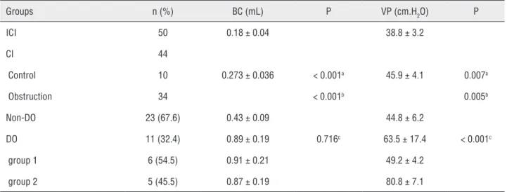
Table 1 - Comparison of cystometric investigation in the control and obstruction rats (Mean ± SD).
Groups n (%) BC (mL) P VP (cm.H2O) P
ICI 50 0.18 ± 0.04 38.8 ± 3.2
CI 44
Control 10 0.273 ± 0.036 < 0.001a 45.9 ± 4.1 0.007a
Obstruction 34 < 0.001b 0.005b
Non-DO 23 (67.6) 0.43 ± 0.09 44.8 ± 6.2
DO 11 (32.4) 0.89 ± 0.19 0.716c 63.5 ± 17.4 < 0.001c
group 1 6 (54.5) 0.91 ± 0.21 49.2 ± 4.2
group 2 5 (45.5) 0.87 ± 0.19 80.8 ± 7.1
a: DO versus control; b: DO versus Non-DO; c: group 1 versus group 2
Figure 5 - The frozen section of detrusor muscle cell (H&E staining, x200). a) control group; b) detrusor overactivity group.
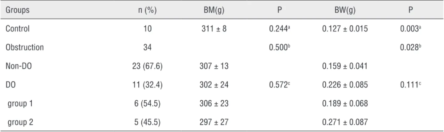
Figure 6 - The frozen section of collagen fiber (H&E staining, x200). a) control group; b) detrusor overactivity group. Table 2 - Comparison of body mass and bladder weight in the control and obstruction rats 6 weeks later (Mean ± SD).
Groups n (%) BM(g) P BW(g) P
Control 10 311 ± 8 0.244a 0.127 ± 0.015 0.003a
Obstruction 34 0.500b 0.028b
Non-DO 23 (67.6) 307 ± 13 0.159 ± 0.041
DO 11 (32.4) 302 ± 24 0.572c 0.226 ± 0.085 0.111c
group 1 6 (54.5) 306 ± 23 0.189 ± 0.068
group 2 5 (45.5) 297 ± 27 0.271 ± 0.087
a: DO versus control; b: DO versus Non-DO; c: group 1 versus group 2
BM = Body mass; BW = Bladder weight; DO= Detrusor overactivity
A B
DISCUSSION
We established an improved model of DO in female Wistar rats. Our results confirmed that the obstruction-related DO rats presented altered voiding patterns. Non-voiding contractions oc-curring prior to voiding were observed in the DO rats. Following voiding, no involuntary detrusor contraction was recorded but the involuntary de-trusor contractions developed slowly with increa-sing amplitude until the next voiding occurred.
In the previous study (3), four types of voiding pattern were described in the DO rats with trans-abdominal approach, and the incidence rate was 54% (type I, non-voiding contractions occur-ring before and after micturition), 26% (type II, non-voiding contractions occurring prior to mic-turition), 13% (type III, similar to that of control) and 7% (type IV, non-voiding contractions asso-ciated with dribbling and no micturition occur), respectively. However, only one type of DO pat-tern was observed in our study and the bladder ca-pacity was low. The differences between our study and the previous study may be due to the different method in establishing and evaluating DO rats.
Interestingly, the DO pattern observed in our study was consistent with one of the patterns (type II) described in the previous study (3) with approximate incidence rate (32.4% versus 26%) and was characterized by an increased involun-tary detrusor contractions during the filling pha-se, which was in agreement with findings in men showing DO associated with an increase in voi-ding pressure or men with prostatic obstruction (11). It was conjectured that the other three types of pattern (type I, type III and type IV) may be induced by the impairment of anatomic structure in the previous study (3). Therefore, the DO model established with our method was more consistent with the natural history of obstruction-related DO.
We summarized the characteristics of the improved DO model as follows:
1. The method was simple and convenient. We used a needle with silk suture and tied perineal urethra to establish obs-truction-related DO model. Moreover, the survival rate after surgical procedu-res was satisfactory (38/40, 95.0%).
2. The method did not need a low ab-dominal incision. The impairment of bladder and its innervation and peri--urethral blood vessel was avoided. Therefore, compared to the previous studies (3,7), the influence of injury on bladder, which might be the etiology of DO, was excluded.
3. Cystometric investigation was perfor-med without anesthesia after ligature removal in obstruction rats, and blad-der was catheterized through urethra. No other operation was performed. The wound and cicatricle was reduced, and the equality and stabilization of obstruction was improved. Moreover, compared to the bladder catheter im-plantation (3,7), cystometric investiga-tion was performed through urethra in the improved model, which was consis-tent with the clinical examination for patients. In the study, although cys-tometric investigation was performed through urethra by catheter, no obs-truction was observed during voiding. 4. The improved model had no
impair-ment of anatomic structure and had satisfactory adaptive capacity to envi-ronment. Therefore, the model can be fed and transported easily, and bulk production is feasible.
5. It was an easily reproducible method for producing obstruction-related DO model in female Wistar rats, which could provide a continuum of tissue and urodynamic data that could be used to further study the pathophysio-logic changes underlying DO. Moreo-ver, the improved model was poten-tially suitable for further evaluation of mechanisms involved in the develop-ment of DO and the responses to phar-macological treatment.
of DO shown in the study was lower than the pre-vious study (3). Moreover, the movement of rats interfered with cystometric investigation when they showed the sign of discomfort or distress, though the interference was not frequent.
In conclusion, we studied an improved model for the establishment and evaluation of DO in female Wistar rats. Ligating perineal urethra and filling cystometry with intra-urethral cannula approach is a simple and easily reproducible me-thod to establish and evaluate the model of DO rats, which can provide a continuum of tissue and urodynamic data that can accurately reproduce the urodynamic changes observed in patients with overactive bladder and may be used to study the pathophysiologic changes underlying DO.
ACKNOWLEDGMENTS
Our study was supported by the National Natural Science Foundation of China (Grant num-ber: 30872565). Our study was supported by the Autonomic Innovative Foundation of Shandong University (Grant number: 2012TS146).
CONFLICT OF INTEREST
None declared.
REFERENCES
1. Abrams P, Cardozo L, Fall M, Griffiths D, Rosier P, Ulmsten U, et al.: The standardisation of terminology of lower urinary tract function: report from the Standardisation Sub-committee of the International Continence Society. Neurourol Urodyn. 2002; 21: 167-78.
2. Haylen BT, de Ridder D, Freeman RM, Swift SE, Berghmans B, Lee J, et al.: An International Urogynecological Association (IUGA)/International Continence Society (ICS) joint report on the terminology for female pelvic floor dysfunction. Neurourol Urodyn. 2010; 29: 4-20.
3. Lluel P, Duquenne C, Martin D: Experimental bladder instability following bladder outlet obstruction in the female rat. J Urol. 1998; 160: 2253-7.
4. Brading AF: A myogenic basis for the overactive bladder. Urology. 1997; 50: 57-67; discussion 68-73.
5. Oh MM, Choi H, Park MG, Kang SH, Cheon J, Bae JH, et al.: Is there a correlation between the presence of idiopathic detrusor overactivity and the degree of bladder outlet obstruction? Urology. 2011; 77: 167-70.
6. Oelke M, Baard J, Wijkstra H, de la Rosette JJ, Jonas U, Höfner K: Age and bladder outlet obstruction are independently associated with detrusor overactivity in patients with benign prostatic hyperplasia. Eur Urol. 2008; 54: 419-26.
7. Malmgren A, Sjögren C, Uvelius B, Mattiasson A, Andersson KE, Andersson PO: Cystometrical evaluation of bladder instability in rats with infravesical outflow obstruction. J Urol. 1987; 137: 1291-4.
8. Elzayat E, Khaled S, Kashiwabara T, Elhilali M, Corcos J: Effect of the potassium channel opener WAY-133537 on the overactive bladder of spinalized rats. Neurourol Urodyn. 2006; 25: 808-14.
9. Abrams P: Describing bladder storage function: overactive bladder syndrome and detrusor overactivity. Urology. 2003; 62: 28-37; discussion 40-2.
10. Dion SB, Zvara P, Tu LM, Richer M, Corcos J: Evaluation of the role of neurolinins and urecholine hypersensitivity in an animal model of infravesical outflow obstruction. Urology. 1998; 52: 909-14.
11. Cucchi A, Achilli MP, Ravasi S, Arrigoni N: Detrusor instability as an energy-saving device in prostatic obstruction. J Urol. 1997; 157: 866-70.
_______________________ Correspondence address:
EDITORIAL COMMENT
I am particularly happy to see a manus-cript that attempts to create an experimental mo-del for the study of overactive bladder. We know that this disease still lacks adequate animal models and previously published models have potential risk of injury to the bladder nerves of experimen-tal animals adopting a transabdominal approach.
As the authors mentioned, the movement of rats interfered with cystometric investigation when they showed the sign of discomfort or
dis-tress, and that can hence loss results reliability. Another fact of concer is the low incidence rate of DO shown in the study.
I believe, however, that even seeming a simple model, only time will tell if this method is easily reproducible in other research centers.