881
Arq Neuropsiquiatr 2008;66(4):881-884 Clinical / Scientiic note
Tumour-like ChagaSiC enCephaliTiS
in aiDS paTienTS
An atypical presentation in one of them and
outcome in a small series of cases
Roberto E.P. Sica
1, Gisella Gargiullo
2, Cristina Papayanis
3enCefaliTiS ChagáSiCa pSeuDoTumoral en paCienTeS Con SiDa: preSenTaCión aTípiCa en uno De elloS e hiSToria De la enfer-meDaD en una pequeña Serie De CaSoS
División Neurología, Hospital Ramos Mejía, Facultad de Medicina, Universidad de Buenos Aires, Buenos Aires, Argentina: 1Emeritus Profesor, Buenos Aires University, School of Medicine, Buenos Aires University; 2Schollar, Consejo Nacional de Ciencia y Técnica (CONICET); 3Schollar, School of Medi-cine, Buenos Aires University. Funding: This investigation has been funded by the Goverment of the City of Buenos Aires.
Received 22 August 2008, received in inal form 29 September 2008. Accepted 10 October 2008.
Dr. Roberto E.P. Sica – Secretaría de Ciencia y Técnica / Facultad de Medicina / Universidad de Buenos Aires - Paraguay 2155 - 1121 Buenos Aires, Argentina. E-mail: [email protected]
Chagas’ disease is an intracellular parasitic infection
owed to a protozoarium, the
Trypanosoma
cruzi
1,
affect-ing a large population in Latinamerica. Within the region
15 to 16 million people are infected
2.
The worldwide pandemia, due to the infection of the
HIV 1 virus, also affects Latinamerican countries. The
num-ber of patients with this condition in Central and South
Americas amounts to 1.6 million persons
3,4. Therefore,
both illnesses overlap in a broad geographical area and
may coincide in the same patient.
The HIV infection, which causes the AIDS syndrome,
impairs the immunological system and predisposes to the
appearance of opportunistic infections, which may have
been hosted unnoticed by the patient until then.
There-fore, Chagas’ disease, which is a dormant infection in most
patients
5, may reactivate if the immunological surveillance
wanes off as the consequence of the viral insult. Along
the last years we
6,7and others
8-10found patients aflicted
by AIDS, who developed brain lesions yielded by the
Try-panosoma cruzi
.
The present communication describes three further
patients with this condition; one of them is unique
be-cause his clinical, radiological and immunological indings
differ from those previously reported in the literature.
CaSeS
Patient 1
A 28-years-old male. Ten days before admission, he noted progressive weakness on the left side of his body followed by seizures on the same side which started on his upper limb and spread to the left lower limb, lasting 30–40 seconds. The sei-zures appeared 3 days before admission with a frequency of
one per day. Simultaneously, he developed a throbbing bilater-al headache. He bilater-also referred a weight loss of about 20 Kg bilater-along the last four months.
On clinical examination he presented a left-sided hemipare-sis with hyperactive relexes. Ordinary laboratory tests were un-remarkable, except for a very low count of CD4+ lymphocytes, which amounted to 70 cells/mm3. Serum ELISA and Western Blot were positive for HIV infection and an indirect immunolu-orescence assay (IIA) for Toxoplasmosis was positive as well. Se-rological tests for Chagas’ disease (IIA, ELISA) and a search for trypomastigotes in blood were negative.
Arq Neuropsiquiatr 2008;66(4)
882
Chagas’ disease: AIDS Sica et al.
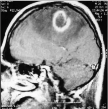
A brain magnetic resonance (MRI) disclosed a rounded im-age located at the parietal lobe, which was surrounded by dig-ital-like oedema pushing aside the lateral ventricle; when gad-olinium was administered an enhanced ring, encompassing the whole lesion, appeared (Fig 1).
Assuming that the lesion was due to toxoplasmosis infection the patient was put on pyrimethamine 100 mg/day, clindamycin 600 mg/qid and leucovorin 15 mg/day. By day 12th after treat-ment, no improvement of his clinical condition was observed; therefore, it was decided to obtain a biopsy of his brain lesion. Histological examination of the specimen showed necrotic tis-sue and macrophages illed with amastigotes.
The diagnosis of Chagas’ disease was done and treatment with Benznidazol, 5 mg/kg/day, and antiretroviral therapy were started. The patient continued to deteriorate and died 50 days after the beginning of the therapy. Autopsy was not permitted.
Patient 2
A 51-years-old male. He came to the clinical ofice complain-ing of language disturbances, visual defects and weakness on the right side of his body. All the symptoms appeared about ten to ifteen days before his assessment and were slowly progressive.
On clinical examination he showed global aphasia, right hemianopia, right mild hemiparesis and spasticity.
Regular laboratory tests disclosed a low number of CD4+ lymphocytes amounting to 46 cells/mm3. Serum ELISA and Western Blot for HIV infection were positive. Serum tests for Toxoplasmosis and Chagas’ disease were positive as well. How-ever, trypomastigotes within the blood were not found.
MRI showed several, spontaneouly enhanced rounded lesions on T1-weighted imaging, probably due to blood in their cores,
distributed along the whole brain, mainly at the boundery be-tween the cortex and the white matter, surrounded by oedema. The assumption of toxoplasmosis was done and appropri-ate treatment was started. At the 4th day after treatment the pa-tient deteriorated, his weakness became worst and partial motor seizures appeared. Biopsy of one of the lesions showed macro-phages illed with amastigotes. Finally, he developed an epilep-tic status and died as a consequence of sepsis. Before dying, a cerebrospinal luid (CSF) sample was obtained, where trypomas-tigotes were found as well.
Patient 3
A 35-years-old male, HIV positive, who had not received an-tiretroviral therapy. He was admitted into the ward because of a sudden weakness on the left side of his body, which had start-ed ten days before his assessment.
Neurological examination disclosed left-sided weakness and mild sensory loss.
Laboratory tests were unremarkable. A computed tomog-raphy (CT) of his brain showed a hemorrhagic lesion of 50 × 40
×26 mm in volume, located at his right parietal lobe. A thor-ough cardiological examination and clotting blood tests were normal.
The patient partially recovered and was discharged with a minimal hemiparesis.
Two months later, he was readmitted because his weakness had became progressively worst along the last 2 weeks, and report-ed a few partial motor seizures and a moderate daily headache. On clinical examination he showed a mild motor deicit and sensory impairment on his left side. Laboratory tests disclosed 551 CD4+ lymphocytes/mm3 and positive serological tests for
Arq Neuropsiquiatr 2008;66(4)
883
Chagas’ disease: AIDS Sica et al.
Toxoplasmosis and Chagas’ disease. A irst blood sample search-ing for parasitemia was negative.
MRI showed that the original hemorrhagic lesion was small-er in size, but a new ring-enhancing lesion, of about 1 cm in di-ameter, appeared at its edge.
Treatment for toxoplasmosis was started; 7 days later the weakness became worst and a new TC disclosed another annu-lar lesion located at the right basal ganglia.
A stereotactic biopsy of the parietal nodular lesion was per-formed; histological examination of the sample showed necrot-ic tissue and macrophages illed with amastygotes (Fig 2). At this time, a new blood examination was able to detect trypo-mastigotes.
The patient was put on Benznidazol 5 mg/kg/day, and anti-retroviral drugs (d4T, 3TC and Nelinavir). He was discharged and had a progressive but sustained improvement. MRI, ive months later, revealed that all lesions had decreased in size. On follow up, 14 months after diagnosis, he showed mild motor sequelae and negative parasitemia. He is still on Benznidazol treatment.
DiSCuSSion
The possibility that a dormant
T. cruzi
infection
reac-tivates in a patient who, simultaneously, is affected by an
HIV infection is quite reasonable knowing that the
para-site is intracellular and becomes the guest of many cells
after its innoculation in the acute period of the disease. It
is noteworthy that the parasites are able to reach the
cen-tral nervous system at the time of their irst contact with
its host
11,12and, thence, most probably, lodge in neurones
and/or glia cells
13,14. In normal conditions, the surveillance
of the immune system takes control of them, but if for
any reason the immune system fails, they will be allowed
to reproduce and leave the host cell
5-9. This sort of
behav-iour has been noted in patients whose immune system has
been weakened by the employment of citotoxic drugs
14,15.
In a previous communication
7we described 10 patients
who had Chagas’ disease and were asymptomatic until
they were infected by the HIV virus. All of them
devel-oped cerebral tumour-like (pseudotumours) lesions when
they had less than 126 CD4+ cells/mm
3.
The present communication describes other three
cases. Two of them had quite a similar behaviour than
those already reported. However, the third patient was
unique regarding his clinical presentation, the type of
le-sions found, the number of CD4+ cells and his response
to treatment.
In regards to the irst two patients, they behaved as
most of the other patients described in the literature
6,7,12,16,17with a very poor outcome in spite of speciic treatment,
and like most of them, died shortly after diagnosis.
Opposite to this behaviour was the third patient. He
disclosed several differences with the others. First, his
clin-ical presentation was a lobar hemorrhagic lesion or,
per-haps, a hemorrhagic infarct, which could not be explained
by any circulatory or blood abnormality. It is worthy to
note that it has been claimed that in chagasic patients
brain trombo-embolism may occur in the absence of
car-diac or arterial damage. Whatever the causes of the
vascu-lar event were, after 2 months of the initial manifestation,
a MRI image suggesting a pseudotumour, was detected.
We do not know how the former lesion may be
re-lated to the later. The only inding that suggests a
con-nection between them is the fact that they were in close
proximity, contacting by their edges; one might speculate
that the parasites, held in the blood of the hemorrhage,
invaded part of the surronding tissues, later developing
the pseudotumour.
The appearance of a third lesion, in the basal ganglia,
strongly suggested an opportunistic disease and provided
the rationale for the biopsy procedure, which made the
di-agnosis of Chagas’ disease. The third difference is the
num-ber of CD4+ cells, being normal in this patient and
suggest-ing a healthy state of his immune system. We do not have
a clear explanation for this, either. Perhaps some type of
impairment of the immune mechanisms, not relected by
the number of CD4+ cells, underlies this behaviour. Finally,
he is the only one who showed an acceptable response
to the speciic treatment, which is currently sustained.
Whether this favourable outcome is connected to the
nor-mal CD4+ cells count or not, is a yet unresolved question.
The cases described here, conirm that dormant
Cha-gas’ disease can reactivate in HIV patients in the form of
brain pseudotumours. They also underline that these
le-sions can be the irst opportunistic infection to appear.
Therefore, Chagas’ disease should be considered an
AIDS-deining illness in Latin America; already some countries,
Brazil and Argentina amongst them, have adopted this
conception. Finally, it seems possible that this disease may
appear in AIDS patients with an apparently normal
immu-nological status, and that the anti-Trypanosoma treatment
in combination with antiretroviral therapy, in this type of
patients, may render more encouraging results.
referenCeS
1. Prata A. Chagas’ disease. Infect Dis Clin N Am 1994;8:61-76. 2. Coura JR. Chagas disease: what is known and what is needed- a
back-ground article. Mem Inst Oswaldo Cruz 2007;102(Suppl 1):S113-S122. 3. World Health Organization. World: estimated number of adults and
children living with HIV/AIDS. Public Health Mapping and GIS. Gene-ve. Map Library, Oct/2005.
4. WHO/UNAIDS database. http://www.unaids.org/en/HIVdata/ 5. Rosa M. Consenso de enfermedad de Chagas. Rev Argent Cardiol 2002;
70:43-51.
6. Sica RE, Pagano M, Segura M. Chagas’ disease in acquired
immunode-iency syndrome. Neurol Croatica 1996;45(Suppl 3):S15-S18.
amer-Arq Neuropsiquiatr 2008;66(4)
884
Chagas’ disease: AIDS Sica et al.
ican trypanosomiasis in acquired immunodeiciency syndrome. Ann
Neurol 1999;45:403-406.
8. Solari A., Saavedra H, Sepúlveda C et al. Successful treatmemt of Try-pamosoma cruzi encephalitis in a patient with hemophilia amd AIDS. Clin Infect Dis 1993;16:255-259.
9. Ferreira M, de Andrade Nishioka S, Rocha A, et al. Acute fatal
Trypano-soma cruzi meningoencephalitis in a human immunodeiciency
virus-positive hemophiliac patient. Am J Trop Med Hyg 1991;45:723-727. 10. Cohen J., Tsai E, Ginsberg HJ, Godes J. Pseudotumoral chagasic
menin-goencephalitis as the irst manifestation of acquired immunodeiciency
syndrome. Surg Neurol 1998;49:324-327.
11. Hoof R, Teixeira R, Carvalho J, Mott KE. Trypanosoma cruzi in the
ce-rebrospinal luid during the acute stage of Chagas’ disease. N Engl J
Med 1978;298:604-606.
12. Silva N, O’Bryan L, Medeiros E et al. Trypanosoma cruzi
meningoen-cephalitis in HIV infected patients. J Acquir Immune Deic Syndrom
Hum Retrovirol 1999;20:342-349.
13. Corti M. AIDS and Chagas’disease. AIDS patient care. STDS 2000;14: 581-588.
14. Ferreira M. Chagas’ disease and immunosuppression. Mem Inst Os-waldo Cruz 1999;94:325-327.
15. Leiguarda R, Roncoroni A, Taratuto A L et al. Acute CNS infection by Trypanosoma cruzi (Chagas’ disease) in immunosuppressed patient. Neurology 1990;40:850-851.