Risk factors related to the intervention with
intravitreal anti-VEGF injection in patients
with diabetic macular edema
Fatores de risco associados à intervenção com injeção intravítrea
de anti-VEGF em pacientes com edema macular diabético
Aline Roseane Queiroz de Paiva Faria¹, Eufrasio de Andrade Lima Neto², Cesar Cavalcanti da Silva
2ABSTRACT
Purpose: To propose a predictive model to aid in the decision to perform the intravitreal anti-VEGF injection, based on the risk factors
quantification and hierarchy presented by diabetic patients. Methods: It is a cross-sectional, observational and inferential study carried out in three institutions in Paraíba from July 2015 to September 2016. The logistic regression model was used to obtain the predictive model and data were analyzed in R® software. Results: Eighty patients with type 1 or 2 diabetes, over 18 years of age, were included,
57.5% of whom had no indication of IIV and 42.5% received an indication of this treatment. In the group with diabetic macular edema (DME), the mean age was 60.65 years, of which 52.94% were female. In this group, the majority presented severe non-proliferative dia-betic retinopathy or proliferative retinopathy (79.41%). The main risk factors for DME were: be retired (OR = 5.22, p-value0.05), had a personal history of diabetic retinopathy (OR = 20.27, p-value 0.006), and previous treatment with anti-VEGF (OR = 23.23, p-value 0.002). Conclusion: The results of the research showed that a diabetic patient with low visual acuity and presenting these three factors should be referred as soon as possible to the specialist, since he presents a risk of presenting DME with need for anti-VEGF IIV, with 91.17% of accuracy. This tool can serve as an adjunct to decision making, especially the nonretinologist, in order to refer individuals with EMD to early diagnosis and treatment, which may be crucial in preventing irreversible visual loss in these patients.
Keywords: Macular edema; Diabetic retinopathy; Intravitreal injections; vascular endothelial growth factor receptors; Logistic
regression models
R
ESUMOObjetivo: Propor um modelo de regressão logística para auxiliar na decisão de realização da injeção intravítrea (IIV) de anti-VEGF, a partir da quantificação e hierarquização dos fatores de risco que compõem o perfil dos indivíduos diabéticos. Métodos: Trata-se de estudo transversal, observacional e inferencial, realizado em três instituições da Paraíba, de julho de 2015 a setembro de 2016. O mo-delo de regressão logística foi utilizado para obtenção do momo-delo preditivo e os dados foram analisados no software R®. Resultados: Foram avaliados 80 pacientes com diabetes tipo 1 ou 2, maiores de 18 anos, dos quais 57,5% não tiveram indicação de IIV e 42,5% receberam indicação deste tratamento. No grupo com edema macular diabético (EMD), a média de idade foi de 60,65 anos, sendo 52,94% do sexo feminino. Ainda nesse grupo, a maioria apresentou retinopatia diabética não-proliferativa severa ou retinopatia proliferativa (79,41%). Foram identificados como fatores de risco para EMD: o indivíduo ser aposentado (OR=5,22; p-valor 0,05), ter histórico pessoal de retinopatia diabética (OR=20,27; p-valor 0,006) e de tratamento prévio com anti-VEGF (OR=23,23; p-valor 0,002). Conclusão: Os resultados da pesquisa evidenciaram que um indivíduo diabético com baixa visual e apresentando esses três fatores deve ser encaminhado o quanto antes ao especialista, pois possui, com 91,17% de acerto, risco de apresentar EMD com ne-cessidade de IIV de anti-VEGF. Essa ferramenta pode servir como coadjuvante na tomada de decisão, sobretudo do não-retinólogo, a fim de encaminhar para diagnóstico e tratamento precoces os indivíduos com EMD, o que pode ser decisivo na prevenção da perda visual irreversível nesses pacientes.
Descritores: Edema macular; Retinopatia diabética; Injeções intravítreas; Receptores de fatores de crescimento do endotélio vascular; Modelos de regressão logística
Received for publication: 24/01/2017 - Accepted for publication: 03/7/2017. The authors declare not having any conflicts of interest
1Hospital Visão, João Pessoa, PB, Brasil. Graduate Program Modelos de Decisão em Saúde-PPGMDS-UFPB 2Universidade Federal da Paraíba, João Pessoa, PB, Brasil.
Institutions where the study was carried out:
Hospital Universitário Lauro Wanderley – HULW-UFPB Hospital Visão – João Pessoa-PB
I
NTRODUCTIOND
iabetic retinopathy (DR) consists of a microvascular complication both of DM1 and DM2. According to WHO data, DR is the leading cause of predictable irreversible blindness in industrialized countries, accounting for 4.8% of the 37 million cases of blindness worldwide(1). After 20 years of illness, more than 90% of diabetics type 1 and 60% of those with type 2 will have some degree of retinopathy(2), and about 30% may evol-ve with diabetic macular edema (DME)(3). Their prevalence and incidence increase both with the time of development of diabetes and the degree of DR(³). When left untreated, this condition leads to loss of more than two lines of visual acuity (VA) in two years in more than 50% of patients(2).Currently, the treatment with intravitreal injection of an-ti-VEGF (vascular endothelial growth factor) associated with focal, immediate or late photocoagulation is undoubtedly the most effective one to preserve and restore vision in cases in which edema involves the macular center and vision is reduced to levels lower than 20/30(4-9).
The gold standard examination to confirm the diagnosis, do-cument and to measure the central macular thickness in the DME is macular optical coherence tomography (OCT). However, it is a high complexity and high cost exam, not yet accessible in many localtions. In addition, a cautious assessment should be made, and potential benefits should be balanced against the risks for each patient (such as the high cost of medication and the difficulty of access, since it is not present in the RENAME list, the National List of Essential Medications).
Although there is well-documented evidence in the lite-rature about the risk factors associated to increased chances of developing DME, there are no studies in the literature statistically quantifying and ranking the contribution of these factors to the intravitreal injection of anti-VEGF in patients with this condition, this being the problem to be overcome based on the findings of this research.
In other words, the objective of the present study is to propose an assessment and prediction model from the investiga-tion of variables that may be determinant for the prescripinvestiga-tion of intravitreal injection of anti-VEGF in patients with DME. This instrument can work as an adjunct to decision-making, especially of the non-retinoptic and in situations where the gold standard examination is not available, in order to refer individuals with DME to early diagnosis and treatment, which may be decisive in the prevention of irreversible visual loss in said patients.
M
ETHODOLOGYThis is an observational, cross-sectional, analytical and descriptive study carried out in three institutions of Paraíba: Re-ference Ophthalmic Center (CEROF) of Hospital Universitário Lauro Wanderley (HULW) and Hospital Visão, located in João Pessoa, and Centro da Visão Genival Barbosa de Lucena, located in Guarabira-PB, during the period from July 2015 to September 2016. After signing the informed consent, 80 patients with diabetes mellitus type 1 or 2 aged above 18 years were enrolled. Patients undergoing intraocular surgery in the last 6 months and those with other low vision pathologies such as advanced cataracts, advanced glaucoma, age-related macular degeneration, degene-rative myopia, and maculopathy from other causes were excluded from the study. There were also those whose data of necessary
complementary exams were incomplete.
The following data was collected and documented: so-ciodemographic characteristics (name, age, gender, color/race, education, marital status, family income, origin), smoking, BMI, SAH, type of DM, time of disease, interval between diagnosis of DM and the first eye fundus exam made by an ophthalmologist, whether or not in use of insulin, family health insurance coverage; previous personal history of diabetic retinopathy, laser photocoa-gulation, macular edema, intravitreal injection of antiangiogenic; posterior vitrectomy via pars plana; family history of DM, DR, ME and intravitreal injection of anti-VEGF; laboratory tests (me-asurement of fasting blood sugar, glycated hemoglobin, presence of proteinuria or not in urine summary, serum measure of total and fraction cholesterol, triglycerides). Subsequently, patients underwent complete ophthalmologic examination, with visual acuity measurement with the best correction, anterior segment biomicroscopy, eye fundus biomicroscopy using lens 78D, intra-ocular pressure measurement with Goldmann’s tonometer, and macula OCT.
The statistical software R® was used for data analysis. Ini-tially, an exploratory data analysis was performed to identify the profile of diabetic patients. Then, the inferential statistical analysis was used to obtain and adequate the binary logistic regression model, considering the significance level of 5%. The dichotomous response variable was represented by presenting diabetic macular edema with prescription of intravitreal injection of anti-VEGF “yes” (1) or “no” (0).
The project was submitted and approved by the research ethics committee - CEP of HULW/UFPB (opinion number 1.143.635). The research followed the standards for conducting research on human beings - Resolution 466/2012 of the National Health Council.
R
ESULTSOf the 80 diabetic patients evaluated, 42.5% had a pres-cription for intravitreal injection (IVI) of anti-VEGF due to the presence of macular edema, whereas 57.5% did not present DME. Laser treatment was indicated to 8 of these individuals (17.4%), and 38 patients (82.6%) were instructed to have periodic ophthalmologic follow-up.
Regarding the classification of retinopathy, among the individuals who had no prescription for intravitreal injection of anti-VEGF, the majority had no retinopathy nor mild non-proli-ferative diabetic retinopathy (NPDR) (69.56% of cases). Among the patients who had a diagnosis of DME and required treatment, the majority presented severe NPDR or proliferative retinopathy (79.41% of cases).
The results of the ophthalmologic exam in relation to the measures of visual acuity (VA), intraocular pressure (IOP), and central macular thickness (CME) are shown in table 1. It was observed that there was a statistically significant difference, with a level of significance of 5%, between the visual acuity averages of the two groups in both eyes (p-value 0.00012 for right eye and 0.01665 for left eye), as well as between the averages of the CME of the two groups, with p-value < 0.001 for both eyes.
Table 1
Distribution of quantitative variables of the ophthalmologic examination (VA, IOP, CME) of patients included in the study, João Pessoa, 2016
Variable Average Average P-value SD SD
Outcome 0 Outcome 1 t test Outcome 0 Outcome 1
Visual acuity RE 0 0.58 0.00012 0.135 0.601
Visual acuity LE 0.19 0.62 0.01665 0.598 0.735
Intraocular pressure RE 13.71 15.05 0.05543 2.535 3.356
Intraocular pressure LE 13.67 15.05 0.134 2.642 4.785
Central macular thickness RE 226.30 387.70 <0.001 27.55 151.96
Central macular thickness LE 228.35 395.85 <0.001 37.23 144.21
Table 2
Signiicant variables, AIC, residual deviance, Chi-square statistics,
and area under the ROC curve of the models under investigation, João Pessoa, 2016
Model Significant variables AIC Residual deviance X² ASC
1 GENDER/RET 107.7 101.7 98.48 0.669
2 PHDR/PHIVI 65.632 59.6 98.48 0.893
3 TGL>150/PTNEAS 102.31 96.3 98.48 0.699
4. 5 and 6 RET/PHDR/PHIVI 63.145 55.15 97.35 0.917
Table 3
Final Model
Explanatory variable Estimator Standard error p-value OR CI for OR
INTERCEPT -4.3494 1.2109 0.000328
RET 1.6523 0.8587 0.054324 5.2189 [0.970; 28.085]
PHDR 3.0091 1.1055 0.006491 20.269 [2.322; 176.966]
PHIVI 3.1455 1.0192 0.002028 23.231 [3.151; 171.259]
Table 4
Contingency Table for the Logistic Regression Model for IVI
Values predicted
Values observed Total Hit Error
Y=0 Y=1 % %
n(%) n(%)
Y=0 35 (43.75) 11 (13.75) 46 76.09 23.91
Y=1 3 (3.75) 31 (38.75) 34 91.17 8.83
38 42 80
was used, in which the level of significance (α) was 10% for this stage. The 16 significant variables considered were included in the multiple binary logistic regression model, divided into 3 groups: sociodemographic characteristics, personal and family history, and laboratory tests. At this stage, the objective was to determine which variables would be statistically significant at a significance level of 5% in relation to the outcome, with the 6 models being tested. Table 3 presents the significant variables, the AIC (Akaike information criterion), the residual deviance, reference statistics X² for each of the 6 models adjusted, and the area under the ROC curve (ASC).
The comparison was based on the AIC, since the lower the value, the closer the research model is to the saturated model
(that is, the model containing all variables possible). In addition, the model with the largest area under the ROC curve was used as the criterion of choice, that is, with a better ability to correctly classify the individual in relation to the outcome. After adjustment and selection of variables, models 4, 5 and 6 resulted in the same final model (Table 2). An analysis was performed by the deviation function to verify the adequacy of the model. The deviation func-tion presented a value of 55.15 (residual deviance), being smaller than the value of the reference chi-square distribution (97.35), so that the model is statistically accepted.
also categorized, and remained significant when this value was above 150 mg/dl (TGL >150). PHDR: personal history of diabetic retinopathy PHIVI: personal history of intravitreal injection of anti-VEGF. PTNEAS: proteinuria in the urine summary.
Table 3 presents the statistically significant variables asso-ciated to the outcome included in the final model, considering a p-value of 5%, as well as the estimates of parameters β, odds ratio (OR), and confidence interval (CI).
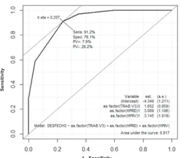
Among the models tested, model 6 was chosen because of its greater area under the curve (0.917), that is, it has better ability to classify the individual correctly, with the highest rate of true positives and lower rate of false positives (figure 1).
Since it is an area close to 1, we concluded that the final model has a good hit rate, both for individuals with prescription for IVI and for individuals who do not need the treatment in question. After the plotting the ROC curve, the cut-off point with the best true positive rate and the lowest false positive rate was chosen. This is a key point for creating the contingency table (or confusion matrix) by establishing the hit and error rates of the final model (Table 4). The cut-off point to create the table was set at 0.577. Thus, the model uses the following decision rule to classify patients: Individuals with probability above the cut-off point will have predicted value Y = 1, and individuals with probability below the cut-off point will have predicted value Y = 0. The final model obtained demonstrated the ability to predict correctly individuals who have a prescription for IVI with a hit rate of 91.17%. This is an important result, since the gold standard test for this diagnosis is expensive and not available in most ophthalmology services.
The analysis of the contingency table shows that the total hit rate of the model was 82.5% (43.75% true negatives and 38.75% true positives), and the error rate was 17.5% % (13.75% false positives and 3.75% false negatives).
D
ISCUSSIONThe present study proposed an investigation regarding the quantification and hierarchy of risk factors for the need for
intra-vitreal injection of anti-VEGF in patients with diabetic macular edema in the State of Paraíba, due to the lack of information about this group of individuals.
Analyzing the risk factors individually, we observed that the variable gender was statistically significant for the outcome in the first step (simple logistic regression analysis at level of significance 10%), with a p-value of 0.545. However, in the multiple logistic regression stage, this was not significant for the development of macular edema and the need for IVI, which corroborates with the literature data(10-13).
Regarding age, studies show that the majority of patients with diabetic macular edema (DME) are older, on average be-tween 64 - 66 years(10,13), similar to that found in this sample (the average age of patients with IVI was 60.65 years). However, as in other studies also using the logistic regression model, the variable age was not considered statistically significant(11,14). In this study, among the patients in the group without treatment indication, the average age was similar (58.02 years). The same happened to the variable color/race, as the majority of patients in both groups (outcome zero and outcome one) were white (43.47% and 52.94%, respectively) or brown (47, 82% and 41.17%), and it was not statistically significant for the need for IVI by DME, which is in agreement with some data found in the literature in which the variable race was also not significant for the outcome in question(11,14).
Analyzing data on marital status, education, income and work situation, no studies were found in the literature to demons-trate these variables as risk factors for the development of DME and the need for anti-VEGF IVI. In this study, most patients with no prescription for IVI lived in a common-law marriage (60.86%), had incomplete primary education (33%), were retired (43%) and belonged to income class B2 (30%). On the other hand, those with prescription for treatment were also living in a common--law marriage (52.94%), had complete high school (38%), were retired (65%), and also belonged to income class B2 (29%). The variables marital status, education, and income were not statisti-cally significant for the outcome in question, which may be due to the similarity of these characteristics between the two groups of the sample in this research, as verified in the results above. The variable work situation, after categorized, showed the category retired as significant, with p-value 0.04, remaining later in the final model. This result may be related to the more advanced age of this group of patients, although the variable age alone was not statistically significant. A study carried out in 2015 to analyze the risk factors for diabetic retinopathy and diabetic macular edema in patients from Austria and Germany reported that individuals in the group macular edema were older(15).
Considering the results of personal history, the following va-riables were statistically significant at level of significance 10% in the stage of simple logistic regression: duration of diabetes mellitus (p-value 0.02), longer interval between the diagnosis and the first ophthalmologist appointment (p-value 0.06), higher rates of sys-tolic blood pressure (p-value 0.03), and body mass index (p-value 0.07). These results are similar to other studies that also detected a higher prevalence of diabetic retinopathy and diabetic macular edema in individuals with longer disease duration(11,12,14-16), and a higher rate of systemic arterial hypertension(10,12,15,17,18). The study by Jew et al. published in 2012 showed that the duration of DM was significantly higher in the group with macular edema (12.72 ± 5.66) than in the group without this complication (8.57 ± 5.66) with p-value = 0.001(11). In the present study, the average duration of DM in the group with macular edema and prescription for IVI
Figure 1. ROC curve for the final model IVI (area 0.917)
Se
n
s
iti
v
ity
was 16.9 ± 9.87, compared with 11.1 ± 10.46 years in the group without treatment indication. Asensio-Sánchez et al. showed that the increase in arterial blood pressure was significantly associated with the DME, with risk 1.55 times greater (CI 1.56-1.78)(10).
Some studies also using the logistic regression analysis de-monstrated that BMI > 35 was significant for the development of diabetic retinopathy, but not for macular edema(11,15). In the stage of multiple logistic regression analyzed along with other variables, the duration of disease, time between diagnosis and the first oph-thalmologist appointment, systolic blood pressure, and BMI were not significant for the final model, with a level of significance 5%. Although insulin therapy is described in some studies as significant for the development of macular edema (10,15), particu-larly in an intensive manner, the use of insulin and the type of diabetes (DM1 or DM2) did not seem to be significant in this study for the development of DME and the need for IVI. This is probably a reflection of the characteristics of this sample, since almost all patients in both groups had DM type 2 (98% and 97%, respectively), and most of them did not use insulin in both groups (54% and 53%, respectively). The same happened for the varia-bles current smoking, past smoking, and physical activity. Most patients in both groups reported being non-smokers (93% and 91%, respectively). Regarding smoking, 50% of the individuals in the group without an prescription for IVI reported being for-mer smokers, compared to only 32% in the group treatment. In the group without prescription for IVI, 52% of the individuals reported regular physical activity, compared to 41% of those who had treatment indication.
Similar to this study, the variable smoking did not show statistical significance for the outcome in other studies(11,15), but a study published in 2008 by Asensio-Sánchez et al. showed a significant association between this characteristic and diabetic macular edema(10).
Also in the analysis of the results related to personal history, the stage of simple logistic regression detected as significant, with level of significance 10%, the presence of personal history of: dia-betic retinopathy, macular edema, intravitreal injection, and laser photopolycoagulation. However, in the multiple logistic regression analysis with level of significance 5%, only the personal history of diabetic retinopathy and intravitreal injection (p-value 0.006 and 0.002, respectively) remained significant for the final model. A strong correlation was found between the variables of macular edema and personal history of intravitreal injection, which was why the former was excluded from the model (considering that this history was dependent on the patient’s memory, it was more likely that the individual remembered having been submitted to intravitreal injection, which is a surgical procedure, than surely remembering whether or not had macular edema, since this condition is not always explained to the patient, or the patient doesn’t understand this complication). No studies were found in the literature analyzing these variables in relation to the outcome in question, with this research being therefore a valid contribution. Regarding the family history, the same limitation was found regarding the socio-economic variables, that is, it is impossible to compare it with data in the literature, since studies evaluating the risk factors for the development of DME do not take this variable into account. In this study, only the family history of diabetic retinopathy was statistically significant in the simple logistic regression stage, with p-value 0.02. However, the stage of analysis of multiple logistic regression was not significant for the final model when analyzed along with the other variables.
Considering the analysis of complementary exams, the data
reflecting the glycemic control are fasting blood sugar (FBS) and glycosylated hemoglobin (HBA1C). In the stage of simple logistic regression with level 10%, fasting blood sugar was significant with a p-value of 0.057, and glycosylated hemoglobin after categoriza-tion was significant when above 0.1 (HBA1C > 10%), presenting p-value 0.04. These results are consistent with data found in the literature, since higher rates of both FBS and HBA1C are consi-dered strongly associated to a higher probability of developing diabetic macular edema(17,18). Studies using logistic regression(11,14,15) showed similar results: a study by Warma et al. published in 2014 found that every 1% increase in HBA1C rate increased by 1.47 times the risk of developing DME (CI, 1.26-1.71; p -value <.0001) (14). Hammes et al. reported in 2015 that individuals with a HBA1C
rate above 8% had a 1.21-fold higher risk of developing DME (CI 1.137-1.279; p-value <0.0001)(15). When categorizing the variable HBA1C above 8% for our study, it was not significant, which occurred only when the category was considered above 10%, as described above.
Regarding dyslipidemia, literature reports that high levels of total cholesterol (TC), LDL cholesterol and triglycerides (TGL) have a significant association to the development of MDE, but not the levels of HDL(10, 11, 15, 17,18). These results were similar to those found in the present study. In the first stage of the simple logistic regression analysis, total cholesterol (p-value 0.04), LDL (p-value 0.05) and TGL were significantly associated to the outcome, the latter being significant after categorization (p-value 0.044 for the category TGL > 150 mg/dL). The variable HDL was not significant (p-value 0.92). In the multiple logistic regression analysis of the models separated by groups and considering a level of significance of 5%, only the variable TGL > 150 remained. And after analysis along with the variables of the other groups, it was not significant for the final model.
Finally, the variable proteinuria in the urine summary (PT-NEAS) was considered statistically significant for the outcome in question when simple logistic regression (p-value 0.015) was carried out, which is in accordance with the literature data on this variable(10,15,19,20). It’s worth pointing out that most studies consider the microalbuminuria in urine of 24h, which has a higher specificity and sensitivity than the urine summary. However, the latter was used due to being a more affordable and simple exam to be carried out, since some patients fail to collect the urine of 24h correctly. In the analysis by groups of variables, this still re-mained significant, with p-value = 0.02. However, when analyzed in the model of multiple logistic regression, this variable was not considered significant for the final model.
As observed in the final model of this study, the variables RET (retired), PHDR (personal history of diabetic retinopathy) and PHIVI (personal history of intravitreal injection) were statis-tically significant in relation to the outcome with prescription for IVI. From the analysis and interpretation of the odds ratio (OR), a diabetic individual who is retired (i.e., older) has an approxi-mately 5-fold greater chance of developing macular edema with prescription for IVI than younger individuals of working age (p-value 0.05). Those individuals who have a personal history of diabetic retinopathy are about 20 times more likely to develop macular edema with prescription for IVI than those who do not have it (p-value 0.006). In addition, there is an approximately 23-fold increase in the risk of diabetic individuals who have already undergone this treatment at least once requiring intravitreal injection than those who have not yet required such procedure (p-value 0.002).
the outcome in question in other studies (such as use of insulin, disease, systolic and diastolic blood pressure, BMI, glycated ha-emoglobin, total cholesterol, LDL and TGL) have not entered the final model of this study may be due to the small number of cases (80 patients) and the features of this sample, which showed similarity in the results of some variables between groups , as described above.
C
ONCLUSIONData from the literature reinforce that the anatomical and visual results for the treatment with intravitreal injection of anti-VEGF in patients with diabetic macular edema are directly related to certain risk factors, clinical and anatomical characteris-tics of the patients treated, and it depends on how early are the diagnosis and treatment of this complication.
The present study reached to the conclusion, based on the logistic regression model proposed and the joint analysis of the risk factors, that a diabetic individual complaining of low visual acuity has the following characteristics: retired (i.e., older), having a personal history of diabetic retinopathy and a positive history for prior treatment with anti-VEGF, should be referred immediately to the ophthalmologist (preferably a retina specialist), because there is a probability of 91.17% of having diabetic macular edema with prescription for intravitreal injection of anti-VEGF. This is an important result, since the gold standard test for this diagnosis of this complication is expensive and not available in most oph-thalmology services.
In isolation, if a diabetic individual is retired (i.e., older) there is an approximately 5-fold greater chance of developing macular edema with prescription for IVI than younger individuals of working age. Those individuals who have a personal history of diabetic retinopathy are about 20 times more likely to develop macular edema with prescription for IVI than those who do not have it. In addition, there is an approximately 23-fold increase in the risk of diabetic individuals who have already undergone this treatment at least once requiring intravitreal injection than those who have not yet required such procedure.
A limitation of this study is related to the small sample size (80 patients) and its characteristics, which may have influenced the fact that certain variables considered significant for the outcome in question in other studies did not enter the final model of this research. This reinforces the need for further studies evaluating the risk factors related to the development of diabetic macular edema and the need for IVI anti-VEGF with a greater number of individuals, in order to guide medical practice, which may contri-bute to the reduction of the prevalence of irreversible blindness due to this complication.
In addition, as a future proposal is very important to add an evaluation of the impact of these risk factors on the response to treatment to this type of study, i.e., to anatomical (central macular thickness in the OCT of the macula) and visual (visual acuity) results, in order to determine those susceptible to prevention or early therapeutic intervention, which may increase the probability of successful treatment.
The evaluation process of the logistic model proposed in this study has aims at helping and not replacing the medical professional in planning and deciding the treatment in question. In summary, we proposed a model of evaluation and prediction to serve as a supporting tool in decision-making, especially for the non-retinal physician, in order to refer patients with diabetic
retinopathy to the early diagnosis and treatment, and their main cause of low visual acuity - diabetic macular edema -, which may be decisive in preventing irreversible visual loss in these patients.
R
EFERENCES1. World Health Organization. Prevention of blindness from diabetes mellitus: report of a WHO consultation in Geneva, Switzerland, 9-11-November, 2005. Geneva: WHO; 2005. p. 2-13.
2. Klein R, Klein BE, Moss SE. Visual impairment in diabetes. Oph-thalmology. 1984; 91:1-9.
3. Photocoagulation for diabetic macular edema. Early Treatment Diabetic Retinopathy Study report number 1. Early Treatment Diabetic Retinopathy Study Research Group. Arch Ophthalmol. 1985; 103(12):1796-806.
4. Nguyen QD, Shah SM, Heier JS, Do DV, Lim J, Boyer D, Abraham P, Campochiaro PA; READ-2 Study Group. Primary end point (six months) results of the ranibizumab for edema of the macula in dia-betes (READ- 2) study. Ophthalmology. 2009; 16(11):2175-81.e1. 5. Nguyen QD. Shah SM, Khwaja AA, Channa R, Hatef E, Do DV,
Boyer D, Heier JS, Abraham P, Thach AB, Lit ES, Foster BS, Kruger E, Dugel P, Chang T, Das A, Ciulla TA, Pollack JS, Lim JI, Eliott D, Campochiaro PA; READ-2 Study Group. Two-year outcomes of the ranibizumab for edema of the macula in diabetes (READ-2) study. Ophthalmology. 2010; 117(11):2146-51.
6. Massin P, Bandello F, Garweg JG, Hansen LL, Harding SP, Larsen M, et al. Safety and efficacy of ranibizumab in diabetic macular edema (RESOLVE Study): a 12-month, randomized, controlled, double-masked, multicentre phase II study. Diabetes Care. 2010; 33(11):2399-405.
7. Mitchell P, Bandello F, Schmidt-Erfurth U, Lang GE, Massin P, Schlingemann RO, Sutter F, Simader C, Burian G, Gerstner O, Weichselberger A; RESTORE study group. The RESTORE study: ranibizumab monotherapy or combined with laser versus laser monotherapy for diabetic macular edema; RESTORE study group. Ophthalmology. 2011;118(4):615-25.
8. American Academy of Ophthalmology. Preferred pratice patterns. Diabetic retinopathy [Internet]. American Academy of Ophthalmo-logy. 2014. [cited 2017 June 24]. Available from: http://www.aao.org/ preferred-practice-pattern/diabetic-retinopathy-ppp--2014 9. Evangelista IW, Leitao KC, Leite EP, Costa CB, Oashi E G,
Almeida SC, et al. Protocolo clínico e diretrizes terapêuticas em oftalmologia. 2a ed. Unidade da Visão, Hospital Universitário Lauro Wanderley, EBSERH-UFPB; 2015.
10. Asensio-Sánchez VM, Gómez-Ramírez V, Morales-Gómez I, Rodrí-guez-Vaca I. Clinically significant diabetic macular edema: systemic risk factors. Arch Soc Esp Oftalmol. 2008; 83: (3):173-6.
11. Jew OM, Peyman M, Chen TC, Visvaraya S. Risk factors for clinically significant macular edema in a multi-ethnics population with type 2 diabetes. Int J Ophthalmol. 2012; 5(4):499-50.
12. Yau JW, Rogers SL, Kawasaki R, Lamoureux EL, Kowalski JW, Bek T, Chen SJ,Dekker JM, Fletcher A, Grauslund J, Haffner S, Hamman RF, Ikram MK, Kayama T,Klein BE, Klein R, Krishnaiah S, Mayurasakorn K, O’Hare JP, Orchard TJ, Porta M, Rema M, Roy MS, Sharma T, Shaw J, Taylor H, Tielsch JM, Varma R, Wang JJ, Wang N,West S, Xu L, Yasuda M, Zhang X, Mitchell P, Wong TY; Meta-Analysis for EyeDisease (META-EYE) Study Group. Global prevalence and major risk factors of diabetic retinopathy. Diabetes Care. 2012 Mar;35(3):556-64. Review.
13. Channa R, Sophie R, Khwaja AA, Do DV, Hafiz G, Nguyen QD, Campochiaro PA; READ-2 Study Group.. Factors affecting visual outcomes in patients with diabetic macular edema treated with ranibizumab. Eye (Lond). 2014;28(3):269-78.
15. Hammes HP, Welp R, Kempe HP, Wagner C, Siegel E, Holl RW; DPV Initiative—German BMBF Competence Network Diabetes Mellitus. Risk Factors for Retinopathy and DME in Type 2 Dia-betes—Results from the German/ Austrian DPV Database. PLOS ONE. 2015;10(7):e0132492.
16. Klein R, Klein BE, Moss SE, Davis MD, DeMets DL. The Wisconsin epidemiologic study of diabetic retinopathy. II. Prevalence and risk of diabetic retinopathy when age at diagnosis is 30 or more years. Arch Ophthalmol. 1984;102(4):527-32.
17. Diabetes Control and Complications Trial Reserach Group, Nathan DM, Genuth S, Lachin J, Cleary P, Crofford O, Davis M, Rand L, Siebert C. The effect of intensive treatment of diabetes on develop-ment and progression of long-term complications in insulin-dependet diabetes mellitus. N Eng J Med. 1993;329(14):977-86.
18. Intensive blood-glucose control with sulphonylureas or insulin compared with conventional treatment and risk of complications in patients with type 2 diabetes(UKPDS 33). UK Prospective Diabetes Study (UKPDS) Group. Lancet. 1998;352(9131):837-53. Erratum in: Lancet 1999 Aug 14;354(9178):602.
19. Skuta GL, Cantor LB, Weis JS. Retina e Vítreo: 2010-2011, revisão 2008-2009 / [organização] Gregory L. Skuta, Louis B. Cantor, Jayne S. Weiss; [edição Carl Regillo...et al; tradução Beatriz Sayuri Takahashi... et al.; revisão científica José Byron Vicente Dias Fernandes]. São Paulo: Santos, 2012. p. 112, 113, 157
20. Cruickshanks KJ, Ritter LL, Klein R, Moss SE. The association of microalbuminuria with diabetic retinopathy. The Wiscosin Epi-demiologic Study of Diabetic Retinopathy. Ophthalmology.1993; 100(6):862-7.
Corresponding author:
Aline Roseane Queiroz de Paiva Faria
Rua Valdemar Chianca, 205, apto 202, Bessa, ZIP Code 58037-255-João Pessoa-PB