(1) Hospital de Reabilitação de Anomalias Craniofaciais- Universidade de São Paulo, HRAC-USP, Bauru, SP, Brasil. (2) Departamento de Fonoaudiologia da Faculdade de Odontologia de Bauru- Universidade de São Paulo, Bauru, SP, Brasil.
Funding source: RUSP- Reitoria da Universidade de São Paulo.
Conlict of interest: non-existent
Orthognathic surgery effect of orofacial sensitivity in individuals
with cleft lip and palate
Efeito da cirurgia ortognática na sensibilidade orofacial
em indivíduos com fissura labiopalatina
Andréia Fernandes Graziani(1) Carla Franciele Souza Garcia(1) Giédre Berretin-Felix(2) Katia Flores Genaro(2)
Received on: November 10, 2015 Accepted on: March 30, 2016
Mailing address: Katia Flores Genaro
Departamento de Fonoaudiologia da Faculdade de Odontologia de Bauru-USP Alameda Dr. Octavio Pinheiro Brisolla 9-75, Vila Universitária - Bauru - SP
CEP: 17012-901 E-mail: [email protected]
ABSTRACT
Purpose: to study changes in the orofacial sensitivity and the recovery time after the completion of this orthognathic surgery in individuals with cleft lip and palate.
Methods: after the approval of the Ethical Committee, began the study which analyzed the examination
reports of orofacial myofunctional performed before and after orthognathic surgery from 2012 to 2014. Was selected the tests contained in 53 medical records of patients who underwent sensitivity testing, aged between 18 and 40 years,the both genders (57% male and 43% female). The sensitivity test was
applied on the lips, tongue, incisive papilla and chin, from the extensiometer (Semmes-Weintein), prior to
surgery (2 to 3 days) and after surgery (3 to 6 months / 9 to 12 months). The results were analyzed using descriptive statistics to verify of frequency the change of sensitivity, and comparisons were performed by
Wilcoxon tests and Chi-square (p<0.05).
Results: before surgery all cases showed adequate sensitivity of the tongue, while in the remaining few tested structures have changed before and after orthognathic surgery. After surgery there was an incre-ase of frequency the sensitivity of change of the incisive papilla (p=0.004). There was no association between the evaluation periods after surgery.
Conclusion: although the prevalence of adequacy of sensitivity after surgery was observed for the incisive papilla increased frequency of change and we found no difference between the cases evaluated before and after 6 months.
Keywords: Cleft Lip; Cleft Palate; Maxillofacial Abnormalities; Orthognathic Surgery; Evaluation
RESUMO
Objetivo: veriicar a ocorrência de alterações na sensibilidade orofacial e o tempo de recuperação desta
após a realização da cirurgia ortognática, em indivíduos com issura labiopalatina.
Métodos: após aprovação do Comitê de Ética em Pesquisa, iniciou-se o estudo o qual analisou os rela
-tórios de exames miofuncionais orofaciais, realizados antes e após a cirurgia ortognática no período
de 2012 a 2014. Foram selecionados 53 prontuários de pacientes que realizaram a prova de
sensibili-dade, com idade entre 18 e 40 anos, de ambos os gêneros (57% masculino e 43% feminino). A prova
de sensibilidade foi aplicada nos lábios, na língua, na papila incisiva e mento, a partir do estesiômetro (Semmes-Weintein), antes da cirurgia (2 a 3 dias) e após a cirurgia (3 a 6 meses/9 a 12 meses). Os
resultados foram analisados por meio de estatística descritiva para veriicar a frequência de alteração da sensibilidade, e as comparações foram realizadas pelos testes de Wilcoxon e Qui-Quadrado (p<0,05).
Resultados: antes da cirurgia todos os casos apresentaram sensibilidade adequada da língua, enquanto nas demais estruturas testadas poucos apresentaram alteração, antes e após a cirurgia ortognática.
Após a cirurgia houve aumento da frequência de alteração da sensibilidade da papila palatina (p=0,004).
Não houve associação entre os períodos de avaliação após a cirurgia.
Conclusão: apesar da prevalência de adequação da sensibilidade, após a cirurgia foi observado para a
papila incisiva aumento da frequência de alteração e não foi constatada diferença entre os casos avalia -dos antes e após 6 meses.
INTRODUCTION
Cleft lip and palate is a malformation with great
frequency in the human species1. This malformation
changes the stomatognathic system, the morphology and the orofacial functions, which requires surgery to correct it2. Among the changes related to orofacial functions, we can mention: occlusion problems, feeding, swallowing, speech and breathing3.
Studies have shown that dental anomalies are prevalent in individuals with cleft lip, the most common dental agenesis is the presence of supernu-merary teeth in the permanent dentition4. In addition, changes related to malocclusion are directly related to issues involving size, shape and position of teeth5. In many cases, the patient does not treat the occlusion disorders during childhood and reaches adulthood with those changes. In other cases, even when the patient performs orthodontic treatment in childhood, this is not
suficient to correct these changes. In both cases, it is not possible to ix the skeletal changes with orthodontic
treatment, there is the need for orthognathic surgery for dentofacial deformity correction6.
The role of speech-language pathologist in cases undergoing orthognathic surgery occurs in two stages: pre-surgery, in which anamnesis, evaluation, orien-tation and speech therapy is performed and are related
to working with the perioral muscles, global posture
and proprioception7, and after surgery, in addition to involving all the items mentioned above, it also includes
working with chewing, swallowing, speech and
orofacial sensitivity8.
The orthognathic surgery is a procedure that aims
to correct maxillomandibular and facial changes9.
Patients with masticatory alteration, respiratory,
speech and even esthetic dificulties, resulting from occlusion or jaw positioning irregularities, can beneit
from this procedure10. Thus, the surgery for dentofacial deformity correction (DDC) leads to improvement of the
functional aspects and facial appearance, which makes the individual happier and satisied11-14.
According to the literature, complications after
orthognathic surgery15 are not common but they can
occur, and the loss of sensitivity is one of the compli-cations16-18. This change is due to the osteotomy site, close to the peripheral branches of the maxillary and mandibular nerve19, which can lead to trauma in this structure. Thus, the sensitivity of orofacial structures, as well as its recovery should be assessed20. Several studies on sensory changes after orthognathic surgery have revealed loss of orofacial sensitivity9,21,22. In
general, this change is transitory23,24; however, the
recovery period can be variable23,25. The change in
orofacial sensitivity can cause discomfort to the patient
and affect the orofacial functions, causing dificulty in
controlling saliva, feeding and speech17.
According to some authors19, there is no standard method to assess changes in sensitivity and it can be performed through electrophysiological and sensory testing and evaluation from the patient’s perception that reports the degree of commitment. A useful tool to assess the sensitivity is the esthesiometer25,26, origi-nally proposed for evaluating the tactile sensitivity in Hansen’s disease and diabetes27. This is a set of
monoilaments27 able to identify changes in sensi-tivity from a light touch in the region to be tested25,26. Regarding oral cavity, some studies have used this tool to assess sensitivity18,22,24.
Individuals with cleft lip presenting dentofacial deformity require surgical procedure for the correction, which in turn may affect the orofacial sensitivity and consequently the performance of orofacial functions. It is believed that before the orthognathic surgery,
sensi-tivity is preserved and in the irst months after surgery,
this would be changed, which would return later. Once the orofacial functions are already affected due to functional changes, due to the structural condition, the change in sensitivity is another aggravating factor.
Thus, the evaluation of this aspect will enable a better understanding of the functional changes and allow the development of guidelines to the stimulation
of sensitivity and preventive actions in the ield of
orofacial disorders.
Thus, the objective of this research was to analyze the orofacial sensitivity in individuals with cleft lip and palate, to verify the occurrence of changes and the recovery time after orthognathic surgery.
METHODS
The study was approved by the Research Ethics Committee of Craniofacial Anomalies Rehabilitation Hospital (HRAC), from University of São Paulo where the study was conducted (No. 543621). This is a retro-spective study that analyzed orofacial myofunctional tests performed before and after orthognathic surgery.
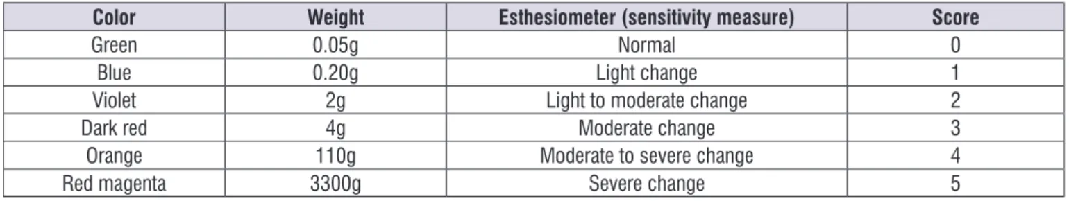
In orofacial myofunctional test, the sensitivity test of the lips, tongue, incisive papilla and the mental region is performed through the esthesiometer, consisting
of a set of six monoilaments (Semmes-Weinstein) of
colored nylon and different diameters, which touch the area to be tested and generates a pressure (Figure 1).
Color Weight Esthesiometer (sensitivity measure) Score
Green 0.05g Normal 0
Blue 0.20g Light change 1
Violet 2g Light to moderate change 2
Dark red 4g Moderate change 3
Orange 110g Moderate to severe change 4
Red magenta 3300g Severe change 5
*Adapted SORRI- Bauru
Figure 1. Esthesiometer score as the color and weight of the ilaments
In the analysis, we considered the result of each of the tested areas before and after surgery: between 2 to 3 days before surgery (pre) as well as in two other moments after surgery: between 2 to 5 months and
between 6 to 12 months. Each monoilament was
assigned a score, in which zero (0) corresponded to the
thinner ilament and which represents the best sensi
-tivity and ive (5) corresponded to the thicker ilament,
indicating worse sensitivity. In this study, the scores 0 and 1 were considered as appropriate outcome, and changed outcomes, the scores higher or equal to 2.
The data obtained were stored in an Excel spread-sheet and analyzed using descriptive statistics and
veriied the sensitivity change frequency. The individual
scores before and after surgery for each region were compared using the t test for paired samples, and comparing the occurrence of adaptation and changes between stages and between the evaluation time after surgery was investigated by Chi-square test, which
were considered signiicant the values of p<0.05.
RESULTS
A total of 53 patients met the inclusion criteria and were enrolled in the institution between 2012 and 2014. The description of the cases studied, according to gender, age and type of cleft lip and palate is shown in Table 1, in which there is a predominance of males (59.09%) in cases with cleft lip and palate, females in isolated cleft palate (66.67%), gender balance in the cleft lip and a single case that had other anomalies was male. The minimum age of the sample was 18 years and the maximum 40 years, with a median of 23 years.
Table 2 shows the result of sensitivity assessment in both study moments. All cases had adequate tongue
sensitivity before surgery, while some showed changes in the lip (2%) in the incisive papilla (4%), and in the mental region (4%). After surgery there was a predomi-nance of adequacy, with changes in some cases of lip (2%), tongue (2%), in the incisive papilla (24%), and in the mental region (2%). Through the comparison of the frequencies of adequacy and changes in sensitivity between the moments, there was a difference only to the incisive papilla (p = 0.004), which showed an increase number of cases after surgery.
The minimum and maximum values, and the median, referring to the sensitivity assessment scores obtained in the pre and post-surgery tests are shown in Table 3. Of the cases that showed changes before surgery, the maximum obtained score was 2 (lip, incisive papilla and mental region) and after surgery, it was 5 (incisive papilla and mental region). The comparison of scores between moments showed difference to the incisive papilla, with worsening of sensitivity after surgery (p=0.003).
To analyze the changes that occurred after surgery in each tested structure, it was considered that sensi-tivity remained adequate or changed, as well as if
modiication occurred (Table 4).
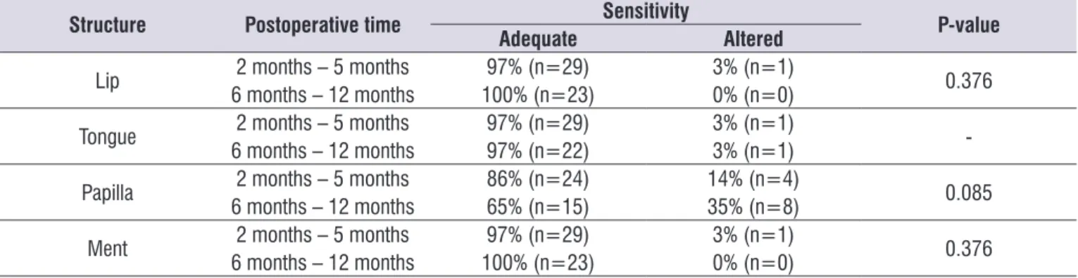
The frequency of change and adequacy of the sensi-tivity observed in post-surgical evaluation, according to the time between surgery and evaluation are shown
in Table 5. It can be veriied predominance of cases
with adequate sensitivity in all structures and periods assessed (2 to 5 months, n=30; and 6 to 12 months, n=23), the incisive papilla structure had the largest number of cases changed in both moments. There was
no signiicant difference between the moments for the
Table 2. Comparison of pre- and post-surgical evaluation of sensitivity
Structure Moment Adequate Altered P-value
Lip Pre 98% (n=52) 2% (n=1) 1.00
Post 98% (n=52) 2% (n=1)
Tongue Pre 100% (n=53) 0% (n=0) 0.315
Post 98% (n=52) 2% (n=1)
Papilla Pre 96% (n=49) 4% (n=2) 0.004ª²
Post 76% (n=39) 24% (n=12)
Ment Pre 96% (n=51) 4% (n=2) 0.558
Post 98% (n=52) 2% (n=1)
ª Signiicant difference by Chi-square test
²2 cases were not considered, no rating post (n=51)
Table 3. Evaluation scores of sensitivity obtained in the studied phases
Structure Moment Scores
Median (minimum- maximum) P-value
Lip Pre 0 (0 - 2) 0.568
Post 0 (0 -2)
Tongue Pre 0 (0 – 0) 0.321
Post 0 (0 – 2)
Papilla Pre 0 (0 – 2) 0.003ª
Post 0 (0 – 5)
Ment Pre 0 (0 – 2) 0.863
Post 0 (0 – 5)
ª Signiicant difference by t test
á 2 cases were not considered, excluded in the post intervention (n=51)
Table 4. Changes of sensitivity after surgery
Results Structure
Lip Tongue Papilla Mental region
Kept adequate 94% (n=50) 98% (n=52) 72% (n=38) 94% (n=50) Kept changes 0% (n=0) 0% (n=0) 2% (n=1) 0% (n=0) Became changed 2% (n=1) 2% (n=1) 20% (n=11) 2% (n=1) Became adequate 4% (n=2) 0% (n=0) 2% (n=1) 4% (n=2) Not assessed after surgery 0% (n=0) 0% (n=0) 4% (n=2) 0% (n=0) TOTAL 100% (n=53) 100% (n=53) 100% (n=53) 100% (n=53)
Table 1. Distribution of the sample according to gender, age and type of cleft lip and palate
Type of cleft lip N Gender Age Median
(minimum- maximum)
Male Female
Lip 3,77% (n=2) 50,00% (n=1) 50,00% (n=1) 29a5m (24a11m-34a) Lip and palate 83,02% (n=44) 59,09% (n=26) 40,91% (n=18) 23a2m (18a2m-40a2m)
Palate 11,32% (n=6) 33,33% (n=2) 66,67% (n=4) 23a4m (21a8m-34a6m) Others 1,89% (n=1) 100,00% (n=1) 0,00% (n=0) 20a5m (20a5m-20a5m) TOTAL 100,00% (n=53) 56,06% (n=30) 43,04%(n=23) 23a3m (18a2m-40a2m)
according to other authors24, sagittal technique has
greater inluence on the tactile feeling of the lip and mental region, one week after surgery, compared to the
vertical technique.
In this study, when mandibular osteotomy was performed this occurred through the sagittal technique, which would justify the result in relation to mental region. In another study30, the authors found that after surgery the most affected structures were mental region, as well as the hard and soft palate; similar to the
indings of this sample, which found greater change in
sensitivity in the incisive papilla and in mental region.
With regard to the time between surgery and evalu
-ation of sensitivity, there was no signiicant difference
between the two periods for the studied structures. However, when analyzing the distribution of relative frequencies in both periods, it is observed sensitivity adequacy predominance, although more cases of changes were shown for the incisive papilla. One
hypothesis for this inding is that patients with cleft lip
and palate can have consequences in the jaw region resulting from surgical procedures which they were
submitted, besides suffering the inluence of bone
and tissue manipulation in this region due to maxillary advancement procedure.
For individuals without craniofacial malforma-tions, some autors21-25 observed variability in sensi-tivity recovery time after surgery, and that can be transitory23,24 and range from 30 days to a period greater than 6 months until total recovery23,25,26. For some authors20, the sensitivity of the lip and mental region
showed signiicant recovery of sensitivity after 30 days
of surgery and at 6 months the results resemble those observed before surgery. According to other authors30, from 3 to 6 months of surgery, 40% of cases still have altered sensitivity in one or more structures.
DISCUSSION
Orthodontic treatment, in many cases, is not enough to correct major maxillomandibular disproportions, justifying the performance of orthognathic surgery. In this study, we analyzed the speech therapy evalua-tions of 53 patients with cleft lip and palate submitted to orthognathic surgery. In the sample studied predomi-nated complete cleft lip and palate, corroborating the
indings28,29 referring to this type of cleft lip and palate with increased demand for orthognathic surgery. Although the literature reports that postoperative
complications15 are not common, impaired orofacial
sensitivity has been reported after orthognathic surgery16-22, which can affect the functional recovery process.
In the assessment performed before surgery, there was predominance of adequacy of the structures sensitivity, but some cases had changes considered mild (score 2) to lip, incisive papilla and mental region. The lip and incisive papilla are regions handled during surgery to correct the cleft lip and palate, and result in scars29, which may inluence the sensitivity; however, it does not apply to the mental region.
After surgery, we also observed predominance of sensitivity adequacy condition, but some cases with change were also found. In relation to the incisive papilla, this structure was the most changed one, with increased number of cases mild and more pronounced changes (scores 3 and 5). In the mental region, we found the score 5 in a single case, and for tongue
score 2. Comparing the scores, there was signiicant
difference after surgery only for the incisive papilla. According to some reports21 in mandibular osteotomy, both in vertical as in the sagittal technique, patients had altered sensation in the postoperative period, with the mental region being the most affected. However,
Table 5. Results of the evaluation of the sensitivity of the structures according to the evaluation time after surgery
Structure Postoperative time Sensitivity P-value
Adequate Altered
Lip 2 months – 5 months 97% (n=29) 3% (n=1) 0.376 6 months – 12 months 100% (n=23) 0% (n=0)
Tongue 2 months – 5 months 97% (n=29) 3% (n=1) -6 months – 12 months 97% (n=22) 3% (n=1)
Papilla 2 months – 5 months 86% (n=24) 14% (n=4) 0.085 6 months – 12 months 65% (n=15) 35% (n=8)
Ment 2 months – 5 months 97% (n=29) 3% (n=1) 0.376 6 months – 12 months 100% (n=23) 0% (n=0)
After surgery, individuals were in the process of adaptation to the new functional condition and thus, identifying and monitoring changes of sensitivity favors the establishment of appropriate therapeutic targets in the pursuit of faster functional recovery and without compromising the stability of the surgical outcome. It is worth noting that the patient should be informed about
the possible risks of temporary change of sensitivity,
which also contributes to face a more favorable postop-erative recovery.
In this study, it was expected that the sensitivity was preserved before orthognathic surgery, however, in some cases changes were found. It was also hoped
that changing the sensitivity only occurred in the irst
months after surgery, with decreased frequency of changed cases after 6 months, which was not found. Thus, new longitudinal studies are useful to better understand the results, as well as a more represen-tative sample, and a comparative study related to the evaluation of orofacial sensitivity with the use of nylon
monoilaments, and other types of instrument to check
results.
CONCLUSION
In this sample, the results showed a predominance of adequate sensitivity to all tested structures, with few cases changed to lip, incisive papilla and mental region, both before and after orthognathic surgery; we
did not ind difference in sensitivity change among the
cases evaluated up to 5 months after surgery and those evaluated after 6 months.
REFERENCES
1. Murray JC. Gene/environment causes of cleft lip and/or palate. Clin Genet. 2002;61:248-56.
2. Bertier CE, Trindade IEK, Silva Filho OM. Cirurgias primárias de lábio e palato. In: Fissuras Labiopalatinas: Uma abordagem interdisciplinar. Trindade IEK, Silva Filho OG, organizadores. São Paulo: Santos; 2007. p. 73-86.
3. Figueiredo MC, Pinto NF, Faustino-Silva DD, Oliveira M. Fissura bilateral completa de lábio e palato: alterações dentárias de má oclusão - relato de caso clínico. UEPG: Ciências Biológicas e da Saúde. 2008;14(1):7-14.
4. Montandon EM, Duarte RC, Furtado PGC. Prevalência de doenças bucais em crianças
portadoras de issuras labiopalatinas. J. Bras.
Odontopediatr. Odontol. Bebê. 2001;4(17):68-73.
5. Neves ACC, Patrocínio MC, Leme KP, UI TR. Anomalias dentárias em pacientes portadores de
issuras labiopalatinas: revisão de literatura. Rev.
Biociênc Taubaté. 2002;8(2):75-81.
6. Lurentt K. Cirurgia ortognática em paciente portador
de issura lábiopalatina. Relato de caso. Rev. Cir.
Traumatol. Buco-Maxilo-Fac. 2012;12(1):47-52.
7. Bianchini EMG. Desproporções maxilomandibulares: atuação fonoaudiológica com pacientes submetidos à cirurgia ortognática. In: Marchesan IQ e colaboradores. Tópicos em Fonoaudiologia. São Paulo: Lovise; 1995. p. 129-45.
8. Berretin-Felix G, Jorge TM, Genaro KF. Intervenção fonoaudiológica em pacientes submetidos à cirurgia ortognática. In: Fernandes FDM, Mendes BCA, Navas ALPGP (Org.). Tratado de Fonoaudiologia. 2ªed.São Paulo: Roca; 2009. p. 545-57.
9. Lima Júnior N, Moro MA, Tanaka FY, Fattah CMRS,
Renon MA. O que signiica cirurgia ortognática?
Arq Ciênc Saúde UNIPAR. 1999;3(3):273-6.
10. Ribas MO, Reis LFG, França BHS, Lima AAS. Cirurgia ortognática: orientações legais aos ortodontistas e cirurgiões bucofaciais. Rev Clin Ortod Dental Press. 2005;10(6):75-83.
11. Alanko OM, Svedström-Oristo AL, Tuomisto MT. Patients’ perceptions of orthognathic treatment, well-being, and psychological or psychiatric status: a systematic review. Acta Odontol Scand. 2010;68(5):249-60.
12. Choi WS, Lee S, McGrath C, Samman N. Change in quality of life after combined orthodontic-surgical treatment of dentofacial deformities. Oral Surg, Oral Med, Oral Pathol, Oral Radiol and Endod. 2010;109(1):46-51.
13. Khadka A, Liu Y, Li J, Zhu S, Luo E, Feng G et al. Changes in quality of life after orthognathic surgery: a comparison based on the involvement of the occlusion. J. Cleft Palate Craniofac. 2011;112(6):719-25.
14. Soh CL, Narayanan V. Quality of life assessment in patients with dentofacial deformity undergoing orthognathic surgery - A systematic review. Int J Oral Maxillofac Surg. 2013;42(8):974-80.
16. Blomqvist JE, Alberius P, Isaksson S. Sensibility following sagittal split osteotomy in the mandible: a prospective clinical study. Plast Reconstr Surg. 1998;102(2):325-33.
17. Marchesan IQ, Bianchini EMG. A Fonoaudiologia e a cirurgia ortognática. In: Araújo A. Cirurgia ortognática. São Paulo: Santos; 1999. p. 353-62. 18. Alves TCNV. Análise da produção de fala nas
correções cirúrgicas da deformidade dentofacial [tese]. Bauru (SP): Hospital de Reabilitação de Anomalias Craniofaciais, Universidade de São Paulo; 2008.
19. Phillips C, Essack G. Inferior alveolar nerve injury following orthognathic surgery: a review of assessment issues. J Oral Rehabil. 2011;38(7):547–54.
20. Monazzi MS. Avaliação clínica do grau de sensibilidade cutânea, na região mentoniana e de lábio inferior, em pacientes tratados por meio de Osteotomia Sagital Bilateral da Mandíbula [tese]. Campinas (SP): Faculdade de Ciências Medicas, Universidade Estadual de Campinas; 2011.
21. Oliveira SR, Santos P, Roberto TN, Grechi TH, Travit LV. Alterações da sensibilidade orofacial em pacientes pós-operatório de cirurgia ortognática. Anais do 16° Congresso Brasileiro de Fonoaudiologia; 2008 set; Campos do Jordão, SP. Campos do Jordão: SBFA; 2008. p. 1142.
22. Passos DCBOF, Alves MM, Zanferrari EO, Berretin-Félix G. Efeitos da Cirurgia ortognática sobre a sensibilidade e a mobilidade mandibular. Anais da XVI Jornada Fonoaudiológica de Bauru; 2009 ago; Bauru, SP. Bauru; 2009. p 64.
23. Geha HJ, Gleizal AM, Nimeskern NJ, Beziat JL. Sensitivity of the inferior lip and chin following mandibular bilateral sagittal split osteotomy using Piezosurgery. Plast Reconstr Surg. 2006;118(7):1598-607.
24. Trawitzki LVV, Germano KS, Picinato-Pirola MNC, Silva JB, Grechi TH, Mello-Filho FV. Sensibilidade orofacial em pacientes com deformidades dentofaciais três meses após cirurgias de mandíbula. Anais 39th Annual Convention International Association of Orofacial Myology – IAOM; São Paulo; 2011.
25. Kobayashi A,. Yoshimasu H, Kobayashi J, Amagasa T. Neurosensory alteration in the lower lip and chin area after orthognathic surgery: bilateral sagittal split osteotomy versus inverted L ramus osteotomy. J Oral Maxillofac Surg. 2006;64(5):778-84.
26. Lemke RR, Clark GM, Bays RA, Rugh JD. Effects of hypesthesia on oral behaviors of the orthognathic surgery patient. J Oral Maxillofac Surg. 1998;56(2):153-7.
27. Lehman LF, Orsini MB, Nicholl AR. The development and adaptation of
Semmes-Weinstein monoilaments in Brazil. J Hand Ther. 1993;6(4):290-7.
28. Raposo-do-Amaral CA, Raposo-do-Amaral CE, Carone DR, Pinheiro AF, Braga EVB, Guidi MC et al. Estudo do avanço maxilar e das complicações em
pacientes issurados e não-issurados submetidos
a cirurgia ortognática. Rev. Bras. Cir. Plást. 2008;23(4):263-7.
29. Freitas RS, Canan Junior LW, Roça GB, Busato LS, Alonso N, D’oro U. Cirurgia ortognática nos
portadores de issuras lábio-palatais: experiência e desaios. Rev. Soc. Bras. Cir. Craniomaxilofac.
2009;12(3):89-93.
30. Zanferrari EO, Berretin-Félix G. Efeitos da cirurgia ortognática sobre a sensibilidade e motricidade orofacial. Anais do 15º Simpósio Internacional de
Iniciação Cientíica- SIICUSP; Ribeirão Preto; 2007