ABSTRACT
http://dx.doi.org/10.1590/1678-775720160029
Ozone therapy as an adjuvant for endondontic
protocols: microbiological –
ex vivo
study and
citotoxicity analyses
Carlos Goes NOGALES1, Marina Beloti FERREIRA1, Antonio Fernando MONTEMOR2, Maria Filomena de Andrade RODRIGUES3, José Luiz Lage-MARQUES1, João Humberto ANTONIAZZI1
1- Universidade de São Paulo, Faculdade de Odontologia, Departamento de Endodontia, São Paulo, SP, Brasil.
2- Instituto de Pesquisas Tecnológicas do Estado de São Paulo - IBL Núcleo de Bionanomanufatura, São Paulo, SP, Brasil. 3- Instituto de Pesquisas Tecnológicas do Estado de São Paulo, Departamento de Biotecnologia , São Paulo, SP, Brasil.
Corresponding address: Carlos Goes Nogales - Rua Joaquim Antunes, 1034 - apto 74 - Pinheiros - São Paulo-SP - Brazil - 05415-001 - Email: c.nogales173@ gmail.com
O
contaminated with Pseudom onas aeruginosa, Ent erococcus faecalis, and St aphylococcus aureusEnt erococcus faecalis, Pseudom onas aeruginosa, and St aphylococcus aureus. Groups were formed: Group I – control; Group II – standard protocol; Group III – standard protocol + ozone gas at 40 μg/mL; and Group IV – standard protocol + aqueous ozone at 8 μg/mL. then ozone was applied as follows: Group I (control) – broth medium; Group II – aqueous ozone at 2 μg/mL; Group III – aqueous ozone at 5 μg/mL; and Group IV – aqueous ozone at 8 μg/mL. Data were submitted to the Kruskal Wallis test and Bonferroni post hoc
analyses to assess microbiology and cytotoxicity, respectively (p<0.05%). Results: The assay showed Groups III and IV to be the most aggressive, providing a decrease in cell viability at hour 0 from 100% to 77.3% and 68.6%, respectively. Such a decrease in cell viability was reverted, and after 72 hours Groups III and IV provided the greatest increase in cell viability, being statistically different from Groups I and II. Conclusion: According to the applied methodology and the limitations of this study, it was possible to conclude that ozone therapy improved the decontamination of the root canal ex vivo. Ozone was toxic that ozone might be useful to improve root canal results.
Keywords: Ozonated water. Ozone. Endodontic. Cytotoxicity. Antimicrobial activity.
INTRODUCTION
In Endodontic therapy, success is closely linked to the removal of debris and the elimination of
this requirement, the clinician must use Endodontic instruments associated with chemical substances. In order to improve the decontamination procedures due to the inaccessible areas in the root canal system, more effective antimicrobial treatment
strategies and substances should be studied22.
Ozone is both antimicrobial2,6,7,10,12-18 and
biocompatible8. Studies have shown the important
efficacy of aqueous ozone18 especially when
associated with ultrasonic activation10,17. The
concentrations of sodium hypochlorite10,14.
by mitochondria, which leads to a metabolic improvement and to healing of inflammatory/ infectious processes1,2,4,8,11,15,17,19.
Ozone therapy is proposed as a coadjuvant to Endodontic treatment in order to improve the
decontamination process6,7,10,12-17. Furthermore
due to the low level of hazardousness11,17 and the high level of biocompatibility8, it might directly affect the healing process. Studies have proven its ability to interact effectively with microbiota in the root canal system and therefore to eliminate microorganisms1,3,5-7,10,12-17. This feature is so remarkable that bacterial resistance has not been
Thus, the aim of this study was to evaluate the
contaminated with Pseu d o m o n as aer u g i n o sa, Ent erococcus faecalis,and St aphylococcus aureus
this study also aimed to evaluate the cytotoxicity
MATERIAL AND METHODS
This study was approved by the Ethics Committee of the Dental School of the University of São Paulo (154/06).
Ozone standardization
Ozone standardization procedures followed the protocol described in a prior study2. An ozone generator (Philozon, Balneario Camboriú, SC, Brazil) was employed in this study. It has a
self-rate of 1 L/min. Ozone was geneself-rated by electrical discharge on pure oxygen (Respirox LTDA, Sao Paulo, SP, Brazil).
A glass flask was attached to the ozone generator, and ozone was bubbled into the cold bi-distilled water for 5 minutes. Then the solution
(CHEMets™ Kit Ozone K-7402, CheMets, Midland, Washington, USA).
Preparation of contaminated specimens En t e r o c o c c u s f a e c a l i s ( AT C C 2 9 2 1 2 ) ,
Pseu d om on as aer u g i n osa (ATCC 27853), and
St aphylococcus aureus (ATCC 6538) were cultivated
(TSB - Oxoid, Cambridge, London, UK) at 37°C for 24 hours in an aerobic chamber. The bacterium concentrations were spectrophotometrically adjusted.
One hundred and eighty extracted teeth were selected. En t er ococcu s f aecalis, Pseu d om on as aeruginosa, and St aphylococcus aureus were grown
was inoculated in 60 teeth. The tooth selection was based on the following inclusion criteria: complete rizogenesis, single root, and single canal. All specimens underwent radiographic testing to verify these criteria. The external surfaces were cleaned with a Gracey curette, and all specimens were kept in saline solution until usage. In order to standardize the intracanal volume, crowns were removed with carborundum discs providing the root length of 17 mm. The apical foramen was
Maillefer, Ballaigues, Swiss). Irrigation with 3 mL of 1% sodium hypochlorite was performed after each instrument was used. Gates-Glidden burs
were applied to the canal entrance. Then, the root
and then with 5 mL of physiologic saline solution. The apical region was sealed with a light-cured resin composite (Z250, 3M Dental Products, St Paul, MN, USA), and the outer root was covered with ethyl-cyanoacrylate (Henkel, Itapevi, SP, Brazil). All of the teeth were sterilized for 15 minutes.
The specimens were randomly added to a polyethylene plate and fastened with acrylic resin. These plaques were separated according to the
microorganism. Then, 10 μL of each microbial
suspension was inoculated into the root canals and incubated for 7 days. The broth was renewed every 48 hours. After incubation, the teeth were then divided into the experimental groups:
Group I (45 teeth) – contamination control: The teeth were immersed individually in 10 mL of 0.1% sodium thiosulfate solution and Vortex. Serial dilution was performed, followed by plating and aerobically incubating for 24 hours at 37°C. Then, CFUs were counted.
Group II (45 teeth) – treatment control: The teeth were mechanically prepared using 25/.06,
(Brasseler USA® Dental, Savannah, GA, USA) in a
Rotary Master rotary device (J. Morita MFG. Corp, Suita City, Osaka, Japan) calibrated to 700 rpm and 3 N torque. The teeth were chemically prepared with 1% sodium hypochlorite associated with Endo-PTC gel (urea peroxide, carbopol, polyethylene glycol, and Tween 80). Each instrument change was followed by a 2 mL rinse with 1% sodium hypochlorite. At the end of the chemo/mechanical procedures, the teeth were submitted to a 10 mL
1% sodium hypochlorite, comprising a total volume of 15 mL of 1% sodium hypochlorite. Following this, the teeth were submitted to the same procedures as described in Group I for CFU counting.
prepared using 25/.06, 30/.06, 35/.06, and 40/.06
EndoSequence files (Brasseler USA® Dental,
Savannah, GA, USA) in a Rotary Master rotary device (J. Morita MFG. Corp, Suita City, Osaka, Japan) calibrated to 700 rpm and 3 N torque. The teeth were chemically prepared with 1% sodium hypochlorite associated with Endo-PTC gel (urea peroxide, carbopol, polyethylene glycol, and Tween 80). Each instrument change was followed by a 2 mL rinse with 1% sodium hypochlorite. At the end of the chemo/mechanical procedures, the teeth were submitted to 10 mL intracanal rinse of 17% EDTA
comprising a total volume of 15 mL of 1% sodium hypochlorite. The root canal was dried with paper points and a 10 mL syringe (Terumo, Shibuya-ku, Tokyo, Japan) was connected to the ozone
of 40 μg/mL and injected into the root canal with
NaviTips® (Ultradent Products Inc, South Jordan,
UT, USA) for 30 seconds. Following this, the teeth were submitted to the same procedures described in Group I for CFU count.
Group IV (45 teeth): The teeth were mechanically prepared using 25/.06, 30/.06, 35/.06, and 40/.06 EndoSequence files (Brasseler, Savannah, GA, USA). The teeth were chemically prepared with 1% sodium hypochlorite associated with Endo-PTC gel (urea peroxide, carbopol, polyethylene glycol, and Tween 80). Each instrument change was followed by a 2 mL rinse with 1% sodium hypochlorite. At the end of the chemo/mechanical procedures, the teeth were submitted to 10 mL of intracanal rinse
with 1% sodium hypochlorite, the root canal was
dried and 10 mL of aqueous ozone at 8 μg/mL,
preparared as decribed above, was applied with an irrigation point (NaviTips®, Ultradent Products Inc, South Jordan, UT, USA), in a forward and backward movement in order to provide an intracanal turbulence. Following this, the teeth were submitted to the same procedures described in Group I for CFU count.
After 24 hours of aerobic incubation, the CFUs were counted and the data was submitted to statistical analysis. First, the data was submitted to a Kolmogorov-Smirnov test and then a Levene test to evaluate their normality and homogeneity, respectively. Considering the data related to the microbiological counts, for all bacteria species, the data did not follow a normal or homogeneous distribution. Therefore, the data was presented as a median and an interquartile range, and comparisons among the four groups were carried out through Kruskal-Wallis tests.
Cytoto[icity assay in ¿EroEOast ceOO
provided by the Basic Research Laboratory at the Restorative Department of Dental School of the University of São Paulo, was employed.
All procedures were performed in a laminar
and room temperature. The entire experiment was performed in triplicate.
A cryogenic tube containing cells was defrosted. Afterwards, the suspended cells were transferred to a centrifuge tube containing 5 mL of DME broth (Dulbecco’s modified Eagle’s medium; Sigma Chemical Co., St Louis, MO, USA), 10% bovine fetal serum (Cultilab, Campinas, SP, Brazil), and 1% of antibiotic-antimycotic (Sigma Chemical Co., St Louis, MO, USA). The cell growth was monitored every 24 hours using an inverted phase microscope, and the broth was exchanged every two days, according to cell metabolism.
Cell viability analyses using direct contact with the tested substances were performed at 0, 24, 48, and 72 hours.
Thus, cells were divided into experimental Groups:
Group I (Control): Broth medium
Group II: Aqueousozone at 2 μg/mL
Group III: Aqueousozone at 5 μg/mL
Group IV: Aqueousozone at 8 μg/mL
2 cells/
well. The experimental groups were comprised of 8 wells each. The ozonized PBS was applied into the cell culture of each group after 24 hours and remained in contact with the cells for 5 minutes. Then, it was removed and replaced by the medium broth. The MTT test was applied at 0, 24, 48, and 72 hours. The results were measured by an ELISA spectrophotometer with a 562 nm wavelength.
The data was recorded and submitted to statistical analysis using a two-way repeated measures analysis of variance with the Bonferroni
post h oc
differences among the groups.
Data related to the cell count presented normality and homogeneity. Therefore, we opted to present the mean and standard deviation to represent these variables. As the samples were submitted to different ozone concentrations, and the cell viability was assessed at different time periods, we performed a two-way repeated measures analysis of
RESULTS
Microbiological assay
Statistical analyses provided comparison between control and experimental groups of the same bacterium.
The results revealed a decrease from 7.60 x 10⁵
CFU/mL in Group I to no CFU count of Ent erococcus faecalis in Groups II, III, and IV. Pseudom onas aeruginosa was detected in Groups I, II, and III and it was not detected in Group IV, thus Group
I (p<0.05). Group IV was effective with no CFU count, statistically different from Groups I, II and III. St aphylococcus aureus showed a decrease from 6.80x10⁵ in Group I to 3.20x10⁴ in Group II. This
Groups III and IV, the CFU count was not detectable. Group IV was the most effective, with no CFU count (Table 1).
Cytotoxicity assay
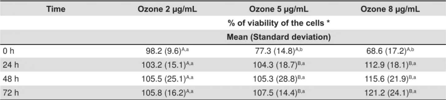
The results are expressed in Table 2. The Group I (control) was considered to be 100%. The results revealed that Groups III and IV provided the greatest decrease in cell viability at 0 hour, from 100% to 77.3% and 68.6%, respectively, which was statistically different from Group I (p<0.05) and different from Group II. After 24 hours, the cell viability increased in Groups II, III, and IV from 98.2%, 77.3%, and 68.6% at 0 hours to 103.2%, 104.3%, and 112.9%, respectively. No statistical
but there was a difference between Groups I and II. After 48 and 72 hours, the cell viability continued
(p<0.05). Groups III and IV provided the greatest decrease in cell viability at 0 hour but the highest increase after 72 hours, with statistical differences among the groups (p<0.05).
Enterococcus faecalis Pseudomonas aeruginosa Staphylococcus aureus
CFU/ml x 105 CFU/ml x 104 CFU/ml x 105 Median
(Interquartile range)
Group I (Control) 7.60A
(1.04 – 10.8)
7.93A (5.83 – 7.70)
6.80A (5.27 – 9.33)
Group II 0.00B
(0.00 – 0.00)
1.44B (0.68 – 6.30)
0.00B (0.00 – 0.32)
Group III 0.00B
(0.00 – 0.00)
1.20B (0.65 – 8.88)
0.00C (0.00 – 0.00)
Group IV 0.00B
(0.00 – 0.00)
0.00C (0.00 – 0.00)
0.00C (0.00 – 0.00)
CFU=colony forming units
'LIIHUHQWXSSHUOHWWHUVLQGLFDWHVWDWLVWLFDOO\VLJQL¿FDQWGLIIHUHQFHVDPRQJWKHJURXSVFRQVLGHULQJWKHVDPHEDFWHULDVSHFLHV (p<0.05 by Kruskal-Wallis test)
Table 1- Counts of colony forming units of Enterococcus faecalis, Pseudomonas aeruginosa, and Staphylococcus aureus from root canals treated with different methods
Time Ozone 2 μg/mL Ozone 5 μg/mL Ozone 8 μg/mL
% of viability of the cells * Mean (Standard deviation)
0 h 98.2 (9.6)A,a 77.3 (14.8)A,b 68.6 (17.2)A,b
24 h 103.2 (15.1)A,a 104.3 (18.7)B,a 112.9 (18.1)B,a
48 h 105.5 (25.1)A,a 105.3 (28.8)B,a 115.6 (21.9)B,a
72 h 105.8 (16.2)A,a 107.5 (14.4)B,a 121.2 (24.1)B,a
* Compared with the viability of the cells in the control group, which was considered to be 100%. Different upper case OHWWHUVLQGLFDWHVWDWLVWLFDOO\VLJQL¿FDQWGLIIHUHQFHVDPRQJWKHGLIIHUHQWSHULRGVFRQVLGHULQJWKHVDPHR]RQHFRQFHQWUDWLRQ S'LIIHUHQWORZHUFDVHOHWWHUVLQGLFDWHVLJQL¿FDQWGLIIHUHQFHVDPRQJGLIIHUHQWR]RQHFRQFHQWUDWLRQVFRQVLGHULQJWKH same period (p<0.05)
DISCUSSION
The scientific literature has already proven that traditional Endodontic therapy is not able to promote the root canal system’s sterilization23-25. Sodium hypochlorite is the most employed solution worldwide due to its extremely potent antimicrobial activity22-25 and tissue dissolution21. However, it has 5,9.
Ozone has been proposed as an alternative for Endodontic treatment due to its potent antimicrobial activity2,6,7,10,12-18,20 as well as its low cytotoxicity within cells, which is the opposite from the highest concentration of sodium hypochlorite, being remarkably cytotoxic22.
In parallel with cytotoxicity assays, ozone’s antimicrobial activity has already been tested17. However, the concentration of ozonated water in the present study was higher than that proposed by the study referred to17. Both studies, Nagayoshi, et at.17
cytotoxicity, but only the present assay evaluated three different concentrations.
The methodological model employed in this study evaluated a single concentration of aqueous ozone and ozone gas in an antimicrobial assay. A prior studyprovided the support to justify this conduct20. This study evaluated three different concentrations of aqueous ozone over En t er ococcu s f aecalis, Pseu d om on as aer u g in osa, and St ap h y lococcu s aur eus, though in planktonic form. Despite the methodological limitations of this previous study,
aqueous ozone in a concentration of 8 μg/mL was
the most effective. Therefore, this information was transferred to the present study.
the effectiveness of the ozonated water against the tested bacteria. The tested microrganisms showed sensitivity to all the applied protocols.
Pseudom onas aeruginosa was the most resistant
(Group I). This data was in accordance with a prior study12 which evaluated the effectiveness of ozone
species. The ozone group provided a decrease in CFU count but it was not able to completely
eliminate Psedom onas aer uginosa. The present
study provided no CFU count in Group IV, employing ozonated water as complement to Endodontic protocol.
standard Endodontic protocols (Group II) for
Pseu d om on as aer u g in osa and St ap h y lococcu s aur eus and the importance of a complementary t h e r a p y t o i m p r o v e r o o t c a n a l s y s t e m
the results achieved by different studies6,12,16,18,20. Rôças and Siqueira23 (2011)studied the complexity
of the Endodontic infection and related it to the necessity of a complementary therapy to optimize the decontamination stage and eliminate the most resistant bacteria located in the root canal system. Thus, ozone was highlighted as a promising tool to
which provided no CFU count for any of the tested microorganisms.
Aqueous ozone retains only 20–25% of the delivered ozone3
prior study20
of 8 μg/mL, the ozone device was calibrated to
produce 40 μg/mL of ozone. According to the
conclusion of this previous study20, this concentration was adopted as a standard for the present assay. The methodological chronology employed first
40 μg/mL and the same concentration was applied
8 μg/mL. The results provided by microbiological
assay encouraged the cytotoxicity analyses over
A better comprehension of ozone’s action mechanism improves the correlation between the results of this study and their clinical appliance. As a variation of oxygen, one of ozone’s greatest features is its oxidative power2,4,19. The oxidative stress, which is caused by its oxidative power, is responsible for the reactive oxygen species (ROS) which are converted into free radicals such as singlet oxygen, hydrogen peroxide, and superoxide anions2,8,19.
Due to the production of free radicals, ozone has a remarkable antimicrobial effect. Singlet oxygen, hydrogen peroxide, and superoxide anions oxidate the polyunsaturated fatty acid of the bacterial cell wall membrane provoking the disruption of the bacterial membrane2,10,13,15,19.
On the other hand, when in contact with cells, ozone delivers full biochemical reactions, and the
mitochondrion is the target1,2. Such a concept
in the present study to evaluate the mitochondrial activity11,17. Previous literature has shown the effects of ozone to produce oxidative stress1,2,1,4,8, which in
vit ro 1,4
viability from 100% to 98.2%, 77.3%, and 68.6% in Groups II, III, and IV, respectively. Free radicals are released to stimulate mitochondrion to produce adenosine triphosphate (ATP). Physiologically, this event represents the cells’ metabolic improvement1 and will improve cell viability17, which has been
These results are in accordance with studies that evaluated the cytotoxicity11,17 and biocompatibility8 of ozone.
Several studies have treated ozone isolated from other chemical substances6,7,10,12-18,20. The aim
therapy but not as a new chemical solution that would substitute any component of the protocol.
statement that ozone is most effective after traditional cleaning, shaping, and irrigation has been completed15.
Pure oxygen is extremely important to improve
the production of ozone1,2,19, thus its clinical
application. The ozone device employed in this study (Philozon, Balneario Camburiú, SC, Brazil)
Odontology. Clinical application in both areas, medicine and Odontology are similar when they cope with ozone as a topical agent. A syringe is
and produce aqueous ozone. Both forms, gas and aqueous are perfectly suitable to apply to clinical sites.
Commercially there are few ozone devices
as feedstock. Therefore the adaptation from medical to dental application must be perfectly suitable due to the simplicity of the technique.
Unfortunatelly, there is a lack of consistent studies to support the clinical application of ozonetherapy due to the lack of clinical trials. The complexity of the Endodontic infection associated to anatomical variations require a complement to Endodontic therapy, as studied23. Thus several doubts surround the therapy. This study, under limitations, opens
6,7,10-18,20 to
purpose ozone as a complementary procedure to improve root canal decontamination as part of the chemicalmechanical preparation.
CONCLUSION
According to the applied methodology and the limitations of this study, it was possible to conclude that in association with standard Endodontic
ozone, improved the decontamination of the root canal ex v iv o. In parallel, ozone was toxic to the gingival fibroblast on first contact, but the cell viability was recovered by the end of the
ozone has the potential to take part in Endodontic treatment, but further studies, especially clinical trials, must be encouraged.
ACKNOWLEDGMENTS
The authors would like to thank São Paulo
support of this study. Process number: 06/04205-2
REFERENCES
1- Bocci V. Ozone as Janus: this controversial gas can be either 2- Bocci V. Physical-chemical properties of ozone. Natural production of ozone. The toxicology of ozone. In:______Ozone, a new medical drug. Italy: Springer; 2005. p. 5-8.
3- Bocci V. Preparation of ozonated water and oil for the topical therapy. Ozone as a drinking water disinfectant. Ozone disinfection to prevent nosocomial infections. In: ______Ozone, a new medical drug. Italy: Springer; 2005. p. 12-18.
4- Bocci V, Zanardia I, Valacchi G, Borrelli E, Travagli V. Validity of oxygen-ozone therapy as integrated medication form in chronic 2015;15(2):127-38.
5- Botton G, Pires CW, Cadoná FC, Machado AK, Azzolin VF, Cruz IB, et al. Toxicity of irrigating solutions and pharmacological associations used in pulpectomy of primary teeth. Int Endod J. 2016;49:746-54.
6- Cardoso MG, Oliveira LD, Koga-Ito CY, Jorge AO. Effectiveness of ozonated water on Candida albicans, Ent erococcus faecalis, and endotoxins in root canals. Oral Surg Oral Med Oral Pathol Oral Radiol Endod. 2008;105:e85-91.
7- Case PD, Bird PS, Kabler WA, Georde R, Walsh LJ. Treatment
Ent erococcus faecalis with ozone gas and passive ultrasound activation. J Endod. 2012;38:523-6. 8- Frascino AV, Mantesso A, Corrêa L, Deboni MC. Aqueous-ozone irrigation of bone monocortical wounds in hyperglycemic rats. Acta Cir Bras. 2013;28(5):327-33.
9- Gomes-Filho JE, Aurélio KG, Costa MM, Bernabé PF. Comparison of the biocompatibility of different root canal irrigants. J Appl Oral Sci. 2008;16(2):137-44.
of aqueous ozone in root canals infected by Ent erococcus faecalis. J Microbiol. 2014:7(7):e11411.
11- Huth KC, Jakob FM, Saugel B, Cappello C, Paschos E, Hollweck R, et al. Effect of ozone on oral cells compared to established antimicrobials. Eur J Oral Sci. 2006;114:435-40.
12- Huth KC, Quirling M, Maier S, Kamereck K, AlKhayer M, Paschos E, et al. Effectiveness of ozone against endodontopathogenic 2009;42:3-13
13- Kaptan F, Güven EP, Topcuoglu N, Yazier M, Külekçi G. I n vit ro
assessment of the recurrent doses of topical gaseous ozone in the removal of Ent erococcus faecalis
Clin Pract. 2014;17(5):573-8.
temperature atmospheric pressure plasma on root canals infected with Ent erococcus faecalis. Lett Appl Microbiol. 2013;58:8-15. 15- Lynch E. Comment on “The application of ozone in dentistry: a systematic review of the literature”. J Dent. 2009;37(5):406-10. 16- Mirhadi H, Abbaszadegan A, Ranjbar MA, Azar MR, Geramizadeh B, Torabi S, et al. Antibacterial and toxic effect of hydrogen peroxide combined with different concentrations of chlorhexidine in comparison with sodium hypochlorite. J Dent. 2015:16(4):349-55.
18- Niewierowski RS, Scalzilli LR, Morgental RD, Figueiredo JA, Vier-Pelisser FV, Borba MG, et al. Bovine pulp tissue dissolution ability of irrigants associated or not to ultrasonic agitation. Braz Dent J. 2015:26(5):537-40.
19- Nogales CG, Ferrari PH, Kantorovich EO, Lage Marques JL. Ozone therapy in medicine and dentistry. J Contemp Dent Pract. 2008;19(4):75-84.
20- Nogales CG, Ferreira MB, Lage-Marques JL, Antoniazzi JH. Comparison of the antimicrobial activity of three different concentrations of aqueous ozone on Pseudom onas aeruginosa,
St aphylococcus aureus and Ent erococcus faecalis - in vit ro study. Rev Espan Ozon. 2014:4(1);9-15.
21- Noites R, Vaz C, Rocha R, Carvalho MF, Gonçalves A, Pina-Vez I. Synergistic antimicrobial action of chlorhexidine and ozone in Endodontic treatment. Biomed Res Int. 2014;2014:592423.
22- Prado M, Silva EJ, Duque TM, Zaia AA, Ferraz CC, Almeida JF, et al. Antimicrobial and cytotoxic effects of phosphoric acid solution compared to other root canal irrigants. J Appl Oral Sci. 2015:23(2);158-63.
23- Rôças IN, Siqueira Jr JF. I n v iv o antimicrobial effects of Endodontic treatment procedures as assessed by molecular microbiologic techniques. J Endod. 2011;37:304-10.
24- Rodrigues E, Albergaria A, Barbosa GL, Santoro M, Pacheco P, Leal Silva EJ. Effectiveness of different formulations of Endo-PTC to promote root canal cleaning. Indian J Dent Res. 2015:26(5):520-3. 25- Saxena D, Saha SG, Saha Mk, Dubey S, Khatri M. An in vit ro
comparison of their activity with 2.5% sodium hypochlorite against