I
ABSTRACT
EVALUATION OF OCULAR PROSTHESIS BIOFILM AND
ANOPHTHALMIC CAVITY CONTAMINATION AFTER USE
OF THREE CLEANSING SOLUTIONS
Regina Márcia Zuccolotto Felippe PARANHOS1, Carlos Henrique BATALHÃO2, Marisa SEMPRINI3, Simone Cecílio Hallak REGALO3, Izabel Yoko ITO4, Maria da Glória Chiarello de MATTOS5
1- DDS, MSc, Graduate student, Department of Dental Materials and Prosthodontics, School of Dentistry of Ribeirão Preto, University of São Paulo, Ribeirão Preto, SP, Brazil.
2- DDS, Undergraduate student, School of Dentistry of Ribeirão Preto, University of São Paulo, Ribeirão Preto, SP, Brazil.
3- DDS, MSc, PhD, Full Professor, Department of Morphology, Stomatology, and Physiology, School of Dentistry of Ribeirão Preto, University of São Paulo, Ribeirão Preto, SP, Brazil.
4- DDS, MSc, PhD, Full Professor of Microbiology and Immunology, Department of Clinical Analysis, Toxicology and Bromatology, School of Pharmaceutical Sciences of Ribeirão Preto, University of São Paulo, Ribeirão Preto, SP, Brazil.
5- DDS, MSc, PhD, Full Professor, Department of Dental Materials and Prosthodontics, School of Dentistry of Ribeirão Preto, University of São Paulo, Ribeirão Preto, SP, Brazil.
Corresponding address: Profa. Dra. Maria da Gloria Chiarello de Mattos - Faculdade de Odontologia de Ribeirão Preto, Universidade de São Paulo - Avenida do Café s/nº Monte Alegre, Cep.: 14040-904 - Ribeirão Preto, SP - Fax: 55 16 3633 0999 - e-mail: [email protected]
Received: January 16, 2006 - Modification: July 18, 2006 - Accepted: February 22, 2007
n addition to an initial socket discomfort, ocular prosthesis (OP) installation may allow the adherence of fungi and/or bacteria due to the superficial characteristics of the prosthesis’ material, use of inadequate cleansing solutions and methods, or because the void located between the internal portion of the prosthesis and the anophthalmic cavity (AC) mucosa. Objective: The aim of this study was to evaluate OP biofilm formation and the level of contamination of the internal portion of the OP and the AC in 24 patients. Material and Methods: Material was collected from the AC at the beginning of the study and 15 days after cleansing of the OP with 3 cleansing solutions: a neutral liquid soap, a multiuse solution for contact lens (Complete) and 0.12% chlorhexidine (Periogard). The collected materials were sowed in Petri dishes containing selective media for aerobic and facultative microorganisms, specifically staphylococci (Hipersalt agar with egg yolk), aerobic microorganisms (Brain Heart Infusion Blood Agar), streptococci (Mitis salivarius Agar), gram-negative bacilli (MacConkey Agar) and yeasts (Chromagar Candida™), incubated at 35oC or 37oC and the number of colony forming units were counted. Data were analyzed statistically
by ANOVA, Friedman’s test and Spearman’s correlation. Results: Aerobic microorganisms, gram-negative bacilli and S. aureus were found in the OP biofilm and in the AC. There was statistically significant difference (p<0.05) between the number of microorganisms before and after the use of the cleansing solutions. Conclusion: There was positive correlation with respect to the microorganisms present in the OP biofilm and AC for the 4 proposed treatments, indicating that the decrease of OP contamination leads to AC contamination as well.
Uniterms: Artificial eye; Eye infections; Prosthesis-related infections.
INTRODUCTION
Ocular prosthesis (OP) is an artificial replacement for the bulb of the eye and has the goal of reestablishing facial esthetics while maintaining the form of the anophthalmic cavity (AC), preserving the palpebral muscle tone, inhibiting palpebral collapse, directing tear drainage, preventing fluid accumulation in the socket and aiding the patient’s social contact7.
OP wearers are preset to infections, inflammations and traumas related to physiological and morphological modifications of the AC; AC colonization by pathogenic
microbiota; impaired mobility to inadequate prosthesis installation; neglected prosthesis cleansing without removal from the socket for months or years and no washing; and accumulation of secretion, which may cause giant papillary conjunctivitis and consequent intolerance to prosthesis use6,9,10,12-14.
MATERIAL AND METHODS
Twenty-four OP wearers of both genders with mean age of 44 years were selected from the Rehabilitation Service for Patients with Mutilations of the Face, Head and Neck Regions of the Department of Dental Materials and Prosthodontics (FORP/USP) and followed-up during a 45-day period. Patients attended four visits (0, 15, 30 and 45 days), in which biofilm was collected from the internal surface of the OP as well as from the AC. For collection of material on day 0 (I – Initial), patients did not receive any hygiene instruction. After collection, the patients received cleansing solutions for the OP and were oriented how to clean it 4 times a day, during 15 days. The first solution used, after the initial material collection, was a neutral liquid soap (LS) (Daterra, Ribeirão Preto, São Paulo, Brazil). The patients were instructed to put the LS on their clean hand palms and dab it on the prosthesis for 1 minute, rinsing in running water thereafter. The second solution used was a multiuse solution (MS) for cleansing of contact lenses (Complete, Allergan, Guarulhos, São Paulo, Brazil) and the third was a 0.12% chlorhexidine solution [Periogard (P), Colgate, São Paulo, SP, Brazil]. The cleansing instructions were similar to those given to LS, except for the fact that MS was not rinsed after application, according to the manufacturer’s instructions. Material was collected from the OP and AC after 15 days of use of each solution. After the last solution was used for 15 days, the patients attended the fourth appointment and the final collections were performed.
In preparation for material collection, the patients were instructed to wash their hands (water and soap) and the antisepsis was made with a 68% alcohol gel. The OP was removed by the patient, placed in a sterile Petri dish (20x100mm) and taken to an aseptic zone (obtained by 2 alcohol lamps). Collection of the AC material and the biofilm from the internal portion of the OP was done by a single operator with a sterile swab (DME – Diagnósticos Microbiológicos Especializados, Araçatuba, São Paulo, Brazil), maintaining the same frequency of movements during 5 minutes. Still in the aseptic zone, the swab was introduced into a test tube containing 2.0 mL of Letheen Broth – Calet (Difco, Detroit, Michigan, USA) and was forwarded to the laboratory packed in ice-filled polystyrene boxes to ensure an adequate conservation.
Thereafter, the OPs were polished with pumice (Vigodent, Rio de Janeiro, RJ, Brazil) and Kaolin (ARJ Chemical do Brazil Ltda, Rio de Janeiro, RJ, Brazil), washed with water and soap, rinsed in running tap water and given back to the patients.
The collected material was agitated for 1 minute in a shaker (Mixtron Toptronix, São Paulo, SP, Brazil) and submitted to decimal dilution up to 10-4. These suspensions
were dropped in equidistant points on the Petri dish in a volume of 50 µL, according to Westergren and Krasse15
(1979) onto MacConkey Agar (Mc; Difco, Detroit, Michigan, USA), Chromagar Candida™ (Cm; CHROMagar, Paris, France), Brain Heart Infusion Blood Agar (As; Difco, Detroit,
Michigan, USA), Hipersalt Agar with egg yolk (Ni) according to Ito, et al.5 (1979) and Mitis salivarius Agar (Ms; Difco,
Detroit, Michigan, USA).
About 4.0 mL of thioglycollate medium without dextrose or indicator (Tio’s; Difco, Detroit, Michigan, USA) were added to the remaining material and incubated at 37ºC for 10 days for detection of less than 20 microorganisms.
Mc, As, and Ni media were incubated at 37ºC for 24 to 48 hours, while Cm medium was incubated at 35ºC for 48 to 72 hours. Ms medium was incubated at 37ºC for 24 to 96 hours under capnophilic conditions by the candle jar system. After incubation, the material was examined with a stereomicroscope (Nikon, Tokyo, Japan) under reflected light and the number of colony forming units (cfu) was counted. Data were analyzed statistically by analysis of variance, Friedman’s test and Spearman’s correlation. Significance level was set at 5%.
RESULTS
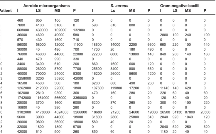
Aerobic microorganisms, S. aureus and gram-negative bacilli were detected in the OP biofilm and in the AC (Table 1 and 2).
The statistical results of ANOVA for aerobic microorganisms are shown on Table 3 while the statistical results of Friedman’s test for S. aureus and gram-negative bacilli are shown on Table 4. The analysis of the results showed that the initial condition (I) was statistically different from the use of the cleansing solutions (LS, MS, and P) (p<0.05). The results of the Spearman’s correlation for the microorganisms present in the OP biofilm and AC for the 4 proposed treatments (I, LS, MS, and P) (Table 5) showed a positive correlation, indicating that as the number of microorganisms on OP surface increased, the number of microorganisms in the AC increased accordingly, in any of the tested conditions. The inverse also occurred. A numerical comparison was made and confirmed this correlation (Table 6).
DISCUSSION
In the present study, the varied microbiota observed in the OP biofilm (aerobic microorganisms, gram-negative bacilli and Staphylococcus aureus) as well as the presence of these microorganisms in the AC are results consistent with the literature1-3,6,9,10,12,13.
OP wearers may present a pathogen microbiota in the AC, mainly those who neglect the cleansing of prosthesis, not removing them for days, months or even years, sometimes leading to an intolerance to prosthesis use1,3,6,9,10,12-14. The results of this study agree with those of
these authors1,3,6,9,10,12-14regarding the fact that, despite the
Aerobic microorganisms S. aureus Gram-negative bacilli
Patient I LS MS P I Ls MS P I LS MS P
1 960 160 250 100 0 0 0 0 0 0 0 0
2 3000 7400 3800 0 1440 4800 3200 0 0 0 0 0
3 54000 21900 1900 17100 0 0 0 0 0 0 0 0
4 12000 850 3300 5900 0 0 0 0 1120 600 760 4260
5 90000 180 840 100 0 0 0 0 0 0 0 0
6 72000 7300 2130 5200 1800 2600 3800 3400 102 60 180 180
7 24000 40 340 80 11800 20 180 40 0 0 0 0
8 44000 10600 22000 980 21000 1600 6000 40 0 0 0 0
9 170 380 20 80 0 0 0 0 0 0 0 0
10 440 220 590 0 430 60 170 0 0 0 0 0
11 28000 58000 74000 3300 15600 3800 800 20 0 0 0 0
12 42000 66000 44000 12000 26000 34400 12000 2800 0 0 0 0
13 38000 790 1840 6000 0 0 0 0 0 0 0 0
14 28000 5000 340 2800 4000 1200 180 1000 0 0 0 0
15 3004000 4658000 402000 80 179000 656400 144200 0 15780 31200 220 0
16 92400 6200 8600 570 1600 1600 980 700 100 960 810 40
17 578000 226000 74000 910000 0 0 0 0 0 0 0 0
18 642000 56000 60000 5300 125000 1400 2660 20 3780 5740 140 760
19 5500 420 80 180 0 0 0 0 0 0 0 0
20 1810000 2132000 2476000 2962000 15700 11000 1800 1800 19940 13580 378000 17020
21 14000 1400 7000 2000 2600 680 1200 80 80 40 1020 60
22 480 790 500 280 0 0 0 0 0 0 0 0
23 14700 720 1110 3000 0 0 0 0 0 0 0 0
24 34000 590 560 330 520 0 0 0 140 80 60 60
TABLE 1- Cfu counting for aerobic microorganisms, S. aureus and gram negative bacilli on the ocular prosthesis
I = Initial; LS = Liquid Soap; MS = Complete Multiuse Solution; P = Periogard
Aerobic microorganisms S. aureus Gram-negative bacilli
Patient I LS MS P I Ls MS P I LS MS P
1 460 650 100 120 0 0 0 0 0 0 0 0
2 7800 4100 3100 0 590 810 800 0 0 0 0 0
3 668000 430000 102000 132000 0 0 0 0 0 0 0 0
4 36000 4600 40000 580 0 0 0 0 2800 100 240 100
5 570 430 1600 710 0 0 0 0 0 0 0 0
6 86000 58000 12000 11900 18600 14000 2200 6600 660 220 100 140
7 30000 40 480 700 1700 20 180 490 0 0 0 0
8 76000 46000 44000 22000 22200 6000 13800 140 840 200 140 180
9 440 470 990 330 0 0 0 0 0 0 0 0
10 3400 3400 610 200 860 1600 600 120 0 0 0 0
11 88000 26000 8700 7700 25600 8400 800 660 0 0 0 0
12 40000 70000 24000 5300 16200 26000 5600 1200 0 0 0 0
13 1258000 3200 35900 42000 0 0 0 0 0 0 0 0
14 16500 4500 560 180 6200 600 490 260 0 0 0 0
15 1262000 212000 22000 1800 107800 119800 17200 0 11140 140 620 0
16 102000 2810 9300 360 470 160 280 20 220 60 40 80
17 1742000 1164000 566000 3360000 0 0 0 0 120 80 40 20
18 28000 3700 1600 6000 6200 370 260 20 300 40 100 220
19 10800 40 380 280 0 0 0 0 0 0 0 0
20 3182000 4178000 4024000 3266000 18680 21200 24600 19000 26500 31130 87800 20500
21 5600 3900 44000 18000 31800 2800 25800 340 2040 920 1040 120
22 20000 9800 36000 18000 580 40 20 20 0 0 0 0
23 32000 1800 1890 9700 0 0 0 0 2040 520 250 620
24 42000 610 500 260 850 60 0 0 1180 20 40 40
I = Initial; LS = Liquid Soap; MS = Complete Multiuse Solution; P = Periogard
the cleansing solutions (neutral liquid soap, multiuse solution and Periogard) were compared to the initial condition (no cleansing).
Few studies have addressee OP cleansing methods, the most common being the use of water and soap11,12. Removal
of the OP, use of solutions indicated for cleansing of contact lenses and periodical examination by a health professional have also been recommended to ensure the proper cleansing and assess the integrity of tissues that cover the AC and the need for changing the prosthesis4,6,8,13.
The findings of the present study showed that the use of a contact lens multiuse solution for cleansing of the OP yielded a decrease in the number of microorganisms on both the prosthesis and the anophthalmic cavity.
The use 0.12% chlorhexidine is widely widespread for
chemical biofilm control because of its bacteriostatic action against gram-positive and gram-negative microorganisms. Periogard was used in this study because it is a readily available product that does not offer risks to patients’ health. After use of Periogard, the biofilm presented a smaller number of colony forming units in comparison to the initial condition. These results suggest that, although it does not differ significantly from the other solutions, Periogard had an evident bacteriostatic effect, given that, after its use, bacterial growth in the biofilm or AC was less intense compared to the use of the other solutions. In some cases, no bacterial growth was observed.
There are no studies referring to the correlation between the presence of microorganisms in the OP or AC and cleansing solutions that could serve as a parameter to the
(H0) Probability Anophthalmic Cavity Ocular Prosthesis
Among Patients 0.0000% 0.0000% *
Among Solutions 0.0009% 0.0009% *
TABLE 3- Results of the analysis of variance for the presence of aerobic microorganisms
*Statistically significant at 1% level
Two-by-two comparisons S. aureus Gram-negative bacilli x
AC OP AC OP
I x LS n s n s 1% n s
I x MS 1% 5% 1% n s
I x P 0.1% 0.1% 1% n s
LS x MS n s n s n s n s
LS x P 0.1% 1% n s n s
MS x P 5% 5% n s n s
TABLE 4- Results of the Friedman’s test for the presence of S. aureus and gram-negative bacilli
Ns = non-significant. I = Initial; LS = Liquid Soap; MS = Complete Multiuse Solution; P = Periogard. AC= Anophthalmic Cavity; OP= Ocular Prosthesis
Two-by-two comparisons Ho Probability
Aerobic microorganisms S. aureus Gram-negative bacilli
I AC X OP 0.0000% 0.0000% 0.0000%
LS AC X OP 0.2800% 0.0000% 0.0400%
MS AC X OP 0.0200% 0.0000% 0.0000%
P AC X OP 0.0000% 0.0000% 0.0000%
TABLE 5- Results of Spearman’s correlation test for the presence of aerobic microorganisms, S. aureus and gram-negative
bacilli
outcomes of the present study. Portellinha, et al.13 (1984)
correlated the presence of secretion with the time of use and the frequency of prosthesis cleansing and found that the bacterial colonization in the AC and the frequency of OP cleansing had no statistically significant correlation. Campos2 (1994) did not find a positive correlation between
the time of use of OP and the presence of microorganisms. In the present investigation, a positive correlation was found between the microorganisms on the prosthesis/ cavities and the four types of treatment (no cleansing and three cleansing solutions), which indicates that the decrease of OP contamination would lead to a decrease of AC contamination. Therefore, OP cleansing is essential to reduce
contamination of AC, improving the comfort of OP wearers and consequently their life quality.
CONCLUSIONS
1. Aerobic microorganisms and gram-negative bacilli were found in OP biofilm as well as in the AC before and after the use of the studied cleansing solutions.
2. All solutions were similarly effective in decreasing the number of aerobic microorganisms in the OP and AC compared to the initial condition;
3. Periogard and MS decreased of the number of S.
Two-by-two comparisons Aerobic microorganisms S. aureus Gram-negative bacilli
OP AC OP AC OP AC
I 24 + 24 + 14 + 15 + 8 + 11 +
10 - 9 - 16 13
-I X LS 17 ↓ 19↓ 8↓ 10↓ 5↓ 10↓
7 ↑ 4 ↑ 4 ↑ 5 ↑ 3 ↑ 1 ↑
1 = 1 = 9 - 16 - 13
11
-I X MS 17 ↓ 19↓ 11 ↓ 12↓ 4↓ 10↓
7↑ 5↑ 2↑ 2↑ 4↑ 1↑
11- 10 - 16 - 13
-LS X MS 11 ↓ 14↓ 7↓ 9↓ 4↓ 5↓
13↑ 10↑ 6↑ 5↑ 4↑ 6↑
11 - 10 - 16 - 13
-P X I 20 ↓ 20↓ 9↓ 11 ↓ 5↓ 10↓
3↑ 2↑ 1↑ 1↑ 2↑ 14 –
1 - 2 - 14 - 12 - 17
-P X LS 16 ↓ 14↓ 8↓ 12↓ 3↓ 5↓
6↑ 9↑ 2↑ 12 – 4 ↑ 4↑
2 - 1 - 14 - 17 - 1 =
14
-P X MS 14 ↓ 16↓ 8↓ 9↓ 4↓ 4↓
8↑ 7↑ 1↑ 2↑ 2↑ 5↑
2 - 1 - 1 = 1 = 1 = 5 ↑
14 - 12 - 17 14
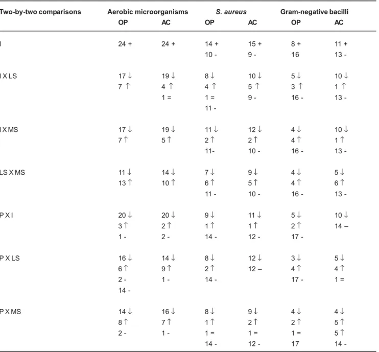
-TABLE 6- Comparison of the number of cfu of aerobic microorganisms, S. aureus and gram-negative bacilli in the OP biofilm
and AC before and after use of the cleansing solutions
aureus in the OP and AC compared to the initial condition; 4. There was no significant difference between the initial condition and the studied solutions regarding the presence of gram-negative bacilli in the OP biofilm; for AC, the three solutions were yielded better outcomes than the initial condition.
5. Under the tested conditions, there was a positive correlation for the presence of aerobic microorganisms, S.
aureus and gram-negative bacilli.
REFERENCES
1- Akman A, Irkec M, Orhan M, Erdener U. Effect of lodoxamide on tear leukotriene levels in giant papillary conjunctivitis associated with ocular prosthesis. Ocul Immunol Inflamm. 1998;6(3):179-84.
2- Campos MS, Campos e Silva LQ, Rehder Jr, Lee MB, O’brien T, McDonnell PJ. Anaerobic flora of the conjunctival sac in patients with AIDS and with anophthalmia compared with normal eyes. Acta Ophthalmol (Copenh). 1994;72(2):241-5.
3- Campos MSQ. Microbiota bacteriana anaeróbica do saco conjuntival humano normal, de cavidades anoftálmicas de usuários de prótese ocular e de pacientes aidéticos [tese]. São Paulo (SP): Escola Paulista de Medicina, Universidade Federal de São Paulo; 1990.
4- Goldfarb HJ, Turtz AI. A detergent- lubricant solution for artificial eyes. Am J Ophthalmol. 1966;16(6):1502-5.
5- Ito Y, Costa A, Barachini O. Emprego da gema de ovo no isolamento deStaphylococus. Ann Microbiol. 1979;16:189-92.
6- Kara José N, Prado J Júnior, Sampaio M W. Intolerância ao uso de prótese ocular pelo desenvolvimento de conjuntivite papilar gigante. Rev Bras Oftalmol. 1980;39(1):51-3.
7- Kohlhaas M, Schulz D. The complex facial prosthesis: the value of bone-anchored maxillofacial prostheses in the treatment of extensive loss of facial tissue. Rev Stomatol Chir Maxillofac. 2001;102(5):261-5.
8- Maimone N, Maimone AL. Avaliação de um novo produto na desinfecção do tonômetro de aplanação de Goldmann. Arq Bras Oftalmol. 2001;64(6):545-9.
9- Morris R, Camesasca FI, Byrne J, John G. Postoperative endophthalmitis resulting from prosthesis contamination in a monocular patient. Am J Ophthalmol. 1993;116(3):346-9.
10- Neves ACC. Avaliação clínica e microbiológica da secreção conjuntival em usuários de prótese ocular em resina acrílica [tese]. São José dos Campos (SP): Faculdade de Odontologia de São José dos Campos, Universidade Estadual Paulista “Júlio de Mesquita Filho”; 2000.
11- Nuti A Sobrinho, Lima EG, Mattos MGC, Watanabe S. A study of ocular prostheses. I. Manufacture and indications for use. Rev Fac Odontol Ribeirão Preto. 1986;23(2):135-43.
12- Nuti A Sobrinho. Contribuição ao estudo de microorganismos encontrados em prótese ocular de resina acrílica e em cavidade anoftálmica [tese]. Ribeirão Preto (SP): Faculdade de Odontologia de Ribeirão Preto, Universidade de São Paulo; 1986.
13- Portellinha WM, Belfort JR, Cai S, Novo NF. Estudo clínico – microbiológico, citológico e de função lacrimal em pacientes com cavidade anoftálmica e uso de prótese ocular de acrílico. Arq Bras Oftalmol. 1984;47(4):159-63.
14- Sarac O, Erdener U, Irkec M, Gungen Y. Tear eotaxin levels in giant papillary conjunctivitis associated with OP. Ocul Immunol Inflamm. 2003;11(3):223-30.