93
CYTOTOXIC, ANTIMIGRATORY, PRO-AND ANTIOXIDATIVE ACTIVITIES
OF EXTRACTS FROM MEDICINAL MUSHROOMS ON COLON CANCER CELL LINES
Dragana S. Šeklić*, Milan S. Stanković, Milena G. Milutinović, Marina D. Topuzović, Andraš Š. Štajn and Snežana D. Marković
Department of Biology and Ecology, Faculty of Science, University of Kragujevac, Kragujevac, Serbia. *Corresponding author: draganadjacic@kg.ac.rs
Received: April 27, 2015; Revised: July 1, 2015; Accepted: July 13, 2015; Published online: November 27, 2015
Abstract:Methanol extracts of five commercially available mushroom species (Phellinus linteus (Berk. et Curt) Teng, Cordy-ceps sinensis (Berk.) Sacc., Lentinus edodes (Berk.) Pegler, Coprinus comatus (O. F. Müll.) Pers. and Ganoderma lucidum (Curtis) P. Karst), traditionally used as anticancer agents, were evaluated in vitro for their total phenol and flavonoid con-tents, cytotoxic and antimigratory activities and antioxidant/prooxidant effects on colon cancer cell lines (HCT-116 and SW-480). Spectrophotometric methods were used for the determination of total phenol content, flavonoid concentrations and DPPH activity of the extracts. Cytotoxic activity was measured by the MTT assay. The antimigratory activity of extracts was determined using the Transwell assay and immunofluorescence staining of β-catenin. The prooxidant/antioxidant status was followed by measuring the superoxide anion radical (O2•–), nitrite and reduced glutathione (GSH) concentrations. Our results show that the highest phenolic and flavonoid content was found in P. linteus, and its DPPH-scavenging capacity was significantly higher than in other samples. The P. linteus extract significantly decreased cell viability of both tested cancer cell lines. All other extracts selectively inhibited SW-480 cell viability, but did not show significant cytotoxic activity. The mushroom extracts caused changes in the prooxidant/antioxidant status of cells, inducing oxidative stress. All extracts tested on HCT-116 cells demonstrated significant antimigratory effects, which correlated with increased production of O2•– and a reduced level of β-catenin protein expression, while only P. linteus showed the same effect on SW-480 cells. The results of the present research indicate that the mushroom extracts causes oxidative stress which has a pronounced impact on the migratory status of colon cancer cell lines.
Key words: colon cancer; cytotoxicity; migration; mushrooms extracts; oxidative stress
INTRODUCTION
Cancer is a major health problem in the world. In this context, some prized mushrooms with cytotoxic properties and their compounds are interesting for research. Meshima (Phellinus linteus (Berk. et Curt.) Teng) – PL, caterpillar fungus (Cordyceps sinensis
(Berk.) Sacc.) – CS, Shiitake mushrooms (Lentinus edodes (Berk.) Pegler) – LE, shaggy ink cap (Coprinus comatus (O.F. Müll.) Pers.) – CC, and lingzhi/reishi
(Ganoderma lucidum (Curtis) P. Karst) – GL, are just
some of the many mushrooms that have played an important role in the treatment of ailments. Medicinal properties of mushrooms included their antitumor,
proapoptotic (Wasser, 2010), immunomodulating (Rowan et al., 2003), antiviral (Cheung et al., 2003), antioxidant and free radical scavenging (Kamilya et al., 2006) effects. Mushrooms accumulate a variety of bioactive primary and secondary metabolites, in-cluding mineral compounds, organic acids, vitamins, polysaccharides, proteins, fats, oils, phenols like flavo-noids and phenolic acids (Wasser, 2010). Mushrooms prevent cancer genesis, show direct antitumor activity and prevent tumor metastasis (Wasser, 2010).
com-pounds to act as antioxidants has been well established (Smith et al., 2003). Also, mushroom phenolics were found to be an excellent antioxidant, but they are not mutagenic (Kamilya et al., 2006). Although phenol compounds may have antioxidative properties, some of them are able to activate the production of reactive oxygen species (ROS) in cells (Kamilya et al., 2006). This is one of the reasons for studying the antioxidant properties of different extracts from various mush-room fruiting bodies.
Cancer cell migration and invasion are initial steps in metastasis, which is a primary cause of cancer-re-lated death. During metastasis, primary tumor cells migrate and invade neighboring tissues, entering the circulation to establish new or secondary tumor sites (Bacac et al., 2008). The signaling pathway involved in the etiology of colon cancer is Wnt/β-catenin, and 90% of colon cancers had mutations that resulted in activation of this pathway (Giles et al., 2003). An important protein in the Wnt/β-catenin pathway is β-catenin, and mutations of this pathway generally effect β-catenin phosphorylation and stability (Liu et al., 2002). Degradation of phosphorylated β-catenin takes place via the ubiquitin pathway. If degradation is not efficient, as is the case in genetic mutations of adenomatous polyposis coli (APC) or β-catenin, the β-catenin accumulates and is transported to the nu-cleus, where it acts as a transcription factor together with T-cell factor/lymphocyte enhancer binding fac-tor (TCF/LEF). The resulting β-catenin-TCF/LEF complex activates TCF target genes, which affect cell proliferation, survival, angiogenesis, invasion and me-tastasis (Behrens et al., 1996).
Reactive oxygen species (ROS) as signaling mole-cules in cells have a significant impact on the migrato-ry properties of cancer cells (Alexandrova et al., 2006). Several studies investigated the effects of ROS on cell migration, but different results have been reported. As Kamilya et al. (2006) concluded, the increased ROS in the tumor microenvironment was associated with increased aggressiveness of cancer cells. In addition, some findings indicated that different compounds from mushrooms promoted cell aggregation, inhib-ited cell adhesion and suppressed cell migration in a
dose-dependent manner in human colon cancer cell lines (Chen et al., 2011).
The aim of this study was to determine the bio-logical effects (cytotoxic, antimigratory and pro/an-tioxidant potential) of methanolic extracts from the fruiting body of five commercially available species of mushrooms on colon cancer cell lines HCT-116 and SW-480. Furthermore, the effects of extracts on the expression of nuclear and cytoplasmic β-catenin were investigated.
MATERIALS AND METHODS
Materials
Methanol and sodium hydrogen carbonate were pur-chased from Zorka Pharma, Šabac, Serbia. 2,2-diphe-nyl-1-picrylhydrazyl (DPPH), gallic acid (Ga), Folin-Ciocalteu reagent, aluminum chloride hexahydrate (AlCl3) and rutin (Ru) were purchased from Fluka Chemie AG, Buchs, Switzerland. 3-[4,5-dimethylthi-azol-2-yl]-2,5-diphenyltetrazolium bromide (MTT), nitroblue tetrazolium (NBT), N-(1-naphthyl) eth-ylenediamine, NaNO3, N-(1-naphthyl) ethylene-diamine, sulfanilic acid, phosphoric acid, dimethyl sulfoxide (DMSO), bovine serum albumin (BSA), Triton X-100, 2-(N-morpholino)ethanesulfonic acid (MES), crystal violet, formaldehyde, acetic acid and cisplatin were obtained from Sigma Chemicals Co., St Louis, MO, USA. Polyvinyl alcohol mounting medi -um was obtained from Fluka Analytical, Switzerland, p-formaldehyde from Merck, Germany. Dulbecco’s modified Eagle medium (DMEM), fetal bovine se -rum (FBS), penicillin and streptomycin were obtained from Gibco, Invitrogen, New York, USA. All other solvents and chemicals were of analytical grade. The colon cancer cell lines HCT-116 and SW-480 were ob-tained from the American Tissue Culture Collection.
Preparation of mushroom extracts
-formed with 250 mL of methanol at room temperature for a period of 24 h. The extracts were filtered using Whatman No.1 filter paper and evaporated in rotary vacuum evaporator at 40°C.
Preparation of extracts solutions
Stock solutions of the mushroom fruiting body meth-anol extract, and cisplatin, as positive control, were made in DMSO and DMEM medium at a concentra -tion of 1 mg/mL filtered through a 0.22-mm Millipore filter before use, and diluted by a nutrient medium to various working concentrations. The concentration of DMSO in the most concentrated working solutions was 0.5% (v/v), and not cytotoxic for cells (Hostanska et al., 2007).
Determination of total phenol content and flavonoid concentrations of the extracts
The phenol content of the extracts was determined spectrophotometrically, using Folin–Ciocalteu reagent (Singleton et al., 1999). Briefly, 0.5 mL of methanolic extract solution (1 mg/mL) was added to 2.5 mL of 1:10 Folin-Ciocalteu reagent and then 2 mL of sodium carbonate (7.5%) were added. After 15 min of incuba-tion at 45°C, the absorbance at 765 nm was measured. The total phenolic concentration of plant extracts was expressed as gallic acid equivalents (GAE)/g of ex -tract. The total flavonoid concentration was evalu-ated using AlCl3 (Quettier et al., 2000). The sample for determination was prepared by mixing 1 mL of methanolic solution (1 mg/mL) of the extract and 1 mL of AlCl3(20%). After 1 h of incubation at room temperature, the absorbance at 415 nm was measured. The flavonoid content of plant extracts was expressed as rutin equivalent (RuE)/g of extract (dry weight).
Evaluation of DPPH-scavenging activity
The ability of the extract to scavenge DPPH free radi-cals was assessed using the adopted method with suit-able modifications (Kumarasamy et al., 2005). The stock solution of the extract was prepared in metha-nol to achieve the concentration of 1 mg/mL. Dilutions
were made to obtain concentrations of 500, 250, 125, 62.5, 31.25, 15.62, 7.81, 3.90, 1.99 and 0.97 µg/mL. Di -luted solutions (1 mL each) were mixed with 1 mL of DPPH methanolic solution. After 30 min in darkness at room temperature (23°C) the absorbance was measured at 517 nm. The control samples contained all the re-agents except the extract. The percentage of inhibition was calculated using the equation: % inhibition = 100 x (A control–A sample)/A control), while IC50 values were estimated from the % inhibition versus concentration sigmoid curve, using a non-linear regression analysis.
Cell preparation and culturing
Cells were propagated and maintained in DMEM supplemented with 10% FBS and antibiotics in a hu-midified atmosphere of 5% CO2 at 37°C. Cells were grown and after a few passages, were seeded in well plates. All experiments were done with cells at 70-80% confluence.
HCT-116 and SW-480 cells were seeded (104 cells per well for MTT, NBT and Griess tests, and 5×10⁴ cells per well for GSH determination) in a 96-well plate and treated after 24 h of preincubation. Un -treated cells served as a negative control. For positive control, cisplatin in a concentration 50 µM was used for all assays, except for the MTT test (defined below). The incubation period for the MTT test was 24 and 72 h, and for all other treatments 24 h.
Cell viability assay (MTT assay)
Transwell migration assay
Transwell migration assay was used for studying the motility of different types of cells, including meta-static cancer cells (Chen, 2011). The HCT-116 and SW-480 cells were cultured in DMEM medium with 10% FBS. Cells were washed in PBS twice, harvested and resuspended in serum-free DMEM medium, and centrifuged on 800 rpm for 3 min. After the centrifu-gation, 105 cells were added to 500 mL DMEM me -dium (without sera) containing different mushroom extracts in two selected concentrations (10 and 50 µg/ mL). The cell suspension in 500 µl of each treatment was added to each 24-well ThinCert™ cell culture (Greiner Bio-One) (insert with 8 µm pores), and the plate with inserts was incubated for 24 h in an incuba-tor. In the lower chambers, 10% FBS was added. For negative control, cells were seeded in the same way as in the treatment, but the medium in the lower cham-bers was without sera (0% FBS). After incubation, cells were washed with PBS, fixed in 4% formaldehyde and then washed two times in PBS. Non-migrated cells were removed from the top of each transwell using a cotton swab. The cells were stained with 0.1% crystal violet in MES buffer for 10 min. Then, membranes from the inserts were cut with a razor. The cut mem-branes were placed in 96-well plates and destained in 10% acetic acid. Finally, the migratory cells were quantified and expressed as the optical density mea-sured at 595 nm on the ELISA reader. The obtained data are presented graphically as (a) optical density and (b) in relation to viable cells.
Immunofluorescence staining for determination of nuclear and cytoplasmic β-catenin protein expression
β-catenin protein expression on HCT-116 and SW-480 cells was detected by immunofluorescence (Song et al., 2011). The cells were cultured in 6-well plates on glass coverslips (Thermo Scientific), 7 × 10⁴ cells/ well. When the cells were at 70-80% confluence, the medium was aspirated and the cells were treated with different mushrooms extracts in a concentration of
50µg/mL. For the positive control, cells were treated with 50 µM cisplatin. After 24 h, the medium was aspirated and the cells were washed with phosphate-buffered saline (pH 7.2). Next, the cells were fixed with 4% p-formaldehyde in PBS for 20 min at 37°C. After the fixation, the cells were washed three times with PBS and then permeabilized with 0.25% Tri-ton-X for 3 min, washed with PBS 3 times and 1% BSA was used for 20 min for the blocking of non-specific binding sites. These fixed cells were stained with 1μg/mL anti-β catenin (nuclear or cytoplasm) specific primary antibody (Invitrogen Corporation, Camarillo, CA) for 1 h at room temperature. Then, sample coverslips were washed twice and incubated with a secondary anti-mouse antibody. Sample cov-erslips with marked nuclear β-catenin were incubated with a secondary anti-mouse, conjugated with Alexa 488 (Thermo Scientific) at a 1:200 dilution in PBS. Sample coverslips with marked cytoplasmic β-catenin were incubated with a secondary anti-mouse antibody conjugated with Cy3 (Thermo Scientific) at a 1:200 dilution in PBS. DAPI (4',6-diamidino-2-phenylin-dole) was used to stain the cell nuclei (blue) at 1:1000 dilutions. Sample coverslips were then washed twice and mounted on glass slides using polyvinyl alcohol mounting medium. The cells were visualized using a Nikon inverted fluorescent microscope (Ti-Eclipse) at 600x magnification.
Determination of superoxide anion radical (NBT assay)
Nitrite measurement (Griess assay)
The spectrophotometric determination of nitrites-NO2– (indicator of the nitric oxide – NO level) was performed using the Griess method (Griess, 1879). The nitrite standard solution (100 mM) was serially diluted from 100-1.6 µM in triplicate in a 96-well, flat-bottomed, microtiter plate. All samples were seeded and treated in the same manner as described in the NBT assay, in a 96-well microtiter plate. Equal vol -umes of 0.1% N-(1-naphthyl) ethylenediamine and 1% sulfanilic acid (solution in 5% phosphoric acid) to form the Griess reagent, were mixed together immedi-ately prior to application to the plate. The absorbance at 550 nm was measured on an ELISA reader follow -ing incubation (usually 5-10 min). The results were expressed in nmolNO2–/mL from a standard curve established in each test, constituted of known molar concentrations of nitrites.
Determination of reduced glutathione
This assay is based on the oxidation of the reduced form of glutathione (GSH) with sulfide reagent 5,5’-dithiobis-(2-nitrobenzoic acid) (DTNB) (Baker et al., 1990). The cells were seeded (5 × 10⁵ cells) in triplicates on a 96-well plate. The cells were treated in the same manner as described in the NBT assay. Color reaction was measured spectrophotometrically on the ELISA reader at 405 nm following incubation for 5 min. The results were expressed in nmol/mL from a standard curve established in each test, constituted of known molar GSH concentrations.
Statistical analysis
The data are expressed as the means ± standard errors (SE). Biological activity was examined in three indi -vidual experiments, performed in triplicate for each dose. Statistical significance was determined using the Student’s t-test or one-way ANOVA for multiple com-parisons. A p <0.05 was considered significant. The magnitude of correlation between variables was per-formed using the SPSS (Chicago, IL) statistical software package (SPSS for Windows, ver. 17, 2008). The effect of each extract was expressed by IC50 (inhibitory dose that inhibited 50% of cell growth) and calculated from the dose curves by computer program (CalcuSyn).
RESULTS
Total phenolic content, total flavonoid content
and in vitro antioxidant activity
The antioxidant potential of the methanol extract of PL, CS, LE, CC and GL was estimated by determining their total phenolic and flavonoid contents, as well as their free radical scavenging activity. The results are shown in Table 1. Total phenolic (7.34-557.11 mg Ga/g) and flavonoid (4.53-1047.53 mg Ru/g) varied in a wide range. The highest phenolic and flavonoid content was found in PL and this extract had a significantly higher level of these compounds than other tested extracts. The observed values of IC50 (the concentration of ex-tract which decreased the initial DPPH concentration by 50%) varied from 5.32 to >1000 μg/mL. The DPPH
Table 1. Total phenolic content, flavonoid concentrations and free radical scavenging activity of mushrooms extracts. All values are mean ± SEM, n = 3.
Analysis total phenolic content
(mg GAE/g
flavonoid concentrations (mg RuE/g)
free radical scavenging activity, IC 50 (µg/mL)
PL 557.11 ± 0.68 1047.53 ± 1.01 5.32 ± 0.85
CS 11.67 ± 0.83 6.59 ± 0.35 > 1000
LE 7.34 ± 0.30 4.53 ± 0.42 > 1000
CC 11.96 ± 0.76 10.00 ± 0.27 > 1000
GL 12.02 ± 0.85 10.83 ± 0.38 521.51 ± 1.75
BHA 5.39 ± 0.31
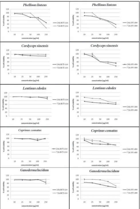
Fig. 1. Effects of methanol extracts of Phellinus linteus, Cordyceps sinensis, Lentinus edodes, Coprinus comatus, Ganoderma lucidum on viability of HCT-116 and SW-480 cell lines after
radical scavenging capacity of PL (5.32 μg/mL) was significantly higher than the capacity of the other four samples, and even than that of the control substances BHA and chlorogenic acid. LE, CC and CS showed the lowest scavenging capacity (>1000.00 μg/mL).
Cytotoxic activities
Extracts form PL, CS, LE, CC and GL were tested for their cytotoxic properties by using the MTT as-say. The PL extract significantly decreased the cell viability of both tested cancer cell lines (Fig. 1), CS decreased only SW-480 cell viability, and both had good cytotoxic effects (IC50 values in Table 2). PL ex -tract induced the best effects on SW-480 cells after 72 h (IC50 = 169.80±2.56 µg/mL). All other extracts had cell-selective effects and decreased the viability of SW-480 cells in a dose- and time-dependent way (Fig. 1), but did not show significant cytotoxic activity (IC50 values were higher than the maximum applied dose). The criterion of cytotoxic activity for the crude extracts was IC50< 30 μg/mL (Itharat et al., 2004), and according to this criterion, the investigated extracts showed no significant cytotoxic effects on HCT-116 and SW-480 cells at the applied concentrations (Table 2).
Antimigratory effects
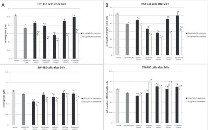
Migration and invasion are critical steps in the initial progression of cancer, and the effects of mushroom extracts on the migration potential of HCT-116 and SW-480 cells were evaluated by using the transwell migration assay. On the basis of obtained optical
den-sity at 595 nm, all mushroom extracts decreased mi-gration of HCT-116 and SW-480 cells in both tested concentrations (Fig. 2A) after 24 h of incubation in comparison with control (non-treated) cells. However, when the data was expressed relative to the number of viable cells (evaluated by MTT assay), all of the tested extracts displayed significant antimigratory effects only on HCT-116 cells in both selected concentra-tions, except GL in the concentration of 10 µg/mL (Fig. 2B). The PL extract in both tested concentra -tions significantly reduced the migratory potential of SW-480 cells, while the CS extract did so only in the concentration 10µg/mL (Fig. 2B). The other tested mushroom extracts had promigratory effects on SW-480 cells. Our results showed that extracts had bet-ter antimigratory effects on HCT-116 cells (Fig. 2A) than on SW-480 cells (Fig. 2B). Cisplatin (50 µM) as a positive control, significantly decreased migration of both treated cell lines (Fig. 2A and 2B) as well. In comparison to cisplatin, all tested extracts in the con-centration of 50 µg/mL and only CS and LE extracts in a dose of 10µg/mL showed higher antimigratory effects on HCT-116 cells. In comparison to cisplatin, PL extracts showed higher antimigratory potential on SW-480 cells in both tested doses. The PL extract showed the best antimigratory effect on SW-480 cells, and extracts of CS and LE on HCT-116 cells.
Protein expression of β catenin
For β-catenin protein expression and localization, the cells were analyzed using immunofluorescence staining. The obtained images demonstrate that both
Table 2. Growth inhibitory effects − IC50 values (μg/mL) of different mushrooms extracts on HCT-116 and SW-480 cell lines after 24- and 72-h exposure. All values are mean ± SEM, n = 3.
Extracts of mushrooms/ chemical drugs IC50 (µg/mL)
HCT-116 SW-480
Time of exposure 24 h 72 h 24 h 72 h
Cisplatin (µM) 263.66±8.02 104.41±9.01 108.40±2.15 59.47±0.48
PL 261.1±11.21 200.58±6.85 170.94±5.11 169.80±2.56
CS >250 >250 174.55±4.02 178.70±4.78
LE >250 >250 >250 >250
CC >250 >250 >250 >250
cell lines express β-catenin (Fig. 3 and Fig. 4). Fluo-rescence microscopy revealed that β-catenin was ex-pressed in the nucleus and cytoplasm in both con-trol and treated cells. β-catenin expression (nuclear and cytoplasmic) was reduced in both cell lines after treatment with cisplatin, and this correlated with its antimigratory effect. In LE- and CS-treated HCT-116 cells, nuclear and cytoplasmicβ-catenin expres-sion was reduced (Fig. 3). Treatments with PL, GL and CC extracts reduced cytoplasmic but not nuclear β-catenin in this cell line compared with the control (Fig. 3). Only in the LE treatment was an increase of β-catenin in intercellular connections observed. These results indicate that a reduction of total (nuclear and cytoplasmic) β-catenin protein expression, but its increase in intercellular connections is responsible
for the antimigratory effects of mushroom extracts in HCT-116 cells (Fig. 2B). Treatments with the tested extracts reduced cytoplasmic β-catenin, while the nuclear pool remained abundant in SW-480 cells (Fig. 4). Nuclear β-catenin was highly expressed in the treated in comparison to control SW-480 cells, which correlated with the promigratory effects of CS, LE, CC and GL extracts (Fig. 2B). Onlyn in the treatment with PL extract was β-catenin observed in intercellular connections and probably mediated the observed antimigratory effects.
Pro/antioxidative effects
Our results revealed that the tested mushroom ex-tracts showed cell-selective cytotoxic and
antimigra-Fig. 2. Effects of methanol extracts of Phellinus linteus, Cordyceps sinensis, Lentinus edodes, Coprinus comatus, Ganoderma lucidum
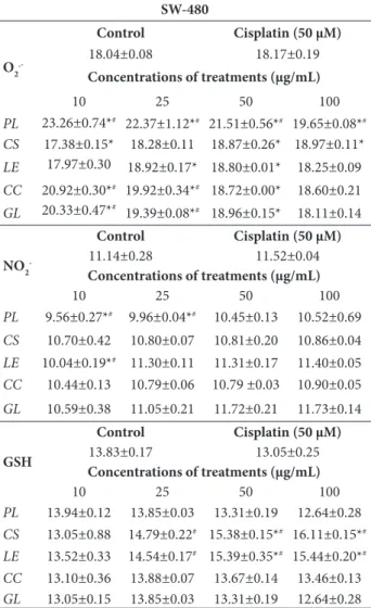
tory effects (cytotoxicity mainly on SW-480, and an-timigratory potential mainly on HCT-166 cells). In order to define the mechanism of the given effects, different parameters of redox status were followed in the treated cells. The presented data (Tables 3 and 4) show levels of reactive species (O2•– and nitrites) and antioxidative capacity (GSH level) in HCT-116 and SW-480 cells after 24 h of treatment. When compared with the control, the production of O2•– significantly increased in all treatments in both cell lines. The high-est production of O2•– was caused by LE and CS ex -tracts on HCT-116, and PL extract on SW-480 cells. The O2•–level was inversely correlated with the doses of extracts, except in the treatment with CS and LE on SW-480 cells (Table 4). These results indicate that mushroom extracts had prooxidative effects in the tested cancer cell lines.
The levels of nitrites, as indicators of NO, were also followed (Tables. 3 and 4). Generally, the tested mushroom methanol extracts increased nitrite levels in HCT-116 cells (PL and CS at all concentrations except the highest, and LE and CC only in the high -est), but the highest values of nitrites were observed in the treatment with cisplatin (Table 3). The treatment with PL and LE (lower doses) significantly decreased the nitrite level in SW-480 cells (Table 4). Other treat-ments had no effects or slightly decreased the nitrite level (non-significant).
The concentration of GSH showed a downward trend in the treatments with mushroom extracts on HCT-116, except in low doses of PL and CS extracts, where GSH levels increased (Table 3). In contrast, GSH had an upward trend in SW-480 cells in the pres-ence of CS and LE extracts (Table 4).
Fig 3. β-catenin expression in HCT-116 cells treated with 50 µg/
mL of methanol extracts of different mushrooms species. Con
-trol (untreated) cells (1) and cells treated with 50 µM cisplatin (2), Phellinus linteus (3), Cordyceps sinensis (4), Lentinus edodes
(5), Coprinus comatus (6), Ganoderma lucidum (7). Cells were incubated for 24 h. The images were obtained using fluorescence microscopy at 600× magnification. Nuclei stained blue, cytoplas-mic β-catenin stained red (a), nuclear β-catenin stained green (b).
Fig. 4. β-catenin expression in SW480 cells treated with 50 µg/
mL of methanol extracts of different mushrooms species. Con
-trol (untreated) cells (1) and cells treated with 50 µM cisplatin (2), Phellinus linteus (3), Cordyceps sinensis (4), Lentinus edodes
DISCUSSION
While the medicinal properties of mushrooms are known (Sullivan et al., 2006), the molecular mecha-nisms of their biological effects remain to be elucidat-ed. Mushrooms accumulate a high content of phenolic compounds and other active substances (Song et al., 2008). These compounds affect the modulation of re-dox status parameters in cancer cells as a first step in the manifestation of their biological effects (Ren et al., 2003). Generally, our study indicates that methanolic
extracts from five commercially available mushrooms had biological effects on HCT-116 and SW-480 colon cancer cell lines. According to the results presented in our study, the methanol extract of PL is abundant with polyphenols, especially flavonoids with signifi-cant scavenging capacity against DPPH radicals and
in vitro antioxidative potential, effects that are usu-ally correlated with quantitative phenolic composition (Alimpic et al., 2014). As our results show, mushrooms with the highest content of phenolics as well as
fla-Table 3. Effects of methanol extracts of Phellinus linteus, Cordyceps sinensis, Lentinus edodes, Coprinus comatus, Ganoderma lucidum
and cisplatin on prooxidative/antioxidative status of the HCT-116 cell line after 24 h of exposure.
HCT-116
O2
.-Control Cisplatin (50 µM)
17.80±0.09 18.46±0.04
Concentrations of treatments (µg/mL)
10 25 50 100
PL 19.02±0.17* 19.61±0.12* 19.48±0.32* 18.98±0.28*
CS 23.79±0.16*# 22.63±0.10*# 21.96±0.09*# 18.81±0.36*
LE 25.38±0.35*# 23.20±0.23*# 22.31±0.05*# 21.05±0.28*#
CC 19.66±0.38*# 19.62±0.22*# 19.59±0.11* 17.27±0.13
GL 21.26±0.83*# 20.74±0.17*# 18.42±0.09* 18.34±0.74*
NO2
-Control Cisplatin (50 µM)
11.83±0.25 14.57±0.40*
Concentrations of treatments (µg/mL)
10 25 50 100
PL 13.26±0.00* 13.14±0.49*# 12.71±1.00*# 10.65±0.06#
CS 13.62±0.29* 13.60±0.21* 12.99±0.11*# 12.31±0.18#
LE 11.10±0.25# 11.59±0.17# 11.91±0.05# 12.65±0.23*#
CC 11.58±0.16# 11.76±0.25# 12.19±0.10# 12.41±0.08*#
GL 11.37±0.23# 11.65±0.09# 12.27±0.14# 12.33±0.02#
GSH
Control Cisplatin (50 µM)
14.04±0.12 13.15±0.15*
Concentrations of treatments (µg/mL)
10 25 50 100
PL 15.44±0.50*# 15.28±0.06*# 13.98±0.60 13.53±1.05
CS 16.09±1.26*# 14.38±0.42 14.10±0.40 14.08±0.12
LE 11.60±0.59*# 12.51±0.19* 14.23±0.23 14.42±0.26#
CC 12.07±0.71* 12.23±0.28* 12.53±0.09* 13.02±0.72*
GL 13.68±0.62 13.76±0.45 14.60±0.65# 15.03±0.51*#
All parameters are expressed as nmol/mL. All values are means±SEM, n
= 3; * p<0.05 as compared with control, #p<0.05 as compared to cisplatin.
Table 4. Effects of methanol extracts of Phellinus linteus, Cordyceps sinensis, Lentinus edodes, Coprinus comatus, Ganoderma lucidum
and cisplatin on prooxidative/antioxidative status SW-480 cell line after 24 h of exposure.
SW-480
O2
.-Control Cisplatin (50 µM)
18.04±0.08 18.17±0.19
Concentrations of treatments (µg/mL)
10 25 50 100
PL 23.26±0.74*# 22.37±1.12*# 21.51±0.56*# 19.65±0.08*#
CS 17.38±0.15* 18.28±0.11 18.87±0.26* 18.97±0.11*
LE 17.97±0.30 18.92±0.17* 18.80±0.01* 18.25±0.09
CC 20.92±0.30*# 19.92±0.34*# 18.72±0.00* 18.60±0.21
GL 20.33±0.47*# 19.39±0.08*# 18.96±0.15* 18.11±0.14
NO2
-Control Cisplatin (50 µM)
11.14±0.28 11.52±0.04
Concentrations of treatments (µg/mL)
10 25 50 100
PL 9.56±0.27*# 9.96±0.04*# 10.45±0.13 10.52±0.69
CS 10.70±0.42 10.80±0.07 10.81±0.20 10.86±0.04
LE 10.04±0.19*# 11.30±0.11 11.31±0.17 11.40±0.05
CC 10.44±0.13 10.79±0.06 10.79 ±0.03 10.90±0.05
GL 10.59±0.38 11.05±0.21 11.72±0.21 11.73±0.14
GSH
Control Cisplatin (50 µM)
13.83±0.17 13.05±0.25
Concentrations of treatments (µg/mL)
10 25 50 100
PL 13.94±0.12 13.85±0.03 13.31±0.19 12.64±0.28
CS 13.05±0.88 14.79±0.22# 15.38±0.15*# 16.11±0.15*#
LE 13.52±0.33 14.54±0.17# 15.39±0.35*# 15.44±0.20*#
CC 13.10±0.36 13.88±0.07 13.67±0.14 13.46±0.13
GL 13.05±0.15 13.85±0.03 13.31±0.19 12.64±0.28
All parameters are expressed as nmol/mL. All values are mean ± SEM, n
vonoids demonstrate the best in vitro antioxidative effect. It is known that PL is active as an antioxidant, and its activity is comparable with vitamin C in free radical scavenging (Song et al., 2003).
Our study showed that the tested mushroom ex-tracts had cell-selective effects – significant decrease of SW480 cells viability, wheras cytotoxic activity (IC50 <200 µg/mL) was only exhibited by PL and CS. As described by Milutinovic et al. (2015), SW-480 cells showed higher sensibility in treatments with natural extracts than HCT-116 cells. Considering the effects on cell viability for all tested mushroom extracts, PL had the highest cytotoxicity in both tested colon can-cer cell lines, probably as a consequence of its high flavonoid and polyphenol content and high produc-tion of superoxide anion. As Li et al. (2004) showed, PL extract had a cytotoxic effect on the SW-480 colon cancer cell line. Also, many studies (Ren et al., 2003; Oak et al., 2005) indicated that the compounds from crude extracts, capable of antioxidant activity, could be responsible for the achieved antitumor effects.
According to our results, the changes in all redox parameters induced by the tested mushroom extracts pointed to the appearance of oxidative stress in co-lon cancer cells, especially in the presence of PL, CS and LE. When the obtained results were expressed with respect to viable cells (on the basis of the MTT test; data not shown), the tested mushrooms signifi-cantly increased all of the examioned redox status parameters in the colon cancer cell lines. High levels of O2•– and nitrites probably result in the production of peroxynitrites (Wink and Mitchell, 1998), which with other reactive nitrogen and oxygen species can induce cell death and thereby participate in the eti-ology of many diseases (Droge, 2002). Also, our re-sults showed higher levels of O2•–, nitrites and GSH (in relation to viable cells) in SW-480 cells than in HCT-116 cells, suggesting one of the main reasons for the mushroom extract-induced decrease in cell viability. On the other hand, the high but obviously physiologically controlled level of the same oxidative stress parameters are probably included in regulation of the antimigratory effects of the tested mushroom extracts in HCT-116 cells.
The results of this study showed that the tested mushroom extracts possessed cell-selective effects on migratory potential: antimigratory effects on HCT-116, and promigratory effects on SW-480 cells. As previously discussed for cell viability, only the PL ex -tract had a strong antimigratory effect on both tested cell lines. According to the presented data, the an-timigratory effects of the tested mushroom extracts were probably the consequence of their prooxidative effects (increased O2•–). Although compounds such as polyphenols, polysaccharides or proteins gener-ally have antioxidative properties, some of them and their interactions are able to activate the intracellu-lar formation of ROS and cause changes in oxidative stress parameters (Smith et al., 2003; Kamilya et al., 2006; Song et al., 2008). Several studies investigated the effects of ROS on cell migration (Alexandrova et al., 2006) and some showed that ROS, in the first in-stance O2•– and H
2O2, had effects on cancer cell motil-ity (Luanpitpong et al., 2010). PL extracts in SW-480, and CS and LE extracts in HCT-116 cells caused the highest O2•–production and induced the highest sup-pression of cells migration. Furthermore, our results showed that treatments of extracts with the highest antimigratory potential significantly reduced the ex-pression of β-catenin, or its relocation in intercellular connections in the tested cells (LE and CS in HCT-116 and PL in SW-480 cells). The literature data con -firmed our result that PL extracts inhibited migratory potential and decreased the expression of β-catenin in SW-480 cells (Song et al., 2011). Also, Wang et al. (2010) showed that lentinan polysaccharides from LE increased β-catenin cell membrane localization in co-lon cancer cells, which correlated with the results of our study.
the presence of LE and CS extracts, points to increased expression of E-cadherin and correlate with the in -hibition of colon cancer cell motility (Albring et al., 2013). The literature data (Kang et al., 2012; Albring et al., 2013) confirm that active substances from natural products such as magnolol from Magnolia officinalis
and/or berberine from Coptis and Berberis species may downregulate the level of cytosolic β-catenin in both observed cell lines. Considering that the tested mushroom extracts suppressed the expression of β-catenin, further experiments were designed to in-vestigate how components from extracts individually affected the proliferation and expression of protein involved in the Wnt/β-catenin pathway.
Obviously, the tested extracts did not effectively inhibit the growth of either HCT-116 or SW-480 cells, which might be the consequence of preparation and storage of the fruiting body or the type of extraction, but the activity of some substances could be preserved and might have significant effects on different mecha-nisms in cells.
CONCLUSION
A general conclusion is that the PL extract is abun -dant in flavonoids and polyphenols, which are re-sponsible for the observed by in vitro antioxidative properties as well as prooxidative potential on cancer cells, which are accompanied by significant cytotoxic and antimigratory effects. The other tested extracts displayed prooxidative effects, inhibited only SW480 cell viability, decreased cell migration and reduced the level of β-catenin in HCT-116 cells. The results of this study show that natural compounds, such as active components from mushroom extracts, may re-duce the migration of cancer cells. Further studies are necessary for detailed chemical characterization of the tested extracts and clarification of the molecular mechanisms of their activities.
Acknowledgments: The Ministry of Education, Science and Technological Development of the Republic of Serbia (III41010) supported this investigation. We wish to thanks to Mr. Dejan Po-slon (Medical Herbs, Novi Sad, Serbia) for providing commercial products of mushrooms.
Authors’ contributions: Study conception and design: Dragana S. Šeklić, Snežana D. Marković; Acquisition, analysis and inter-pretation of data: Dragana S. Šeklić, Milan S. Stanković; Drafting of manuscript: Dragana S. Šeklić, Milena G. Milutinović; Critical revision: Marina D. Topuzović, Andraš Š. Štajn and Snežana D. Marković.
Conflict of interest disclosure: We certify that there is no conflict of interest with any financial organization regarding the material discussed in the manuscript.
REFERENCES
Albring, K.F., Weidemüller, J., Mittag, S., Weiske, J., Friedrich, K., Geroni, M.C., Lombardi, P. and O. Huber (2013). Berberine acts as a natural inhibitor of Wnt/β-catenin signaling-iden-tification of more active 13-arylalkyl derivatives. Biofactors.
39(6), 652-662.
Alexandrova, A.Y., Kopnin, P.B., Vasiliev, J.M., and B.P. Kopnin
(2006). ROS up-regulation mediates Ras-induced changes of cell morphology and motility. Exp. Cell. Res. 312(11), 2066-2073.
Alimpic, A., Oaldje, M., Matevski, V., Marin, P.D., and S. Duletic-Lausecic (2014). Antioxidant activity and total phenolic and flavonoid contents of Salvia amplexicaulis Lam. extracts. Arch. Biol. Sci. 66(1), 307-316.
Auclair, C. and E. Voisin (1985). Nitroblue tetrazolium reduction. In: Handbook of Methods for Oxygen Radical Research (Eds.
RA Greenwald), 122-132. CRC Press, Inc: Boka Raton.
Bacac, M., and I. Stamenkovic (2008). Metastatic cancer cell. Ann Rev Pathol. 3, 221-247.
Baker, M.A., Cerniglia, G.J. and A. Zaman (1990). Microtiter plate assay for the measurement of glutathione and glutathione disulfide in large numbers of biological samples. Anal. Bio-chem. 190, 360-365.
Behrens, J., Von Kries, J.P., Kuhl, M., Bruhn, L., Wedlich, D., Gross-chedl, R. and W. Birchmeier (1996). Functional interaction of beta-catenin with the transcription factor LEF-1. Nat.
382(6592), 638–642.
Chen, N.H. and J.J. Zhong (2011). p53 is important for the anti-invasion of ganoderic acid T in human carcinoma cells.
Phytomed. 18, 719-725.
Cheung, L.M., Cheung, P.C.K. and V.E.C. Ooi (2003). Antioxidant activity and total phenolics of edible mushroom extracts. Food Chem. 81, 249-255.
Dröge, W. (2002). Free radicals in the physiological control of cell function. Physiol. Rev. 82, 47-95.
Giles, R.H., Van Es, J.H. and H. Clevers (2003). Caught up in a Wnt storm: Wnt signaling in cancer. Biochim. Biophys. Acta. 1653(1), 1-24.
Griess, P. (1879). Bemerkungen Zu der Abhandlung der HH. Weselsky und Benedikt Uebereinige Azoverbindungen.
Hostanska, K., Jürgenliemk, G., Abel, G., Nahrstedt, A. and R. Sal-ler (2007). Willow bark extract (BNO1455) and its fractions suppress growth and induce apoptosis in human colon and lung cancer cells. Cancer. Detect. Prev. 31(2), 129-139.
Ilyas, M., TomLinson, I.P., Rowan, A., Pignatelli, M. and W.F. Bod-mer (1997). β-Catenin mutations in cell lines established from human colorectal cancers. Proc. Natl. Acad. Sci. USA.
94, 10330-10334.
Itharat, A., Houghton, P.J., Eno-Amooquaye, E., Burke, P.J., Samp-son, J.H. and A. Raman (2004). In vitro cytotoxic activity of Thai medicinal plants used traditionally to treat cancer.
J.Ethnopharmacol. 90, 33-38.
Kamilya, D., Ghosh, D., Bandyopadhyay, S., Mal, B.C. and T.K. Maiti (2006). In vitro effects of bovine lactoferrin, mush-room glucan and Abrus agglutinin on Indian major carp, catla (Catla catla) head kidney leukocytes. Aquacul. 253, 130-139.
Kang, Y.J., Park, H.J., Chung, H.J., Min, H.Y., Park, E.J., Lee, M.A., Shin, Y. and S.K. Lee (2012). Wnt/β-catenin signaling medi-ates the antitumor activity of magnolol in colorectal cancer cells. Mol. Pharmacol. 82(2), 168-177.
Kumarasamy, N., Solomon, S., Chaguturu, S.K., Cecelia, A.J., Val-labhaneni, S., Flanigan, T. and K. Mayer (2005). The chang-ing natural history of HIV disease: Before and after the introduction of generic antiretroviral therapy in southern India. Clin. Infect. Dis. 41(10), 1525-1528.
Li, G., Kim, D.H., Kim, T.D., Park, B.J., Park, H.D., Park, J.I., Na, M.K., Kim, H.C., Hong, N.D., Lim, K., Hwang, B.D. and
W.H. Yoon (2004). Protein-bound polysaccharide from
Phellinus linteus induces G2/M phase arrest and apoptosis in SW480 human colon cancer cells. Cancer. Lett. 216(2), 175-181.
Liu, C., Li, Y., Semenov, M., Han, C., Baeg, G.H., Tan, Y., Zhang, Z., Lin, X. and X. He (2002). Control of beta-catenin phos-phorylation/degradation by a dual-kinase mechanism. Cell.
108(6), 837-847.
Luanpitpong, S., Jo Talbott, S., Rojanasakul, Y., Nimmannit, U., Pongrakhananon, V., Wang, L. and P. Chanvorachote (2010). Regulation of lung cancer cell migration and invasion by reactive oxygen species and caveolin-1. J Biol. Chem.
285(50), 38832-3840.
Milutinovic, M., Stankovic, M., Cvetkovic, D., Maksimovic, V., Smit, B., Pavlovic, R. and S. Markovic (2015). The molecu-lar mechanisms of apoptosis induced by Allium flavum L.
and synergistic effects with new-synthesized Pd(II) com-plex on colon cancer cells. J. Food. Biohem. 39, 238-250.
Mosmann, T. (1983). Rapid colorimetric assay for cellular growth and survival: application to proliferation and cytotoxicity assays. J. Immunol. Methods. 65, 55-63.
Oak, M.H., Bedoui, J.E. and V.B. Schini-Kerth (2005). Anti-angio-genic properties of natural polyphenols from red wine and green tea. J. Nutr. Biochem. 16, 1-8.
Quettier, D.C., Gressier, B., Vasseur, J., Dine, T., Brunet, C., Luyckx, M.C., Cayin, J.C., Bailleul, F. and F. Trotin (2000). Phenolic compounds and antioxidant activities of buckwheat ( Fago-pyrum esculentum Moench) hulls and flour. J. Ethnophar-macol. 72, 35-42.
Ren, W., Qiao, Z., Wang, H., Zhu, L. and L. Zhang (2003). Fla -vonoids: promising anticancer agents. Med. Res. Rev. 23, 519-534.
Rowan, N.J., Smith, J.E. and R. Sullivan (2003). Immunomodula-tory activities of mushroom glucans and polysaccharide– protein complexes in animals and humans. Int. J. Med. Mushr. 5, 95-110.
Singleton, V.L., Orthofer, R. and R.R.M. Lamuela (1999). Analysis of total phenols and other oxidation substrates and antioxi-dants by means of Folin-Ciocalteu reagent. Meth. Enzymol.
299, 152-178.
Smith, J.E., Sullivan, R. and N.J. Rowan (2003). The role of poly-saccharides derived from medicinal mushrooms in can-cer treatment programs: current perspectives. Int. J. Med. Mushr. 5, 217-34.
Song, Y.S., Kim, S.H., Sa, J.H., Jin, C., Lim, C.J. and EH. Park
(2003). Anti-angiogenic, antioxidant and xanthine oxidase inhibition activities of the mushroom Phellinus linteus. J. Ethnopharmacol. 88(1),113-116.
Song, T.Y., Lin, H.C., Yang, N.C. and M.L. Hub (2008). Antiprolif-erative and antimetastatic effects of the ethanolic extract of
Phellinus igniarius (Linnearus: Fries) Quelet. J Ethnophar-macol. 115, 50-56.
Song, W. and L.D. Van Griensven (2008). Pro- and antioxidative properties of medicinal mushroom extracts. Int J Med Mushr. 10(4), 315-324.
Song, K.S., Li, G., Kim, J.S., Jing, K., Kim, T.D., Kim, J.P., Seo, S.B., Yoo, J.K., Park, H.D., Hwang, B.D., Lim, K. and W.H. Yoon
(2011). Protein-bound polysaccharide from Phellinus linteus inhibits tumor growth, invasion, and angiogenesis and alters Wnt/β-catenin in SW480 human colon cancer cells. BMC Can. 11, 307.
Williams, T.M., Hassan, G.S., Li, J., Cohen, A.W., Medina, F., Frank, P.G., Pestell, R.G., Di Vizio, D., Loda, M. and M.P. Lisanti (2005). Restoration of caveolin-1 expression sup-presses growth and metastasis of head and neck squamous cell carcinoma. J. Biol. Chem. 99(10), 1684-1694.
Wink, D.A. and J.B. Mitchell (1998). Chemical biology of nitric oxide: insights into regulatory, cytotoxic, and cytoprotec-tive mechanisms of nitric oxide. Free. Radic. Biol. Med. 25, 434-456.


