Inactivation of the Progesterone Receptor in
Mx1+ Cells Potentiates Osteogenesis in
Calvaria but Not in Long Bone
Zhendong A. Zhong1, Weihua Sun1, Haiyan Chen1, Hongliang Zhang1,2, Nancy E. Lane1, Wei Yao1*
1Center for Musculoskeletal Health, Department of Internal Medicine, University of California Davis Medical Center, Sacramento, CA, 95817, United States of America,2Department of Emergency Medicine, The Second Xiangya Hospital of Central-South University, Hunan, Changsha, China
*yao@ ucdavis.edu
Abstract
The effect of progesterone on bone remains elusive. We previously reported that global pro-gesterone receptor (PR) knockout mice displayed high bone mass phenotype, suggesting that PR influences bone growth and modeling. Recently, Mx1+ cells were characterized to be mesenchymal stem cell-like pluripotent Cells. The aim of this study was to evaluate whether the PR in Mx1+ cells regulates osteogenesis. Using the Mx1-Cre;mT/mG reporter mouse model, we found that the calvarial cells exhibited minimal background Mx1-Cre activity prior to Cre activation by IFNαtreatment as compared to the bone marrow stromal cells. IFNαtreatment significantly activated Mx1-Cre in the calvarial cells. When the PR gene was deleted in the Mx1-Cre;PR-flox calvarial cellsin vitro, significantly higher levels of
expression of osteoblast maturation marker genes (RUNX2, Osteocalcin, and Dmp1) and osteogenic potential were detected. The PR-deficient calvariae exhibited greater bone vol-ume, especially in the males. Although Mx1-Cre activity could be induced on the bone sur-facein vivo, the Mx1+ cells did not differentiate into osteocytes in long bones. Bone
volumes at the distal femurs and the bone turnover marker serum Osteocalcin were similar between the Mx1-Cre;PR-flox mutant mice and the corresponding wild types in both sexes. In conclusion, our data demonstrates that blocking progesterone signaling via PRs in cal-varial Mx1+ cells promoted osteoblast differentiation in the calvaria. Mx1+ was expressed by heterogeneous cells in bone marrow and did not differentiate into osteocyte during long bone developmentin vivo. Selectively inactivating the PR gene in Mx1+ cells affected the
membrane bone formation but did not affect peripheral skeletal homeostasis.
Introduction
There is a sexual dimorphism in bone mass acquisition; males typically achieve greater peak bone mass (PBM) and greater bone size than females. Females experience an acceleration of bone loss due to declining estrogen levels with menopause and aging [1]. Thus, females are at
a11111
OPEN ACCESS
Citation:Zhong ZA, Sun W, Chen H, Zhang H, Lane NE, Yao W (2015) Inactivation of the Progesterone Receptor in Mx1+ Cells Potentiates Osteogenesis in Calvaria but Not in Long Bone. PLoS ONE 10(10): e0139490. doi:10.1371/journal.pone.0139490
Editor:Jung-Eun Kim, Kyungpook National University School of Medicine, REPUBLIC OF KOREA
Received:April 30, 2015
Accepted:September 13, 2015
Published:October 2, 2015
Copyright:© 2015 Zhong et al. This is an open access article distributed under the terms of the
Creative Commons Attribution License, which permits unrestricted use, distribution, and reproduction in any medium, provided the original author and source are credited.
Data Availability Statement:All relevant data is available via Figshare (http://dx.doi.org/10.6084/m9. figshare.1481100).
Funding:This work was funded by NIH grant P50 AR063043 (to NEL), R01 AR061366 (to WY), and SCOR Pilot Award (to ZAZ).
Competing Interests:The authors have declared that no competing interests exist.
sex hormone receptor knockout models do not necessarily recapitulate the phenotypes of sex hormone deficiency, the functions of estrogen receptors (ERs) and androgen receptors (ARs) on bone homeostasis have been extensively studied using animal models with germ-line or tis-sue-specific knockouts. These genetically modified mouse models have provided crucial infor-mation about the complex functions of sex hormone receptors in relation to bone homeostasis. For example, estrogen receptors display either stimulatory or inhibitory functions on bone for-mation depending on ligand availability and target skeletal location [5].
The effect of progesterone, another important sex hormone, on bone homeostasis remains unclear. Most of the studies that have evaluated the effect of progesterone on bone have utilized either agonists or antagonists in preclinical or clinical studies, and the results are conflicting. However progesterone agonist and antagonist treatments may alter the levels of other hor-mones, such as estrogen, androgens, and follicle-stimulating hormone (FSH), which makes it difficult to dissect the true progesterone responses [6,7]. There are two major PR isoforms—
PR-B and PR-A—in mammals. PR-A and PR-B exhibit different biological functions that are specific to particular cell types and promoter contexts [3]. Although majority of research indi-cates that PR expression alteration or gene mutation is closely related to tumorigenesis and cancer progression, PR dysfunction is also associated with cardiovascular defects (aortic aneu-rysm), neurological defects (migraine, vertigo) and reproductive conditions (endometriosis, infertility) [8–12]. Our research group and others have reported a high-bone-mass phenotype in global progesterone receptor knockout (PRKO) mice age two to 12 months of age, and this phenotype appears to result from a greater bone formation rate in females and a reduced bone resorption rate in males measured at three months of age [13,14]. These data suggest that pro-gesterone signaling through PR may suppress bone mass acquisition in mice. However, it is unclear how PRs affect bone homeostasis; PRs are differentially expressed in a wide range of tissues, including those of the reproductive system and central nervous system. Because PRs are expressed in the osteoblasts, and PRKO bone marrow stromal cells exhibit increased osteo-genic potentialin vitro[15–17], we hypothesized that PRs may directly regulate osteoblastic cells. In this study we utilized the Cre-Lox system to selectively delete both PR isoforms from osteoprogenitor cells to investigate its function in skeletal system.
Mx proteins are the main effectors of the antiviral innate immune response mediated by type I interferon (IFN I) [18]. The Mx1 promoter is active in bone marrow stromal cells and has been used to drive Cre recombinase expression to study gene function in bone and other tissues [19, 20]. Recently, Mx1+ cells were characterized to be mesenchymal stem cell-like pluripotent Cells, which can differentiate into an osteoblastic lineagein vivo, but cannot differentiate into chondro-cytes and adipochondro-cytes [21,22]. The purpose of this study was to use Mx1-Cre to drive PR gene knockout in osteoblastic lineagein vivoandin vitroto study the effects of PRs on osteogenesis.
Results
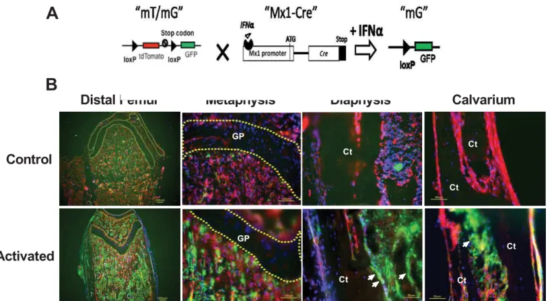
“pI-pC”) [23]. There is ~1% background recombination in mice that are not treated with inter-feron, and this level varies with tissue type presumably due to the numbers of interferon-responsive cells that are present or the availability of interferon in each organ [http://jaxmice. jax.org/strain/005673.html]. However, the Mx1-cre promoter’s specificity in bone tissue has not been elucidated in detail. We generated a Mx1-Cre;mT/mG reporter model to study the activity of Mx1-Cre in bone tissues and primary cultured osteoblasts. The mT/mG reporter mice constitutively express the tomato protein (red) unless they have been exposed to Cre recombinase. Exposure to Cre recombinase results in the deletion of the tomato cassette and the induction of GFP (green) expression [24] (Fig 1A).
We treated 1-month-old Mx1-Cre;mT/mG double transgenic mice with pI-pC, and col-lected the calvariae and distal femurs for cryosection (Fig 1B). We observed small numbers of cells within the bone marrow that were GFP-positive, which corresponded to Cre activity. In contrast, the calvariae exhibited relatively fewer GFP-positive cells prior to pI-pC treatment. One week after pI-pC treatment, we observed a robust induction of GFP expression in the bone marrow and bone surfaces of the distal femurs and calvariae. The growth plate region remained GFP-negative prior to and after the pI-pC treatment, indicating Mx1-Cre did not affect the chondrocytesin vivo(Fig 1B).
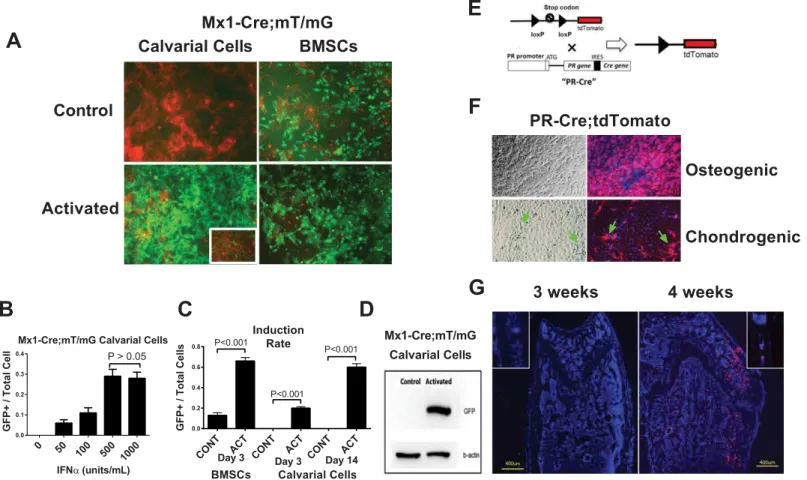
Calvarial cells and bone marrow stromal cells (BMSCs) are two commonly used sources of osteoprogenitor or pre-osteoblast cultures.In vitroexperiments were mostly performed in cells derived from male mice to exclude the potential estrogen and progesterone effects during men-strual cycles in females. We collected BMSCs from 1-month-old male Mx1-Cre:mT/mG mice and calvarial cells from 3-day-old Mx1-Cre:mT/mG pups, and treated the cells with IFNα(500 units/mL) for 72 hours to activate the Mx1-Cre promoter. Similar to its expressionin vivo, we observed substantial basal Cre activity in the BMSCs (13% were GFP-positive prior to activa-tion), and 66% of the BMSCs became GFP-positive after IFNαtreatment (Fig 2A and 2B). In contrast, the calvarial cells exhibited extremely low background Cre activity prior to IFNα treatment. Following IFNαtreatment, approximately 30% of the calvarial cells became GFP-positive three days after IFNαtreatment, and ~60% became GFP-positive two weeks after IFNαtreatment (Fig 2B). We also determined that the minimal IFNαdose required to induce robust Mx1-Cre activity in the calvarial cells was 500 units/mL (Fig 2B). These findings were confirmed by western blot; i.e., no GFP band was detected prior to activation, and a GFP band was present three days after induction in the Mx1-Cre;mT/mG calvarial cells (Fig 2C). Thus, we concluded that the calvarial cells were superior to the BMSCs forin vitrostudies due to the low basal Mx1-Cre activity prior to activation.
2. PR knockout in Mx1+ calvarial cells and calvariae increased
osteoblast differentiation
in vitro
We next generated an inducible PR conditional knockout mouse model by crossing Mx1-Cre and PR-flox mice. Following IFNαtreatment, the Mx1 promoter drives Cre recombi-nase expression, which recombines the loxP sites and deletes exon 2 of the PR gene resulting in PR inactivation in the Mx1+ cells (Fig 3A). We then collected calvarial cells from the Mx1-Cre; PR-flox double transgenic pups and treated the cells with or without IFNα(500 units/mL) for three days. The calvarial cells were then differentiated into osteoblasts in osteogenic media without IFNαtreatment. RNA was collected at 0, 7 and 14 days post differentiation. Real-time PCR revealed significantly greater osteogenic marker gene expression in the IFNα-treated cells on day 14. Specifically, the RUNX2, osteocalcin (Ocn) and DMP1 gene expressions were increased by 4-fold, 9-fold, and 9-fold, respectively, compared with the control groups (Fig 3B). Additionally, the Mx1-Cre-mediated PR knockout significantly increased osteoblast activ-ity as measured by the alkaline phosphatase activactiv-ity (ALP) at 10 days post-differentiation and mineralized nodule formation (alizarin red, AR) at 21 days post-differentiation (Fig 3C). IFNα treatment by itself had no effect on osteoblast differentiation in the PR-flox/flox (no-Cre con-trol) calvarial cells (Fig 3C). These data suggest that inactivation of the PR gene in the Mx1 + calvarial cells accelerated osteoblasts maturation.
To study the functions of PRs in the calvarial bone tissue, we developed anex vivocalvarium organ culture system. First, we wanted to confirm the activation of Mx1-Cre in theex vivo cul-tured calvarium. The Mx1-Cre;mT/mG double transgenic calvariae were induced with IFNα -containing BGJb medium (500 units/mL) for three days and were then cultured in BGJb
Distal Femur
Metaphysis
Diaphysis
Calvarium
Control
Activated
Ct
Ct
Ct
Ct Ct
Ct GP
GP
Fig 1. Characterization of Mx1-Cre in the long bones and calvariaein vivo. (A)A schematic diagram showing that mT/mG is crossed to Mx1-Cre and that IFNαcan activate Cre expression to delete the tdTomato (red fluorescent) cassette and initiate GFP (green fluorescent,“mG”) expression.(B)Mx1-Cre;mT/ mG mice at the age of 5 weeks were injected intraperitoneally with PBS (control) or pI-pC (activated). The distal femurs and calvariae were collected for cryosection after three days. The nuclei were stained with DAPI (blue). The arrows indicate bone surface GFP expression in the pI-pC-treated bones. The dotted lines circle the cartilage regions in the distal femur. GP, growth plate cartilage; Ct, cortical bone.
medium without IFNαthereafter. A significant proportion (~40%) of calvarial cells appeared GFP-positive after three days of IFNαtreatment, and significantly more cells (~80%) became GFP-positive by nine days (Fig 3D), indicating that the Mx1+/GFP+ calvarial cells were capa-ble of proliferating. In separate experiment, we obtained calvariae from the Mx1-Cre;PR-flox/ flox double transgenic mice and treated the calvarial tissue following a similar protocol. Using PR allele-specific PCR, we were able to detect the deleted PR band after PR-flox calvarial cells were treated with IFNα(Fig 3E). In line with these data from thein vitroexperiments, we found that bone volume of the calvariae in male Mx1-Cre;PR-flox knockout mice was 67% higher than their WT littermates (P<0.05) at five months after PR was selectively knocked
Fig 2. Comparison of BMSCs and calvarial cells in terms of Mx1-Cre activation and PR expressionin vitro.(A) BMSCs or calvarial cells were obtained from Mx1-Cre;mT/mG double transgenic mice and treated with IFNα(500 units/mL) for three days. Fluorescent images were taken three days (BMSCs) or 14 days (Calvarial cells) after the IFNαtreatment. The image of calvarial cells three days post IFNαtreatment is shown in the insert. The levels of Cre activation were quantitated by GFP (green) expression. (B) A quantitative histogram showing the induction rates (the GFP+ cells versus the total cells) of the BMSCs and calvarial cells shown in Figure A. CONT, control; ACT, activated. (C) Western blotting was performed to detect GFP protein in the total cell lysates from the calvarial cells with (activated) or without (control) IFNαtreatment on day 3.β-actin was used as an internal control. (D) Mx1-Cre;mT/mG calvarial cells were treated with different concentrations of IFNαfor three days. The ratios of GFP+ cells to total cells under each IFNαconcentration were quantified (A). A PR-Cre;tdTomato schematic diagram showing that the Cre gene is inserted downstream of the endogenous PR gene after an internal ribosome entry site (IRES) sequence such that Cre is expressed simultaneously with PR. When Cre is expressed (together with PR), it recombines loxp sites to remove the stop codon before the tdTomato cassette and activates tdTomato (red fluorescence) expression. (E) A diagram of PR-Cre; tdTomato. The tdTomato expression can be activated following the upstream stop codon removal by Cre, which is expressed simultaneously with endogenous PR gene. So that PR+ cells will express tdTomato (red fluorescent). (F) PR-Cre;tdTomato calvarial cells were differentiated into osteoblasts with osteogenic media or chondrocytes with chondrogenic media for 14 days. The majority of the cells cultured in the osteogenic medium were red. However, when the cells were cultured in the chondrogenic medium, only a small percentage of the fibroblast-like cells (arrows) turned red. The nuclei were stained with DAPI (blue). (G) Distal femurs or calvariae (inserts) from 3-week-old or 4-week-old PR-Cre;tdTomato mice were sectioned for fluorescent microscopy. The nuclei were stained with DAPI (blue). The red fluorescence indicated PR promoter activity.
Fig 3. PR inactivation in the Mx1+ calvarial cells and calvariaein vitro.(A) A schematic diagram showing that PR-flox is crossed to Mx1-Cre. Mx1-Cre can be activated by IFNαto delete exon 2 of the PR gene to generate PR mutants (ΔPR). (B) Mx1-Cre;PR-flox/flox calvarial cells were treated with IFNα(500 units/mL) or without IFNα(control) for three days. The cells were then differentiated into osteoblasts in osteogenic medium without IFNαfor 14 days. The relative expressions of RUNX2, Osteocalcin (Ocn) and DMP1 were evaluated by real-time PCR at day 14 and normalized to endogenousβ-actin. (C) The cells were collected for alkaline phosphatase (ALP) activity assays on day 10 and alizarin red staining (AR) on day 21. The optical density (OD) values were normalized to the corresponding total protein concentrations. Calvarial cells from the PR-flox/flox (without Cre) mice were used as a negative control to exclude the effect of INFαitself. (D) The calvariae obtained from Mx1-Cre;mT/mG double transgenic pups exhibited significant numbers (~40%) of cells that became GFP-positive after three days of IFNα(500 units/mL) treatment, and significantly more cells (~80%) became GFP-positive after an additional six days of culture without IFNα. (E) Genomic DNA was isolated from PR-flox/flox calvarial cells three days after IFNαtreatment, and subjected to PR allele-specific PCR. The deleted PR band (ΔPR) indicated Cre-mediated DNA recombination. (F) Five-weeks-old PR-flox/flox or Mx1-Cre;PR-flox/flox mice were injected with pI-pC. Calvariae were collected five months later for microCT analysis for bone volume and (G) representative calvarial images from microCT scans.
out in the Mx1+ cells from five weeks of age (Fig 3F and 3G). Calvarial bone thickness was sim-ilar between the WT and Mx1-Cre;PR-flox/flox mice in both sexes (data on file).
3. Mx1+ cell fate mapping in long bones
We next asked whether Mx1+ cells represent“osteoprogenitors”that can terminally differenti-ate into osteocytes in long bonesin vivo. We aged the pI-pC-treated Mx1-Cre;mT/mG mice to evaluate whether their Mx1+/GFP+ cells could eventually differentiate into osteocytes. We found some green positive cells were detected in mice that were not treated with pI-pC, sug-gesting a baseline leaky nature of Mx1-Cre in bone marrow (Fig 4A). After 30 or 60 days post pI-pC treatment (Mx1 activation), Mx1+ cells were found very heterogeneous expressed by both the hematopoietic and mesenchymal lineages and while most of Mx–1+ cells were observed within bone marrow, some were observed at the trabecular bone surface. We failed to detect any GFP+ osteocytes that were embedded in the trabecular or cortical bone (Fig 4B and 4C). We used Col1a1-CreERT2;mT/[mG as a positive control and we identified some GFP + osteocytes two weeks after tamoxifen treatment in these tamoxifen-inducible Col1a1--CreERT2;mT/mG mice (Fig 4B, insert). Bone marrow cells culture suggested Mx1 was expressed by multinuclear cells (Fig 4E). These data indicate that the Mx1+ cells were
expressed by mononuclear and multinuclear cells within bone marrow, osteoblasts at bone sur-face but were not able to differentiate into osteocytes in the long bonesin vivo.
4. Mx1-Cre-driven PR knockout did not affect long bone phenotype
The Mx1-Cre;PR-flox/flox mice were viable, exhibited body weights and sizes that were similar to those of their wild-type littermates and exhibited no obviously abnormal behavior (data not shown). To characterize thein vivoskeletal phenotypes of the Mx1-Cre;PR-flox mice, we treated the Mx1-Cre;PR-flox/flox mice with pI-pC at five weeks of age to delete the PR gene. PR-flox/flox (without Cre) mice were used as controls. At one, two and five months post-treat-ment, the distal femurs were collected and subjected to microCT analyses. A baseline scan was performed at five weeks of age before the pI-pC treatment. Serum osteocalcin levels between wild types and mutants were similar (Fig 4D) and serum CXT1 level (Fig 4E) was lower in the Mx1-Cre;PR-flox/flox mice as compared to their WT littermates at two months post pI-pC treatment. Additionally, we did not observe significant BV/TV changes at one, two and five-months post-PR inactivation in Mx1+ cells in both sexes (Fig 4F).Discussion
and plays minimum role in the endochodral ossification in long bones. However, additional studies are required to support our observations.
In the present study, calvarial cells were characterized as a good model for Mx1-Crein vitro because they exhibited minimal basal Mx1-Cre activation prior to IFNαtreatment, and the Cre activity could be robustly activated by IFNα. However, the Mx1-Cre in BMSCs exhibited signif-icant basal activation prior to IFNαtreatment. As IFN secretion increases with age and microbe exposure [25], the basal Mx1 activity in the BMSCs from 1-month-old mice might have been induced by endogenous IFN.
Fig 4. Skeletal phenotypes of Mx1-Cre-driven PR inactivationin vivo.(A) A distal femur from non-pI-PC treated Mx1-Cre;mT/mG mouse. (B—C) Mx1-Cre;mT/mG mice were injected with pI-pC intraperitoneally to induce Cre at one month of age and then sacrificed one (B) or two (C) months later. The distal femurs (D.F.) were collected and sectioned to observe the GFP (green) and tdTomato (red) fluorescence. Green indicates the Mx1+ cells that expressed Cre, and red indicates the Cre-negative cells. The nuclei were stained with DAPI (blue). The femoral trabecular bones from the Col1a1-CreERT2; mT/mG mice that received 4 days of tamoxifen injections were used as a positive control and are shown in the insert in (B). The white arrows indicate the green osteocytes that were observed in the trabecular bone in the Col1a1-mT/mG mice but were absent in the trabecular or cortical bone of the Mx1-mT/mG mice (the white arrowheads indicate the GFP-/tdTomato+ osteocytes). There were no green osteocytes observed in the Mx1-mT/mG bones on either day 30 or 60. (D) Bone marrow cells were collected from 1-month-old Mx1-Cre;mT/mG mice. Some multinuclear cells turned green indicating Mx1-Cre activation in these cells (yellow arrows). (E) Serum osteocalcin and (F) serum CTX1 levels were measured by ELISA two months post pI-pC injection (n = 8/group). (G) Five-week-old male and female Mx1-Cre;PR-flox/flox or PR-flox/flox (control) mice were injected with pI-pC intraperitoneally to induce Cre activity. Baseline microCT scans (BL) of the distal femurs were performed on five-week-old mice without receiving the pI-pC injection. The animals were sacrificed at one, two or five months post-pI-pC injection, and the hind limbs were collected for microCT analysis (n = 8/group).
We found that the calvarial cells exhibited a significantly greater potential to differentiate into osteoblasts after the PR expression was ablated from the Mx1+ cells. The observation of significantly greater levels of osteogenic markers 14 days after differentiation indicates that PRs might play a role in the later stages of osteoblast differentiation. These observations are consis-tent with our previous finding that BMSCs from PRKO mice exhibit increased osteoblastic dif-ferentiationin vitro[13]. In contrast, a number ofex vivostudies have reported that
supplementation of the culture medium with progesterone can stimulate cell proliferation and osteoblastic differentiation possibly through the action of insulin-like growth factors (IGFs). This anabolic effect of progesterone on osteoblast differentiation is more robust in female-derived osteoblasts than those from males [26–29]. PR-mediated progesterone signaling acts through specific progesterone response elements (PRE) within the promoter regions of target genes to regulate transcription, and this process is referred to as the“classical”mechanism. Moreover, some of progesterone’s effects may also result from the“non-genomic”effects of PRs that are mediated by PR [30]. One example of progesterone’s non-genomic effects is the activation of the extracellular signal–regulated kinase (ERK)–mitogen-activated protein kinase (MAPK) pathway; this pathway is critical for osteoblast differentiation and skeletal develop-ment [31]. Based on our results and those of others [30], we speculate that PR inactivation in Mx1+ cells might not block progesterone’s non-genomic effects or recapitulate progesterone antagonism. In our study, PR inactivation did not exhibit an effect on osteoblast differentiation that was exactly opposite to the reported effect of progesterone supplementationin vitro. Addi-tionally, the timing and levels of PR expression in osteoprogenitor cells vary depending upon the source of the derived osteoprogenitor cells. PR expression was detected inin-vitro-cultured calvarial cells from 3-day-old pups. However, PR expression in mouse bones (including the cal-varium)in vivowas not detectable until one month of age (data on-file). This lack of PR expression may have contributed to the discrepancy that we observed between thein vitroand in vivodata. Moreover, Mx1+ cells are found to be expressed by both the hematopoietic as well
as the mesenchymal lineage cells in bone marrow. Mx1-Cre is active in multinuclear cells and PR deletion in Mx1+ cells might affect the function of osteoclast as suggested by lower bone resorption in the male Mx1-Cre; PR-flox mice. Most importantly, Mx1+ cells do not differenti-ate into osteocytes in long bones, which may explain the lack of a peripheral skeletal phenotype in the Mx1-Cre;PR-flox mutant micein vivo. Additional studies using different Cre drivers are underway to pinpoint the effects of PRs on the different stages of osteoblastogenesis, bone development, and the PR-mediated downstream effectors.
In summary, PR inactivation in the Mx1+ cells derived from calvarium enhanced osteoblast differentiationin vitro and in vivo. In contrast, Mx1-Cre is leaky and expressed by heteroge-neous cell populations within bone marrow. The Mx1+ cells did not differentiate into osteo-cytes in the long bonesin vivo, which perplexed the interpretation of the peripheral bone phenotype in our Mx1-Cre-driven PR knockout model. Mx1-Cre may not be a best cre to be used to study the contribution of“mesenchymal”lineage to osteogenesis.
Experimental Procedures
Mouse Lines
trolled environmental conditions (12-hour light/dark cycle, room temperature 22°C), and fed ad libidum (food and water). The animal protocol was approved by the Institutional Animal Care and Use Committee of the University of California Davis (animal protocol #18322). All surgeries were performed under anesthesia, and all efforts were made to minimize animal suffering.
MicroCT measurements
The right distal femur from each animal was scanned and analyzed using VivaCT 40 (Scanco Medical, Bassersdorf, Switzerland) with a voxel resolution of 10μm in all three spatial dimen-sions and a mono-energetic (70 KeV) X-ray source. For the distal femurs, we evaluated 200 slices that were situated approximately 0.2 mm from the distal end of the growth plate. The slides covered a total metaphyses tissue volume of 2–3.5 mm3for each scan and were used to obtain the cancellous bone volume/total volume (BV/TV) ratio [13]. The calvariae was col-lected from PR-flox/flox and Mx1-Cre; PR-flox/flox mice at 4 months post pI-pC injection. Scans and evaluations were imitated from the anterior suture and included 30 slices (0.3 mm) at the frontal region and 200 slices (2mm) at the parietal region but excluded the squamous, sphenoid and occipital bones (Fig 3G).
Calvarium and Calvarial Cell Culture
Calvariae (5 per group) were collected from 3-day old pups. A cut was made through the sagittal suture to separate the calvarium into two halves. One half was used as a control, and the other half was used for the IFNαtreatment. BGJb medium (Life Technologies, Carlsbad, CA) supple-mented with 0.1% bovine serum albumin and 100 U/mL each of penicillin, streptomycin and amphotericin was used for the calvarium culture. The calvarial tissues were cultured in 24-well plates with 8-μm hanging PET inserts (Millipore, Billerica, Massachusetts) as described previously [33]. The calvarial cells were isolated from the calvariae by serial digestion at 37°C with constant agitation in 0.2 mg/ml collagenase type I (Worthington, Lakewood, NJ). The digestion solution was changed and collected an additional five times. Digestions 2–6 (containing the osteoprogeni-tor and osteoblasts cells) were centrifuged and washed withα-MEM containing 10% FBS and 1% penicillin/streptomycin and cultured for 48 hours at 37°C before use in the further experiments.
IFNαwas used to activate Mx1-Crein vitro. In brief, the calvariae or calvarial cells were incubated in medium containing IFNα(R&D System, Minneapolis, MN) for three days, after which the IFNαmedium was replaced with fresh IFNα-free medium. A dose-response study was first performed using the Mx1-Cre;mT/mG calvarial cells to optimize the IFNαdose. The induction rate was determined by the ratio of the GFP-positive cell number to the total cell number based on 250 cells from five different areas in the wells. The calvarial cells were differ-entiated into osteoblasts in osteogenic media (changed every two days) containing 50μg/ml ascorbic acid and 10 mMβ-glycerol phosphate (βGP).
Quantitative Real-time RT-PCR Analysis
Kit and random primers (QIAGEN, Venlo, Netherlands). The cDNA samples were subjected to PCR analysis using Fast SYBR PCR Master Mix and an ABI 7500 Real-time PCR system (Applied Biosystems, NY). The primers used were as follows: RUNX2 (TGGCTTGGGTTTCAGG TTAG,CCTCCCTTCTCAACCTCTAATG); Osteocalcin (TGTGACGAGCTATCAAACCAG,GAGG
ATCAAGTTCTGGAGAGC); and the DMP1 primers (HP226170) were ordered from the
ORI-GENE company. The mRNA level of each gene was normalized to the endogenousβ-actin level.
Staining and Immunohistochemistry
The bones from the Mx1-Cre;mT/mG mice were collected, fixed in 4% formaldehyde with 10% sucrose (W/V) at 4°C overnight, and embedded in Optimal Cutting Temperature (O.C.T.) medium. Cryosections were cut using a CryoJane tape transfer system for a cryostat (Leica Bio-system, IL) and were and observed using a fluorescent microscope. For the quantification of the alkaline phosphatase (ALP) activity, the cell lysate was incubated with One-Step NBT-BCIP solution (Thermo Scientific, Waltham, MA) at 37°C and read at 570 nm. The optical density (OD) values were normalized to the corresponding protein concentrations. For the Alizarin Red staining, the cells were rinsed with PBS and fixed for one hour with 100% ethanol and stained for 30 minutes with Alizarin Red Solution (40 mM solution prepared in dH2O, pH 4.1). The Alizarin Red was resolved in a solution of 20% methanol and 10% acetic acid in water and read on a spectrophotometer at 450 nm. The quantity was normalized to the protein con-tent. A BCA assay (Thermo Scientific, Waltham, MA) was performed to quantify the protein concentration. The calvarium tissues were fixed in 4% paraformaldehyde for 24 hours and then decalcified in 10% EDTA for two days. The decalcified tissues were paraffin-embedded, and 4-μm sections along the sagittal suture were adhered to glass slides as previously described [33]. Immunohistochemistry was performed with anti-RUNX2 antibody (Cell Signaling, Dan-vers, MA.), and an ImmPACT DAB kit (Vector Labs, Burlingame, CA) was used to develop the signal.
Statistical Analysis
Results are expressed as mean ± SEM. One-way ANOVA followed by the Bonferroni test was used to measure statistically significant differences between groups. A value of P<0.05 was considered statistically significant. Data were analyzed using the Gaphpad Prism software package (La Jolla, CA).
Acknowledgments
This work was funded by NIH grant P50 AR063043 (to NEL), R01 AR061366 (to WY), and SCOR Pilot Award (to ZAZ). We thank Ms. Lorna Ringwood and Mr. Joshua Carlucci for writ-ing assistances. We also thank the SCOR External Advisory Board member Dr. Mark Johnson and the Internal Advisory Board members Dr. Robert Nissenson and Dr. Edward Hsiao for their technical supports.
Author Contributions
Conceived and designed the experiments: WY ZAZ. Performed the experiments: ZAZ WS HC HZ. Analyzed the data: ZAZ WS WY. Contributed reagents/materials/analysis tools: NEL, WY. Wrote the paper: ZAZ WY.
References
and disease. Nature reviews Endocrinology. 2013; 9(12):699–712. doi:10.1038/nrendo.2013.179 PMID:24042328; PubMed Central PMCID: PMC3971652.
6. Barengolts EI, Lathon PV, Lindh FG. Progesterone antagonist RU 486 has bone-sparing effects in ovariectomized rats. Bone. 1995; 17(1):21–5. PMID:7577154.
7. Liu JH, Muse KN. The effects of progestins on bone density and bone metabolism in postmenopausal women: a randomized controlled trial. American journal of obstetrics and gynecology. 2005; 192 (4):1316–23; discussion 23–4. doi:10.1016/j.ajog.2004.12.067PMID:15846228.
8. Cui X, Schiff R, Arpino G, Osborne CK, Lee AV. Biology of progesterone receptor loss in breast cancer and its implications for endocrine therapy. J Clin Oncol. 2005; 23(30):7721–35. Epub 2005/10/20. 23/ 30/7721 [pii] doi:10.1200/JCO.2005.09.004PMID:16234531.
9. Thompson AR, Drenos F, Hafez H, Humphries SE. Candidate gene association studies in abdominal aortic aneurysm disease: a review and meta-analysis. Eur J Vasc Endovasc Surg. 2008; 35(1):19–30. Epub 2007/10/09. S1078-5884(07)00496-0 [pii] doi:10.1016/j.ejvs.2007.07.022PMID:17920311. 10. Lee H, Sininger L, Jen JC, Cha YH, Baloh RW, Nelson SF. Association of progesterone receptor with
migraine-associated vertigo. Neurogenetics. 2007; 8(3):195–200. Epub 2007/07/05. doi:10.1007/ s10048-007-0091-3PMID:17609999.
11. Attia GR, Zeitoun K, Edwards D, Johns A, Carr BR, Bulun SE. Progesterone receptor isoform A but not B is expressed in endometriosis. J Clin Endocrinol Metab. 2000; 85(8):2897–902. Epub 2000/08/18. doi:10.1210/jcem.85.8.6739PMID:10946900.
12. Tesarik J, Mendoza C. Defective function of a nongenomic progesterone receptor as a sole sperm anomaly in infertile patients. Fertil Steril. 1992; 58(4):793–7. Epub 1992/10/01. PMID:1426326. 13. Yao W, Dai W, Shahnazari M, Pham A, Chen Z, Chen H, et al. Inhibition of the progesterone nuclear
receptor during the bone linear growth phase increases peak bone mass in female mice. PloS one. 2010; 5(7):e11410. Epub 2010/07/14. doi:10.1371/journal.pone.0011410PMID:20625385; PubMed Central PMCID: PMC2895664.
14. Rickard DJ, Iwaniec UT, Evans G, Hefferan TE, Hunter JC, Waters KM, et al. Bone growth and turnover in progesterone receptor knockout mice. Endocrinology. 2008; 149(5):2383–90. doi: 10.1210/en.2007-1247PMID:18276762; PubMed Central PMCID: PMC2329269.
15. Wei LL, Leach MW, Miner RS, Demers LM. Evidence for progesterone receptors in human osteoblast-like cells. Biochem Biophys Res Commun. 1993; 195(2):525–32. doi:10.1006/bbrc.1993.2077PMID: 7690554.
16. MacNamara P, O'Shaughnessy C, Manduca P, Loughrey HC. Progesterone receptors are expressed in human osteoblast-like cell lines and in primary human osteoblast cultures. Calcif Tissue Int. 1995; 57 (6):436–41. PMID:8581876.
17. Rickard DJ, Waters KM, Ruesink TJ, Khosla S, Katzenellenbogen JA, Katzenellenbogen BS, et al. Estrogen receptor isoform-specific induction of progesterone receptors in human osteoblasts. J Bone Miner Res. 2002; 17(4):580–92. doi:10.1359/jbmr.2002.17.4.580PMID:11918216.
18. Gonzalez-Mariscal JA, Gallardo-Galvez JB, Mendez T, Alvarez MC, Bejar J. Cloning and characteriza-tion of the Mx1, Mx2 and Mx3 promoters from gilthead seabream (Sparus aurata). Fish & shellfish immunology. 2014; 38(2):311–7. doi:10.1016/j.fsi.2014.03.031PMID:24704419.
19. Ambrogini E, Almeida M, Martin-Millan M, Paik JH, Depinho RA, Han L, et al. FoxO-mediated defense against oxidative stress in osteoblasts is indispensable for skeletal homeostasis in mice. Cell Metab. 2010; 11(2):136–46. doi:10.1016/j.cmet.2009.12.009PMID:20142101; PubMed Central PMCID: PMC2819984.
20. Dishowitz MI, Mutyaba PL, Takacs JD, Barr AM, Engiles JB, Ahn J, et al. Systemic inhibition of canoni-cal Notch signaling results in sustained canoni-callus inflammation and alters multiple phases of fracture heal-ing. PLoS One. 2013; 8(7):e68726. doi:10.1371/journal.pone.0068726PMID:23844237; PubMed Central PMCID: PMC3701065.
2012; 10(3):259–72. Epub 2012/03/06. doi:10.1016/j.stem.2012.02.003PMID:22385654; PubMed Central PMCID: PMC3652251.
22. Zaidi M, Sun L, Blair HC. Special stem cells for bone. Cell stem cell. 2012; 10(3):233–4. Epub 2012/03/ 06. doi:10.1016/j.stem.2012.02.012PMID:22385649.
23. Kuhn R, Schwenk F, Aguet M, Rajewsky K. Inducible gene targeting in mice. Science. 1995; 269 (5229):1427–9. Epub 1995/09/08. PMID:7660125.
24. Zhong Z, Zylstra-Diegel CR, Schumacher CA, Baker JJ, Carpenter AC, Rao S, et al. Wntless functions in mature osteoblasts to regulate bone mass. Proceedings of the National Academy of Sciences of the United States of America. 2012; 109(33):E2197–204. Epub 2012/06/30. doi:10.1073/pnas.
1120407109PMID:22745162; PubMed Central PMCID: PMC3421196.
25. Schloot NC, Hanifi-Moghaddam P, Goebel C, Shatavi SV, Flohe S, Kolb H, et al. Serum IFN-gamma and IL–10 levels are associated with disease progression in non-obese diabetic mice. Diabetes/metab-olism research and reviews. 2002; 18(1):64–70. PMID:11921420.
26. Tremollieres FA, Strong DD, Baylink DJ, Mohan S. Progesterone and promegestone stimulate human bone cell proliferation and insulin-like growth factor–2 production. Acta endocrinologica. 1992; 126 (4):329–37. PMID:1375800.
27. Boonyaratanakornkit V, Strong DD, Mohan S, Baylink DJ, Beck CA, Linkhart TA. Progesterone stimula-tion of human insulin-like growth factor-binding protein–5 gene transcription in human osteoblasts is mediated by a CACCC sequence in the proximal promoter. The Journal of biological chemistry. 1999; 274(37):26431–8. PMID:10473602.
28. Scheven BA, Damen CA, Hamilton NJ, Verhaar HJ, Duursma SA. Stimulatory effects of estrogen and progesterone on proliferation and differentiation of normal human osteoblast-like cells in vitro. Biochem Biophys Res Commun. 1992; 186(1):54–60. PMID:1632789.
29. Ishida Y, Heersche JN. Progesterone stimulates proliferation and differentiation of osteoprogenitor cells in bone cell populations derived from adult female but not from adult male rats. Bone. 1997; 20 (1):17–25. PMID:8988343.
30. Singh M, Su C, Ng S. Non-genomic mechanisms of progesterone action in the brain. Frontiers in neuro-science. 2013; 7:159. doi:10.3389/fnins.2013.00159PMID:24065876; PubMed Central PMCID: PMC3776940.
31. Ge C, Xiao G, Jiang D, Franceschi RT. Critical role of the extracellular signal-regulated kinase-MAPK pathway in osteoblast differentiation and skeletal development. J Cell Biol. 2007; 176(5):709–18. doi: 10.1083/jcb.200610046PMID:17325210; PubMed Central PMCID: PMC2064027.
32. Fernandez-Valdivia R, Jeong J, Mukherjee A, Soyal SM, Li J, Ying Y, et al. A mouse model to dissect progesterone signaling in the female reproductive tract and mammary gland. Genesis. 2010; 48 (2):106–13. Epub 2009/12/24. doi:10.1002/dvg.20586PMID:20029965; PubMed Central PMCID: PMC2819579.